. 2020 Aug 25;8(8):e3034.
doi: 10.1097/GOX.0000000000003034. eCollection 2020 Aug.

Two-stage Neoscrotum Reconstruction Using Porcine Bladder Extracellular Matrix after Fournier's Gangrene

Affiliations

Two-stage Neoscrotum Reconstruction Using Porcine Bladder Extracellular Matrix after Fournier's Gangrene

Jacob Thayer et al. Plast Reconstr Surg Glob Open. .

Abstract

Fournier's gangrene is a life-threatening infection. Survivors can be left with significant deformity of their external genitalia. We present our technique for restoring a more normal appearance to the scrotum.

Methods: A 2-stage orchiopexy and scrotoplasty are performed. At the first stage, the testicles are delivered to their anatomic place and sutured together. Xenograft powder and wound matrix are used to stimulate a granulation response. After 2-3 weeks, split-thickness skin grafting is performed to create a neoscrotum. This is protected for 1 week with negative pressure wound therapy. Postoperatively, the scrotum is protected with nonstick dressings to prevent synechiae to the perineum.

Results: Two to three weeks after product application, a robust granulation tissue bed can be seen, which is very receptive to a meshed skin graft scrotal pouch. Circumferential negative pressure wound therapy is safe and prevents synechiae of the scrotum to perineum. The scrotum healed without issue and demonstrated an acceptable aesthetic result.

Conclusions: This technique produces a near-normal appearing scrotum in the normal anatomic position for the testicles. The porcine xenograft material incites an intense granulation reaction, producing a wound bed amenable to accept a skin graft at 2-3 weeks. This 2-stage procedure to create a neoscrotum can be considered for the reconstruction of disfigured genitalia from Fournier's gangrene wounds.


Figures

Fig 1.
Fig 1.
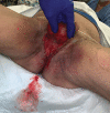
Wound appearance on the day of initial presentation with Fournier’s gangrene infection. The patient was 75 years old with a history of hypertension and hyperlipidemia who presented with 3 days of scrotal pain and swelling. Workup demonstrated a leukocytosis of 12.9, elevated lactic acid at 4.2, hyponatremia to 131, hyperglycemia of 199 with periscrotal subcutaneous gas evident on CT (computed tomographic) pelvis imaging. The patient was taken for emergent surgical debridement after this picture was obtained.
Fig. 2.
Fig. 2.
Testicles within medial thigh subcutaneous pockets following debridement (A). This was a 56-year-old patient who had undergone multiple rounds of excisional debridement by the Urology service and was approximately 2 weeks out from initial presentation. The examiner’s hand demonstrates the buried testicle between their fingers, which was placed in the patient’s right thigh at the time of initial debridement. The white arrow demonstrates the location of the left buried testicle in the medial inner thigh. Testicles were removed from subcutaneous thigh pockets and returned to their (B) anatomic position in preparation for orchiopexy, porcine ECM matrix application, and neoscrotal reconstruction.
Fig. 3.
Fig. 3.
Porcine bladder extracellular matrix powder is applied to the healthy wound bed. Once the majority of infectious tissue burden is debrided, the product is evenly distributed throughout the wound, with attention being paid to filling crevices and depressions. This product has bacteriostatic properties and can be used for treating slightly contaminated wounds without sacrificing product loss. The next step includes application of a 2-layer regenerative matrix sheet, which is then covered with nonstick dressings and placed in negative pressure wound therapy for 1 week.
Fig. 4.
Fig. 4.
A 56-year-old patient with robust granulating wound bed 2 weeks out following orchiopexy and application of porcine bladder ECM product with negative pressure dressing. Wound vacs are left in place for 1 week after product application and then changed bi-weekly on the floor until the wound bed is deemed appropriate for skin grafting.
Fig. 5.
Fig. 5.
Scrotal wound immediately following split-thickness skin graft application, which was harvested from the anterolateral thigh, meshed, and secured with staples. A negative pressure dressing is then applied over the skin graft to serve as a bolster and to optimize skin graft adherence. A nonstick dressing is used as a barrier layer between the skin graft and vac sponge. The wound vac bolster was kept in place for 5 days before takedown.
Fig. 6.
Fig. 6.
Scrotal wound in a 75-year-old patient, at differing time points after split-thickness skin graft reconstruction. A, B, Scrotal wound 1 week after skin graft application, with no evidence of graft loss or nonadherence. C, D, Scrotal wound 3 weeks later, with complete skin graft take and fully healed skin graft. E, F, Scrotal wound 8 months out after neoscrotum reconstruction, with complete healing and Foley catheter removal. This patient did have a reconstructive loss at the penile base resulting in (E) an asymptomatic passageway at the superior aspect of the scrotum. This may be prevented by allowing more time for the wound bed to granulate and by securing the split-thickness skin graft to the penile base with dissolvable suture and staples. Protection and support is afforded thereafter with negative pressure bolster application.
Fig. 7.
Fig. 7.
A healed neoscrotal reconstruction in a 44-year-old patient, 5 months out after split thickness skin grafting. Restored near-anatomic normalcy (A) without evidence of skin graft loss, contracture, or synechiae at the penile base (B, C). The patient was asymptomatic and was back to work at the time of this clinic visit. He denied any negative implication the reconstruction had on his quality of life.

References

    1. Fournier AJ. Gangrene foudroyante de la verge. Semaine Med. 1883;3:345
    1. Sarani B, Strong M, Pascual J, et al. Necrotizing fasciitis: current concepts and review of the literature. J Am Coll Surg. 2009;208:279. - PubMed
    1. Paty R, Smith AD. Gangrene and Fournier’s gangrene. Urol Clin North Am. 1992;19:149–162 - PubMed
    1. Chabak H, Rafik A, Ezzoubi M, et al. Reconstruction of scrotal and perineal defects in Fournier’s gangrene. Modern Plast Surg. 2015;5:23–27
    1. Ferreira PC, Reis JC, Amarante JM, et al. Fournier’s gangrene: a review of 43 reconstructive cases. Plast Reconstr Surg. 2007;119:175–184 - PubMed