Review
. 2020 Aug 28:7:506.
doi: 10.3389/fmed.2020.00506. eCollection 2020.

Purinergic System Signaling in Metainflammation-Associated Osteoarthritis

Affiliations
Review

Purinergic System Signaling in Metainflammation-Associated Osteoarthritis

Paula Gratal et al. Front Med (Lausanne). .

Abstract

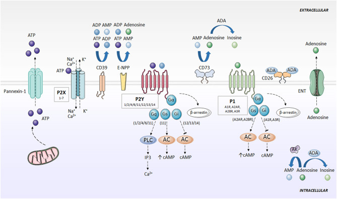
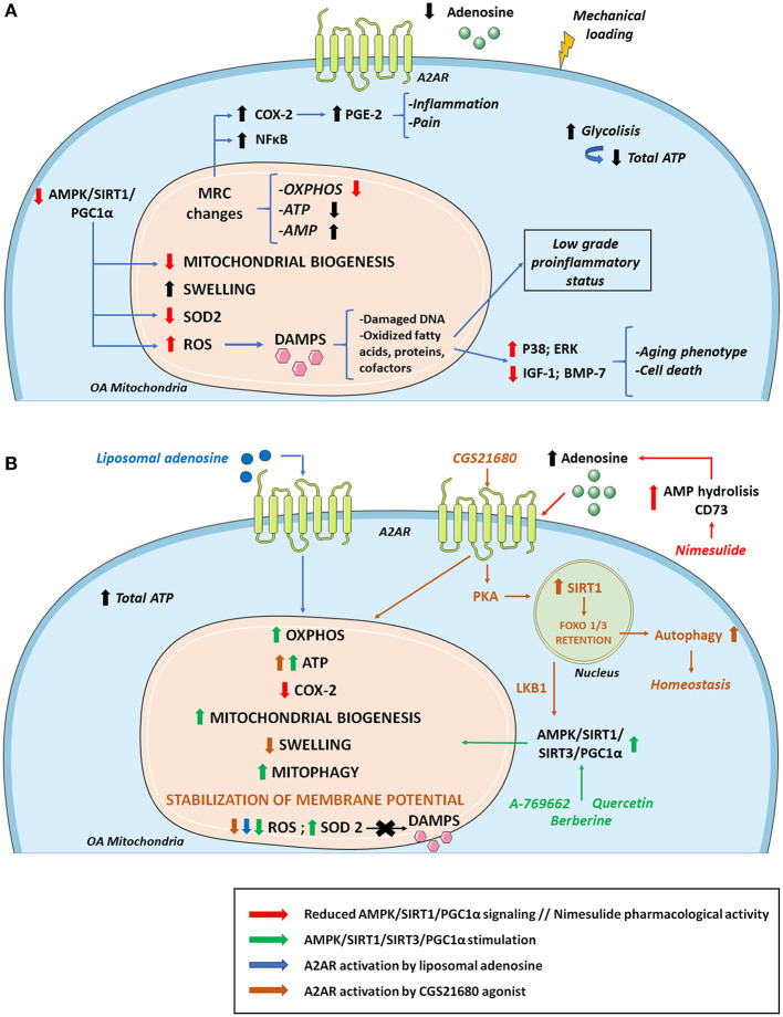
Inflammation triggered by metabolic imbalance, also called metainflammation, is low-grade inflammation caused by the components involved in metabolic syndrome (MetS), including central obesity and impaired glucose tolerance. This phenomenon is mainly due to excess nutrients and energy, and it contributes to the pathogenesis of osteoarthritis (OA). OA is characterized by the progressive degeneration of articular cartilage, which suffers erosion and progressively becomes thinner. Purinergic signaling is involved in several physiological and pathological processes, such as cell proliferation in development and tissue regeneration, neurotransmission and inflammation. Adenosine and ATP receptors, and other members of the signaling pathway, such as AMP-activated protein kinase (AMPK), are involved in obesity, type 2 diabetes (T2D) and OA progression. In this review, we focus on purinergic regulation in osteoarthritic cartilage and how different components of MetS, such as obesity and T2D, modulate the purinergic system in OA. In that regard, we describe the critical role in this disease of receptors, such as adenosine A2A receptor (A2AR) and ATP P2X7 receptor. Finally, we also assess how nucleotides regulate the inflammasome in OA.

Keywords: A2AR; Inflammasome; P2X7 receptor; metainflammation; mitochondrial metabolism; osteoarthritis; purinergic system; rheumatic diseases.


Figures

Figure 1
Figure 1
Purinergic signaling and specific receptors for extracellular nucleotides. ATP is released to the extracellular space mainly through pannexins (e.g., Pannexin-1). Then, ATP is rapidly degraded to ADP, AMP, and adenosine by the action of CD39, E-NPP and CD73. Adenosine can be converted to AMP through AK action, or degraded to inosine by ADA. Adenosine is also directly released outside the cell through ENT. P2X receptors activated by ATP, forming an ion channel for Na+, K+, and Ca2+. P2Y receptors are activated by ATP and ADP, and associate with G proteins, promoting intracellular second messengers' cascades, including Ca2+ and cAMP. Adenosine activates P1 receptors, coupled to G-proteins. A1R and A3R interaction with Gi, and A2AR and A2BR with Gs. P1 and P2Y receptors desensitized through internalization by β-arrestins. AC, adenylate cyclase; ADA, adenosine deaminase; ADP, adenosine diphosphate; AK, adenosine kinase; AMP, adenosine monophosphate; ATP, adenosine 5′-triphosphate; cAMP, cyclic adenosine monophosphate; CD26, dipeptidyl peptidase IV; CD39, ecto-nucleoside triphosphate diphosphohydrolase; CD73, ecto-5′-nucleotidase; E-NPP, ecto-nucleotide pyrophosphatase/phosphodiesterase; ENT, nucleoside transporter; G, G protein; IP3, inositol trisphosphate; P1, adenosine receptors; P2X, ionotropic nucleotide receptors; P2Y, metabotropic nucleotide receptors; PLC, phospholipase C.
Figure 2
Figure 2
Metainflammation in the pathogenesis of osteoarthritis (OA). Mechanical stress contributes, together with the low-grade chronic inflammation associated with aging and the components of the metabolic syndrome (MetS), to the chronic activation of innate immunity in the joint, mainly affecting articular cartilage, during OA progression. Aging-associated factors and senescence together with metabolic factors, such as an increase in adipokines, maintain a systemic low-grade chronic inflammatory status that contributes to the pathogenesis of OA, increasing the expression of cartilage catabolic enzymes (MMPs, ADAMTS), and pro-inflammatory cytokines (IL-1β, IL-6, IL-18, and TNF-α), thus perpetuating inflammation in the joint. ADAMTS, a disintegrin and metalloproteinase with thrombospondin motifs; COL-II, type II collagen; DAMPs, danger-associated molecular patterns; DNA, deoxyribonucleic acid; IL, interleukin; MMP, matrix metalloproteinases; MetS, metabolic syndrome; NF-kB, nuclear factor kappa B; NLRP3, NOD-, LRR- and pyrin domain-containing protein 3; ROS, reactive oxygen species; SASP, senescence-associated secretory phenotype; T2D, type 2 diabetes; TNF, tumor necrosis factor.
Figure 3
Figure 3
Osteoarthritis (OA) mitochondrial metabolism and its modulation by A2AR and AMPK stimulation. (A) During the development of OA, cellular metabolic alterations occur. There is an increase in glycolysis activity to compensate the energy requirement necessary to repair damaged cartilage. However, total ATP levels decrease due to mitochondrial defects together with sustained mechanical loading. Changes in the MRC and its complexes impair ATP and AMP production. As a result, there are lower extracellular levels of ATP and adenosine which diminish A2AR signaling. These alterations in the MRC cause an increase in proinflammatory molecules and transcription factors such as COX-2 and NF-κB, respectively, aggravating inflammation and pain. Reduced AMPK/SIRT1/PGC-1α signaling compromises mitochondrial biogenesis and OXPHOS. There is also a decrease in antioxidant enzymes such as SOD2 that cause increases in ROS production, contributing to the release of DAMPS that aggravate the inflammatory status, stimulating catabolic pathways (p38; ERK) and reducing anabolic pathways (IGF-1; BMP-7), which supposes cell death and an aging phenotype. (B) Mitochondrial OA impairment can be partly reversed by AMPK and A2AR stimulation. Quercetin, berberine and pharmacological AMPK activator A-769662 can activate AMPK/SIRT1/PGC-1α and AMPK/SIRT3 pathways increasing OXPHOS, ATP, mitochondrial biogenesis, mitophagy, and SOD2 activity. AMPK stimulation also promotes a decrease in ROS production with reduced DAMPs secretion diminishing inflammation and aging phenotype. Regarding ROS level reduction, similar effects are found when A2AR is activated by agonists like liposomal adenosine. Moreover, A2AR agonist CGS21680 stimulates SIRT1 via PKA allowing FOXO1/3 retention and increased autophagy. CGS2168 indirectly activates AMPK through LKB1. Nimesulide increases extracellular adenosine levels favoring a reduction in the expression of COX-2 and the associated inflammation and pain. AMP, adenosine monophosphate; AMPK, AMP activated protein kinase; BMP-7, bone morphogenetic protein 7; DAMPS, damage associated molecular patterns; IGF-1, insulin growth factor 1; LKB1, liver kinase B1; MRC, mitochondrial respiratory chain; OXPHOS, oxidative phosphorylation; PGC-1α, peroxisome proliferator-activated receptor-γ coactivator-1α; PKA, protein kinase A; SIRT1, sirtuin 1; SIRT3, sirtuin 3; SOD2, superoxide dismutase.
Figure 4
Figure 4
Purinergic system implication in NLRP3 inflammasome activation. DAMPS or PAMPs present in the microenvironment are recognized by TLR receptors, which in turn, upregulate pro-IL-1β and pro-IL-18 expression through NF-κB activity. BCP and CPPD crystals can be both recognized by TLRs and phagocytized by the cell. Impaired particle or crystal phagocytosis by phagosomes leads to cathepsin B release and mitochondrial dysfunction, which induce elevated ROS production and consequent NLRP3 activation. Additionally, frustrated phagocytosis can lead to massive ATP release through Pannexin-1, activatingP2X7 and causing ionic cytoplasmic imbalance. Excessive K+ efflux, especially through TWIK2, and Na+ and Ca2+ influx through P2X7 activates NLRP3 inflammasome. Activation of different P2Y by ATP or ADP increases intracellular Ca2+ concentration. Particles can also impede induction of purinergic AC-cAMP pathway allowing NLRP3 activity. A2AR activation by extracellular adenosine induces HIF-1α upregulation, which activates NLRP3. AC, adenylate cyclase; AK, adenosine kinase; BCP, basic calcium phosphate; CPPD, calcium pyrophosphate dehydrate; DAMP, danger-associated molecular patterns; ENT2, equilibrative nucleoside transporter 2; GSMD, gasdermin D; IP3, inositol 1,4,5-trisphosphate; NLRP3, NOD-like receptor pyrin domain containing 3; PAMP, pathogen-associated molecular patterns; PKA, protein kinase A; PLC, phospholipase C; ROS, reactive oxygen species; TLR, toll like receptor; TWIK2, two-pore domain weak inwardly rectifying K+ channel 2.

Similar articles

Cited by

References

    1. Burnstock G. Purinergic nerves. Pharmacol Rev. (1972) 24:509–81. - PubMed
    1. Burnstock G. Purinergic receptors. J Theor Biol. (1976) 62:491–503. 10.1016/0022-5193(76)90133-8 - DOI - PubMed
    1. Idzko M, Ferrari D, Eltzschig HK. Nucleotide signalling during inflammation. Nature. (2014) 509:310–7. 10.1038/nature13085 - DOI - PMC - PubMed
    1. Chekeni FB, Elliott MR, Sandilos JK, Walk SF, Kinchen JM, Lazarowski ER, et al. . Pannexin 1 channels mediate “find-me” signal release and membrane permeability during apoptosis. Nature. (2010) 467:863–7. 10.1038/nature09413 - DOI - PMC - PubMed
    1. Eltzschig HK, Eckle T, Mager A, Küper N, Karcher C, Weissmüller T, et al. . ATP release from activated neutrophils occurs via connexin 43 and modulates adenosine-dependent endothelial cell function. Circ Res. (2006) 99:1100–8. 10.1161/01.RES.0000250174.31269.70 - DOI - PubMed

LinkOut - more resources