Retinal findings in patients with COVID-19: Results from the SERPICO-19 study
- PMID: 32984785
- PMCID: PMC7502280
- DOI: 10.1016/j.eclinm.2020.100550
Retinal findings in patients with COVID-19: Results from the SERPICO-19 study
Abstract
Background: Coronavirus disease 2019 (COVID-19) has been associated to microvascular alterations. We screened the fundus of patients with COVID-19 to detect alterations of the retina and its vasculature and to assess possible correlations with clinical parameters.
Methods: Cross-sectional study. The presence of retinal alterations in patients with COVID-19 and subjects unexposed to the virus was assessed using fundus photographs and their prevalence was compared. Mean arteries diameter (MAD) and mean veins diameter (MVD) were compared between patients and unexposed subjects with multiple linear regression including age, sex, ethnicity, body mass index, smoking/alcohol consumption, hypertension, hyperlipidaemia, diabetes as covariates. The influence of clinical/lab parameters on retinal findings was tested in COVID-19 patients.
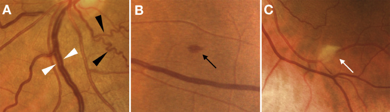
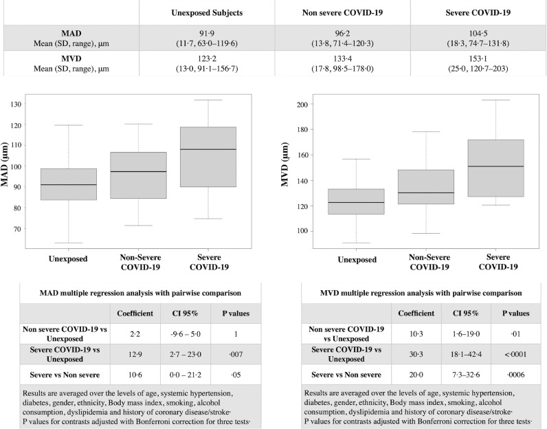
Findings: 54 patients and 133 unexposed subjects were enrolled. Retinal findings in COVID-19 included: haemorrhages (9·25%), cotton wools spots (7·4%), dilated veins (27·7%), tortuous vessels (12·9%). Both MAD and MVD were higher in COVID-19 patients compared to unexposed subjects (98·3 ± 15·3 µm vs 91·9 ± 11·7 µm, p = 0.006 and 138·5 ± 21·5 µm vs 123·2 ± 13·0 µm, p<0.0001, respectively). In multiple regression accounting for covariates MVD was positively associated with COVID-19 both in severe (coefficient 30·3, CI95% 18·1-42·4) and non-severe (coefficient 10·3, CI95% 1·6-19·0) cases compared to unexposed subjects. In COVID-19 patients MVD was negatively correlated with the time from symptoms onset (coefficient -1·0, CI 95% -1·89 to -0·20) and positively correlated with disease severity (coefficient 22·0, CI 95% 5·2-38·9).
Interpretation: COVID-19 can affect the retina. Retinal veins diameter seems directly correlated with the disease severity. Its assessment could have possible applications in the management of COVID-19.
Funding: None.
Keywords: Arteries; COVID-19; Coronavirus; Endothelium; Eye; Retina; SARS-CoV-2; Uveitis; Vasculature; Veins; Vessels.
© 2020 The Authors.
Conflict of interest statement
Dr. Invernizzi reports personal fees from Novartis, personal fees from Bayer, outside the submitted work. Dr. Giacomelli reports personal fees from Mylan, non-financial support from Gilead, outside the submitted work. Dr. Rizzardini reports personal fees and non-financial support from Gilead, personal fees and non-financial support from ViiV, personal fees and non-financial support from MSD, personal fees and non-financial support from AbbVie, outside the submitted work. Dr. Galli reports personal fees and non-financial support from Gilead, personal fees and non-financial support from BMS, personal fees and non-financial support from ViiV, personal fees and non-financial support from MSD, personal fees and non-financial support from AbbVie, personal fees and non-financial support from Janssen, personal fees and non-financial support from Roche, outside the submitted work. Dr. Antinori reports personal fees and non-financial support from Pfizer, personal fees and non-financial support from Merck Sharp & Dome, outside the submitted work. Dr. Staurenghi reports grants and personal fees from Heidelberg Engineering, grants from Optos, other from Ocular Instruments, grants from Optovue, grants from Quantel Medical, grants and personal fees from Centervue, grants from Carl Zeiss Meditec, grants and personal fees from Nidek, personal fees from Apellis, personal fees from Allergan, personal fees from Bayer, personal fees from Boheringer, grants from Topcon, personal fees from Genentech, personal fees from Novartis, personal fees from Roche, personal fees from Chengdu Kanghong Biotechnology Co, personal fees from Kyoto Drug Discovery & Development Co, outside the submitted work. All other authors have nothing to disclose.
Figures