. 2020 Nov;104(3):567-580.
doi: 10.1111/tpj.14994. Epub 2020 Oct 15.

Identification of tannic cell walls at the outer surface of the endosperm upon Arabidopsis seed coat rupture

Affiliations

Identification of tannic cell walls at the outer surface of the endosperm upon Arabidopsis seed coat rupture

Lara Demonsais et al. Plant J. 2020 Nov.

Abstract

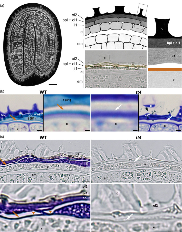
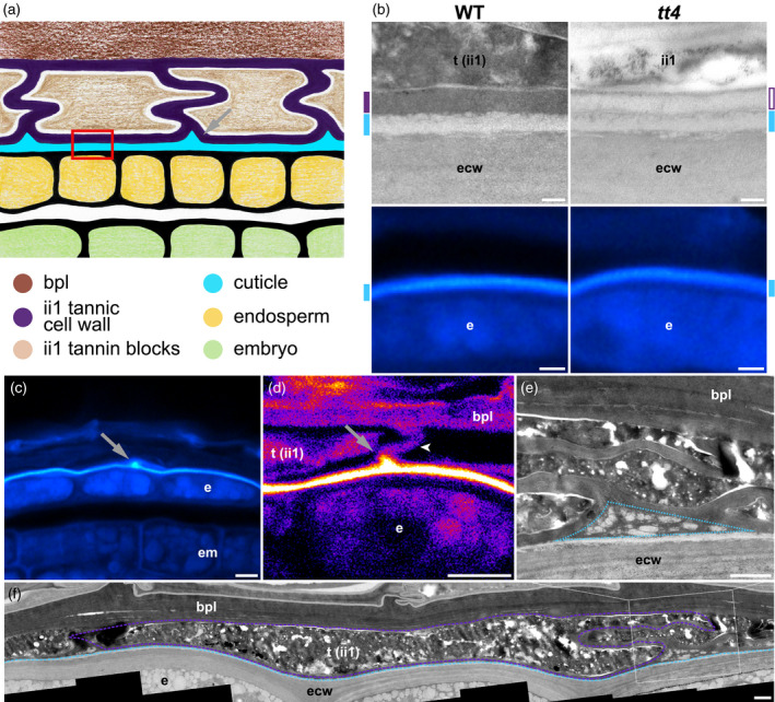
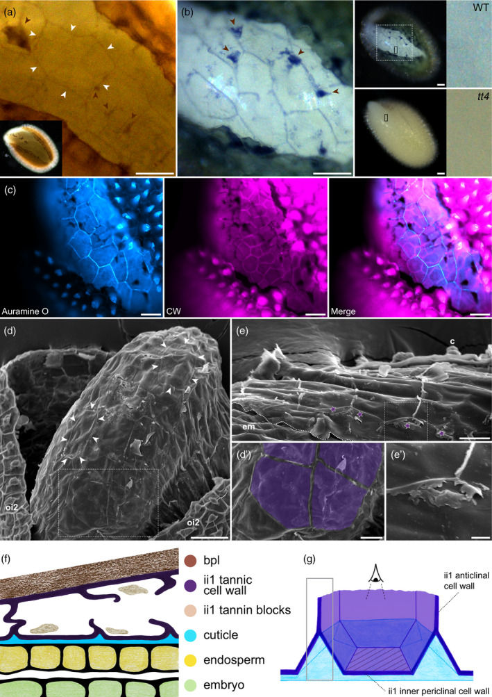
The seed coat is specialized dead tissue protecting the plant embryo from mechanical and oxidative damage. Tannins, a type of flavonoids, are antioxidants known to accumulate in the Arabidopsis seed coat and transparent testa mutant seeds, deficient in flavonoid synthesis, exhibit low viability. However, their precise contribution to seed coat architecture and biophysics remains evasive. A seed coat cuticle, covering the endosperm outer surface and arising from the seed coat inner integument 1 cell layer was, intriguingly, previously shown to be more permeable in transparent testa mutants deficient not in cuticular component synthesis, but rather in flavonoid synthesis. Investigating the role of flavonoids in cuticle permeability led us to identify periclinal inner integument 1 tannic cell walls being attached, together with the cuticle, to the endosperm surface upon seed coat rupture. Hence, inner integument 1 tannic cell walls and the cuticle form two fused layers present at the surface of the exposed endosperm upon seed coat rupture, regulating its permeability. Their potential physiological role during seed germination is discussed.

Keywords: Arabidopsis thaliana; apoplastic barrier; endosperm; seed; seed coat; tannic cell wall.


Conflict of interest statement

The authors declare that they have no competing interests.

Figures

Figure 1
Figure 1
Presence of tannins at the outer surface of the endosperm. (a) Left, semi‐thin section of a wild‐type (WT) seed (autofluorescence). Center top, schematic drawing illustrating the organization of the seed peripheral area (corresponding to the white‐boxed region in the left panel). Center bottom, micrograph of an unstained semi‐thin section showing the seed peripheral area; ii1, the bpl, oi1 and oi2 form the seed coat. Right panel, schematic drawing of an enlarged seed coat area (gray‐boxed region in the center top panel), the brown line indicates the localization of tannins along the endosperm outer surface (arrows in b,c, and in Figure S1a,c). Drawings were not made to scale to visualize the positions of the different cells and structures of interest better. (b) Semi‐thin sections of WT seeds (left panels) and transparent testa (tt)4 seeds (right panels) stained with toluidine blue; in each case, a global view of the seed coat and an enlargement of the region around the arrow is shown. (c) Semi‐thin sections of WT seeds (left panels) and tt4 seeds (right panels) stained with 4‐dimethylaminocinnamaldehyde; in each case, a global view of the seed coat and an enlargement of the region around the arrow is shown below. Brown arrowheads in bottom left panel indicate the reticulated structure formed by ii1 tannic cell walls. In (b) and (c), brown arrows (left panels) indicate the detection of tannins along the endosperm outer cell wall; white arrows (right panels) indicate no detection of tannins along this cell wall. Scale bars = (a) 50 μm (left) and 5 μm (center bottom); (b) 5 and 1 μm for general views and magnified insets, respectively; (c) 5 μm. bpl, brown pigment layer; c, columella; e, endosperm; em, embryo; ii1, inner integument 1; oi1, outer integument 1; oi2, outer integument 2; t, tannin blocks.
Figure 2
Figure 2
ii1 cell walls are tannified and form a continuous reticulated tannic apoplastic barrier tightly bound to the endosperm‐associated cuticle. (a) Schematic drawing illustrating the organization of the endosperm outer surface area. The drawing is not made to scale to visualize the positions of the different structures of interest better. The red‐boxed region indicates the surface of the endosperm where the cuticle and inner tannified cell wall are tightly associated. The gray arrow indicates the location of cuticular accumulation. (b) Transmission electron microscopy (TEM) micrographs of the endosperm outer surface region (top row) and paraffin sections stained with Auramine O (bottom row) in wild‐type (WT) and tt4 mutants, corresponding to the red‐boxed region shown in (a). Cyan marks indicate the position of the cuticle and purple marks indicate the position of the ii1 inner periclinal cell wall (filled in purple and in white when it is electron‐dense and electron‐lucent, respectively). (c) Auramine O staining of a WT seed paraffin section; the arrow indicates a local signal accumulation corresponding to the region indicated by the gray arrow in (a). (d) Confocal micrograph of a WT seed paraffin section stained with Auramine O. The arrow indicates a local signal accumulation corresponding to the region indicated by the gray arrow in (a); the arrowhead indicates an anticlinal cell wall between two adjacent ii1 cells. (e) TEM micrograph of a junction between ii1 cells in a WT seed (enlargement of the white‐boxed region in f). The dashed blue triangle indicates the region where droplet‐like electron‐lucent material accumulates, between neighboring ii1 cells and the cuticle. (f) TEM micrograph presenting a global view of the WT seed inner integuments. The purple dashed line indicates the cell wall of one ii1 cell (electron‐dense material) and the light blue dashed line indicates the cuticle (electron‐lucent material). The original picture is in Figure S5a. Scale bars = (b) 200 nm and 2 μm, top and bottom rows, respectively; (c,d) 5 μm; (e,f) 1 μm. bpl, brown pigment layer; e, endosperm; ecw, endosperm cell wall; em, embryo; ii1, inner integument 1; t, tannin blocks.
Figure 3
Figure 3
The ii1 inner periclinal tannic cell wall remains attached to the endosperm after seed coat rupture. (a) Unstained seed coat‐ruptured wild‐type (WT) seed showing a polygonal pattern present at the outer surface of the endosperm. White arrowheads delineate a particular polygonal contour. (b) 4‐Dimethylaminocinnamaldehyde staining of seed coat‐ruptured WT and tt4 seeds. The left panel is an enlargement of the dashed‐boxed area shown in the WT seed picture of the middle panel. The right panels are enlargements of the small black boxes shown in the middle panel. In (a,b) brown arrowheads indicate remnants of ii1 tannin blocks (see Discussion). (c) Seed coat‐ruptured WT seed co‐stained with Auramine O and CW. (d,e) Scanning electron microscopy micrographs of seed coat‐ruptured (d) and endosperm‐ruptured (e) WT seeds. White arrowheads indicate the visible remnants of ii1 cell walls. (d′) Enlargement of the boxed region in (d), where ii1 inner periclinal wall surfaces were colorized in purple. (e′) Enlargement of the boxed region in (e), where remnants of ruptured ii1 anticlinal cell walls are visible (indicated by purple stars). Note that scanning electron microscopy was performed on fresh (non‐fixed) material; because of the microscope vacuum, ruptured seed coats would close themselves, hiding zones of interest (see Figure S8b for an example); to remedy this, we had to increase the aperture of seed coat‐ruptured seeds manually, thus uncovering a wider space than that corresponding to a normal seed coat‐ruptured seed. (f) Schematic drawing of seed coat organization during seed coat rupture (outer integuments are not represented). (g) Schematic drawing in 3D of one ii1 cell during seed coat rupture. The gray‐boxed region corresponds to the signal given by DMACA (b) and Auramine O (c) along broken anticlinal ii1 walls. The hatched area indicates the ii1 inner periclinal cell wall surface, which becomes the first barrier facing the environment during seed coat rupture. Drawings (f,g) were not made to scale to better visualize the structures of interest. Scale bars = (a–d), 50 μm; (d′,e) 20 μm; (e′) 5 μm. c, columella; em, embryo; ii1, inner integument 1; oi2, outer integument 2

Comment in

References

    1. Abrahams, S. , Tanner, G.J. , Larkin, P.J. and Ashton, A.R. (2002) Identification and biochemical characterization of mutants in the Proanthocyanidin pathway in Arabidopsis. Plant Physiol. 130, 561–576. - PMC - PubMed
    1. Ancillotto, L. , Sozio, G. and Mortelliti, A. (2015) Acorns were good until tannins were found: Factors affecting seed‐selection in the hazel dormouse (Muscardinus avellanarius). Mamm Biol. 80, 135–140.
    1. Andème Ondzighi, C. , Christopher, D.A. , Cho, E.J. , Chang, S.‐C. and Staehelin, L.A. (2008) Arabidopsis protein disulfide isomerase‐5 inhibits cysteine proteases during trafficking to vacuoles before programmed cell death of the endothelium in developing seeds. Plant Cell, 20, 2205–2220. - PMC - PubMed
    1. Auger, B. , Marnet, N. , Gautier, V. , Maia‐Grondard, A. , Leprince, F. , Renard, M. , Guyot, S. , Nesi, N. and Routaboul, J.‐M. (2010) A Detailed survey of seed coat flavonoids in developing seeds of Brassica napus L. J. Agric. Food Chem. 58, 6246–6256. - PubMed
    1. Beeckman, T. , De Rycke, R. , Viane, R. and Inzé, D. (2000) Histological study of seed coat development in Arabidopsis thaliana. J. Plant Res. 113, 139–148.

Publication types

MeSH terms

Substances