

[Early differential diagnosis between COVID-19 and mycoplasma pneumonia with chest CT scan]
- PMID: 32985160
- PMCID: PMC8800730
- DOI: 10.3785/j.issn.1008-9292.2020.07.04
[Early differential diagnosis between COVID-19 and mycoplasma pneumonia with chest CT scan]
Abstract
Objective: To early differentiate between coronavirus disease 2019 (COVID-19) and adult mycoplasma pneumonia with chest CT scan.
Methods: Twenty-six patients with COVID-19 and 21 patients with adult mycoplasma pneumonia confirmed with RT-PCR test were enrolled from Zibo First Hospital and Lanshan People's Hospital during December 1st 2019 and March 14th 2020. The early chest CT manifestations were analyzed and compared between the two groups.
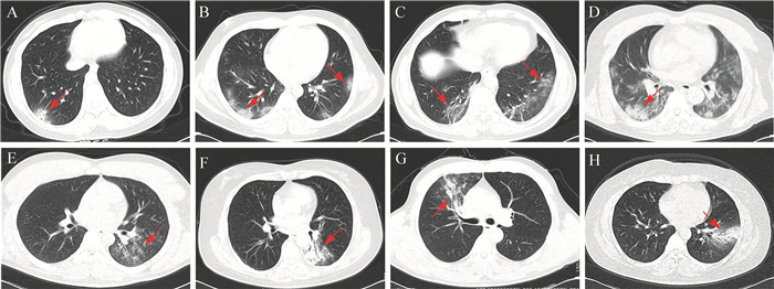
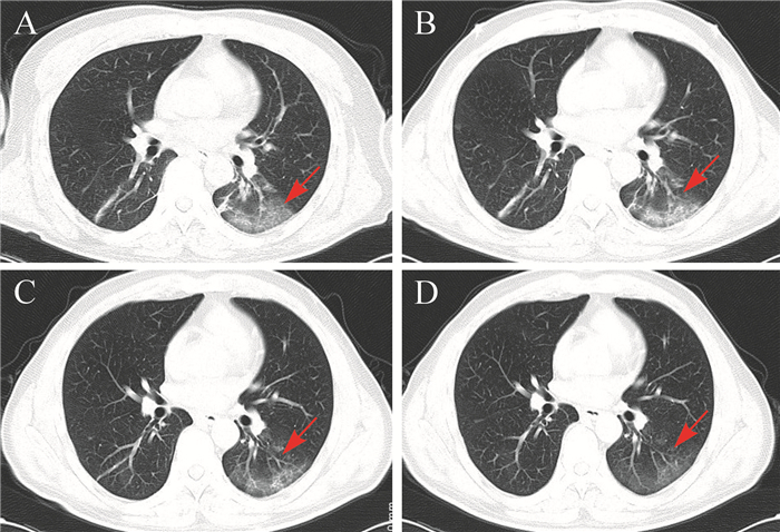
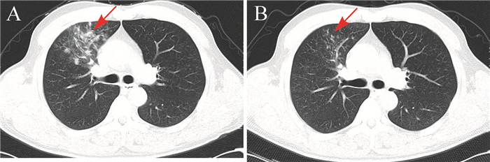
Results: The interstitial changes with ground glass density shadow (GGO) were similar in two groups during first chest CT examination (P>0.05). There were more lung lobes involved on the first chest CT in COVID-19 patients, which were mostly distributed in the dorsal outer zone (23/26, 88.5%), and nearly half of them (12/26, 46.2%) were accompanied by crazy-paving sign; while the lesions in adult mycoplasma pneumonia patients were mostly distributed along the bronchi, and the bronchial wall was thickened (19/21, 90.5%), accompanied with tree buds / fog signs (19/21, 90.5%). The above CT signs were significantly different between the two kinds of pneumonia (all P<0.01). COVID-19 had a longer course compared with mycoplasma pneumonia, the disease peaks of COVID-19 patients was on day (10.5±3.8), while the disease on CT was almost absorbed on day (7.9±2.2) in adult mycoplasma pneumonia. The length of hospital stay in COVID-19 patients was significantly longer than that of mycoplasma pneumonia patients [(19.5±4.3) d vs (7.9±2.2) d, P<0.01].
Conclusions: The lesions of adult mycoplasma pneumonia are mostly distributed along the bronchi with tree buds/fog signs, while the lesions of COVID-19 are mainly distributed in the dorsal outer zone accompanied by crazy-paving sign, which can early distinguish two diseases.
目的: 探讨胸部CT在2019冠状病毒病(COVID-19)与支原体肺炎患者早期鉴别诊断中的价值。
方法: 回顾性分析2019年12月1日至2020年3月14日山东省淄博市第一医院和山东省临沂市兰山区人民医院收治的26例成人COVID-19患者和21例成人支原体肺炎患者,比较两组早期胸部CT表现,并通过动态CT纵向评估疾病的转归。
结果: COVID-19患者和支原体肺炎患者首次胸部CT检查示胸部病变均表现为磨玻璃影的间质性改变( P>0.05)。COVID-19患者累及肺叶数较多,且多分布于背侧外带(23/26,88.5%),46.2%(12/26)的患者伴有铺路石征;支原体肺炎患者累及肺叶数较少,多沿支气管分布,90.5%(19/21)的患者伴有支气管管壁增厚征象,周围多伴有树芽/雾征(19/21,90.5%),两组在病灶分布和影像学征象上差异有统计学意义(均 P < 0.01)。COVID-19患者病程较长,平均(10.5±3.8)d达到峰值,而支原体肺炎患者(7.9±2.2)d病变明显好转。COVID-19组和支原体肺炎组住院时长分别为(19.5±4.3)d和(7.9±2.2)d,差异有统计学意义( P < 0.01)。
结论: COVID-19患者病灶多分布于外围背侧,常伴有铺路石征,而支原体肺炎患者病灶多沿支气管分布,支气管管壁增厚并伴有树芽/雾征,这些早期影像学征象有利于两种疾病的鉴别诊断。
Keywords: Computed tomography; Coronavirus disease 2019; Crazy-paving sign; Mycoplasma pneumoniae; Tree-bud sign.
Figures
Similar articles
-
[Dandelion clock-like sign on CT for diagnose of COVID-19].Nan Fang Yi Ke Da Xue Xue Bao. 2020 Feb 29;40(2):159-163. doi: 10.12122/j.issn.1673-4254.2020.02.03. Nan Fang Yi Ke Da Xue Xue Bao. 2020. PMID: 32376543 Free PMC article. Chinese.
-
Differential diagnosis between the coronavirus disease 2019 and Streptococcus pneumoniae pneumonia by thin-slice CT features.Clin Imaging. 2021 Jan;69:318-323. doi: 10.1016/j.clinimag.2020.09.012. Epub 2020 Oct 6. Clin Imaging. 2021. PMID: 33045476 Free PMC article.
-
Imaging and clinical features of patients with 2019 novel coronavirus SARS-CoV-2.Eur J Nucl Med Mol Imaging. 2020 May;47(5):1275-1280. doi: 10.1007/s00259-020-04735-9. Epub 2020 Feb 28. Eur J Nucl Med Mol Imaging. 2020. PMID: 32107577 Free PMC article.
-
Similarities and Differences of Early Pulmonary CT Features of Pneumonia Caused by SARS-CoV-2, SARS-CoV and MERS-CoV: Comparison Based on a Systemic Review.Chin Med Sci J. 2020 Sep 30;35(3):254-261. doi: 10.24920/003727. Chin Med Sci J. 2020. PMID: 32972503 Free PMC article.
-
CT Manifestations and Clinical Characteristics of 1115 Patients with Coronavirus Disease 2019 (COVID-19): A Systematic Review and Meta-analysis.Acad Radiol. 2020 Jul;27(7):910-921. doi: 10.1016/j.acra.2020.04.033. Epub 2020 May 5. Acad Radiol. 2020. PMID: 32505599 Free PMC article.
Cited by
-
Clinical Features of Long COVID Patients Coinfected With Mycoplasma pneumoniae.Can J Infect Dis Med Microbiol. 2024 Dec 7;2024:7213129. doi: 10.1155/cjid/7213129. eCollection 2024. Can J Infect Dis Med Microbiol. 2024. PMID: 39679212 Free PMC article.
-
Application of CT images based on the optimal atlas segmentation algorithm in the clinical diagnosis of Mycoplasma Pneumoniae Pneumonia in Children.Pak J Med Sci. 2021;37(6):1647-1651. doi: 10.12669/pjms.37.6-WIT.4860. Pak J Med Sci. 2021. PMID: 34712299 Free PMC article.
-
Distinguishing types and severity of pediatric pneumonia using modified lung ultrasound score.Front Pediatr. 2024 Aug 5;12:1411365. doi: 10.3389/fped.2024.1411365. eCollection 2024. Front Pediatr. 2024. PMID: 39161635 Free PMC article.
-
Imaging of Pulmonary Superinfections and Co-Infections in COVID-19.Curr Probl Diagn Radiol. 2022 Sep-Oct;51(5):768-778. doi: 10.1067/j.cpradiol.2021.09.009. Epub 2021 Nov 11. Curr Probl Diagn Radiol. 2022. PMID: 34903396 Free PMC article. Review.
References
-
- XIE X, ZHONG Z, ZHAO W, et al. Chest CT for typical 2019-nCoV pneumonia: relationship to negative RT-PCR testing. Radiology. 2020:200343. doi: 10.1148/radiol.2020200343. [XIE X, ZHONG Z, ZHAO W, et al. Chest CT for typical 2019-nCoV pneumonia: relationship to negative RT-PCR testing[J]. Radiology, 2020:200343. DOI:10.1148/radiol.2020200343. ] - DOI - PMC - PubMed
-
- FANG Y, ZHANG H, XIE J, et al. Sensitivity of chest CT for COVID-19: comparison to RT-PCR. Radiology. 2020:200432. doi: 10.1148/radiol.2020200432. [FANG Y, ZHANG H, XIE J, et al. Sensitivity of chest CT for COVID-19: comparison to RT-PCR[J]. Radiology, 2020:200432. DOI:10.1148/radiol.2020 200432. ] - DOI - PMC - PubMed
-
- HUANG G, GONG T, WANG G, et al. Timely diagnosis and treatment shortens the time to resolution of coronavirus disease (COVID-19) pneumonia and lowers the highest and last CT scores from sequential chest CT. AJR Am J Roentgenol. 2020:1–7. doi: 10.2214/AJR.20.23078. [HUANG G, GONG T, WANG G, et al. Timely diagnosis and treatment shortens the time to resolution of coronavirus disease (COVID-19) pneumonia and lowers the highest and last CT scores from sequential chest CT[J]. AJR Am J Roentgenol, 2020:1-7. DOI:10.2214/AJR.20.23078. ] - DOI - PubMed
-
- ZHOU Z, GUO D, LI C, et al. Coronavirus disease 2019: initial chest CT findings. Eur Radiol. 2020;30:4398–4406. doi: 10.1007/s00330-020-06816-7. [ZHOU Z, GUO D, LI C, et al. Coronavirus disease 2019: initial chest CT findings[J]. Eur Radiol, 2020, 30:4398-4406. DOI:10.1007/s00330-020-06816-7. ] - DOI - PMC - PubMed
-
- XU X, YU C, QU J, et al. Imaging and clinical features of patients with 2019 novel coronavirus SARS-CoV-2. Eur J Nucl Med Mol Imaging. 2020;47(5):1275–1280. doi: 10.1007/s00259-020-04735-9. [XU X, YU C, QU J, et al. Imaging and clinical features of patients with 2019 novel coronavirus SARS-CoV-2[J]. Eur J Nucl Med Mol Imaging, 2020, 47 (5):1275-1280. DOI:10.1007/s00259-020-04735-9. ] - DOI - PMC - PubMed
