. 2020 Sep 28;11(1):4885.
doi: 10.1038/s41467-020-18689-x.

CLR01 protects dopaminergic neurons in vitro and in mouse models of Parkinson's disease

Affiliations

CLR01 protects dopaminergic neurons in vitro and in mouse models of Parkinson's disease

Nora Bengoa-Vergniory et al. Nat Commun. .

Abstract

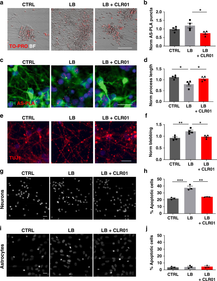
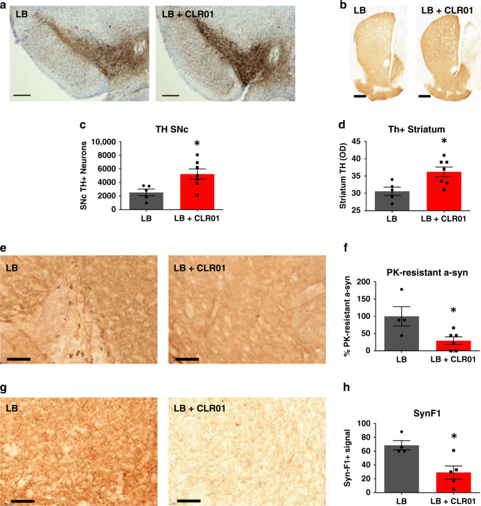
Parkinson's disease (PD) affects millions of patients worldwide and is characterized by alpha-synuclein aggregation in dopamine neurons. Molecular tweezers have shown high potential as anti-aggregation agents targeting positively charged residues of proteins undergoing amyloidogenic processes. Here we report that the molecular tweezer CLR01 decreased aggregation and toxicity in induced pluripotent stem cell-derived dopaminergic cultures treated with PD brain protein extracts. In microfluidic devices CLR01 reduced alpha-synuclein aggregation in cell somas when axonal terminals were exposed to alpha-synuclein oligomers. We then tested CLR01 in vivo in a humanized alpha-synuclein overexpressing mouse model; mice treated at 12 months of age when motor defects are mild exhibited an improvement in motor defects and a decreased oligomeric alpha-synuclein burden. Finally, CLR01 reduced alpha-synuclein-associated pathology in mice injected with alpha-synuclein aggregates into the striatum or substantia nigra. Taken together, these results highlight CLR01 as a disease-modifying therapy for PD and support further clinical investigation.


Conflict of interest statement

The chemical composition of CLR01 is protected by International Patent No. PCT/US2010/026419, USA patent No. 8,791,092, and European patent No. EP2403859 A2.

Figures

Fig. 1
Fig. 1. CLR01 reduces α-syn aggregation and toxicity in iPSC-derived dopaminergic and primary rat cortical cultures.
a, c, e Representative brightfield and immunofluorescent images of live and dead cells (labeled by To-pro), dopaminergic cells (TH-positive), AS-PLA (red puncta), and neuronal processes of four independent control cell lines analyzed. Cells were treated with LB extracts, which had been pretreated with CLR01 or PBS as a negative control. LB treatment caused an abnormal deposition of material in treated wells, accompanied by increased AS-PLA signal process degeneration, which were reduced by CLR01 pre-treatment. Scale bars = 100, 25, 25 µm, respectively. b, d, f Quantification of indicated parameters (from c and e) of four independent control cell lines analyzed using a one-way ANOVA (Sidak’s). b F(2, 9) = 5.154, p = 0.0213. d F(2, 9) = 7.309, p = 0.0112, p = 0.0356. f F(2, 9) = 10.67, p = 0.0038, p = 0.0126. g, i Representative images of neuronal (top) and astrocytic (bottom) primary rat cultures stained with DAPI (n = 3 independent cultures and treatments). Scale bars 20 µm. h, j Quantification of apoptotic cells (expressed as % from total) analyzed using a one-way ANOVA (Sidak’s, n = 3 independent cultures and treatments). k F(2, 6) = 35.09, p = 0.0005, p = 0.0012. *p < 0.05, **p < 0.01, ***p < 0.001. For all appropriate panels, data are presented as mean values ± SEM. BF brightfield, Norm normalized.
Fig. 2
Fig. 2. CLR01 prevents α-syn aggregation in microfluidic chambers.
a, c Schematic representation of the experimental design for the microfluidic-chamber experiments. b, d Representative immunofluorescence images of dopaminergic neurons grown in the microfluidic devices insulted with oligomers and/or treated with CLR01 for 5 days (25 µg/ml and/or 10 µM, respectively). Images were taken at least one field of view away from the microgrooves to avoid artifacts due to contact with the silicone. Scale bars = 25 µm. Quantification of normalized puncta per field of three independent control cell lines analyzed using a one-way ANOVA (Tukey). Oligo: oligomer. b F(2, 6) = 24.31, p = 0.0009 and F(2, 6) = 39.09, p = 0.0005, p = 0.0006. d F(2, 6) = 71.45. *p < 0.05, **p < 0.01, ***p < 0.001. For all appropriate panels, data are presented as mean values ± SEM.
Fig. 3
Fig. 3. Glial activation and α-syn aggregation in SNCA-OVX mice.
a, c, e Representative images of immunofluorescence of GFAP (astrocytes), IBA1 (microglia), and AS-PLA in dopaminergic neurons, respectively. Scale bars = 200, 25, and 25 µm, respectively. b, d, f Quantification of normalized stained area, morphology (expressed as a % from total), and puncta per TH+ cell analyzed using a two-way ANOVA (Dunnett’s for age and Sidak’s for genotype, for age all values compared to 3 months), n = 3–4 animals per group. b F(3, 20) = 1.211, p = 0.0177 for age and F(1, 20) = 0.1572, p = 0.0106 for genotype. d F(1, 22) = 2.216, p = 0.0287 for genotype and F(3, 22) = 1.764, p = 0.0209, p = 0.0104, p = 0.0192 for age. f F(3, 21) = 4.528 for age, p = 0.0058, p = 0.0016, p = 0.0024 and F(1, 21) = 33.41, p = 0.0043, p = 0.0015, p = 0.0061 for genotype. *p < 0.05. For all appropriate panels, data are presented as mean values ± SEM. OVX: SNCA-OVX.
Fig. 4
Fig. 4. CLR01 restores motor behavior and reduces early pathology in vivo at 12 months of age.
ad Behavior analysis of rotarod (a) and catwalk gait analysis (bd) after 2 months of subcutaneous administration of 40 µg/kg/day drug or PBS analyzed using a one-way ANOVA (Sidak), n = 11–14 animals per group. e, f Representative images and quantification of AS-PLA puncta per TH+ cell. Scale bars = 25 µm. gj Representative images and quantification of GFAP-stained area per field and microglial morphology (expressed as a % from total). Scale bars = 100 and 50 µm, respectively. aj were analyzed using one-way ANOVA (Holm–Sidak), ej n = 8/8. CLR: CLR01 *p < 0.05, **p < 0.01. a F(2, 35) = 4.138, p = 0.0376, p = 0.0205. b F(2, 37) = 4.000, p = 0.0058. c F(2, 38) = 4.884, p = 0.0078, p = 0.0451. d F(2, 38) = 3.982, p = 0.0151. f F(2, 19) = 6.057, p = 0.0072, p = 0.0338. h F(2, 21) = 8.797, p = 0.0091, p = 0.0012. j F(2, 23) = 4.750, p = 0.0106. For all appropriate panels, data are presented as mean values ± SEM.
Fig. 5
Fig. 5. CLR01 reduces α-syn aggregation in vivo at 18 months of age.
ae Representative images and quantification of AS-PLA puncta per TH+, GFAP+, and Iba1+ cells after osmotic mini-pump implantation of 40 µg/kg/days CLR01 or PBS, analyzed using a two-tailed t-test, n = 3/4 independent animals. Scale bars = 50 µm. c p = 0.0117, d p = 0.0319, e p = 0.0179. f, g Western blottings of PGC1α and Lamp2a, analyzed using a one-tailed Mann–Whitney U-test, n = 3–4 independent animals. f p = 0.0286, g p = 0.0286. h, i Native western blottings of Triton X-100 and SDS-soluble α-syn, analyzed using a one-tailed Mann–Whitney U-test, n = 3–4 independent animals. i p = 0.0286. *p < 0.05. AV average, CLR CLR01. For all appropriate panels, data are presented as mean values ± SEM.
Fig. 6
Fig. 6. CLR01 protects dopaminergic neurons from LB-induced cell death through reduction of α-syn aggregation in mice.
a, b Representative photomicrographs of TH-immunostained SNc and striatum (respectively) in LB-inoculated mice after osmotic mini-pump implantation for CLR01 (or PBS) delivery (40 µg/kg/h). c Stereological cell counts of SNc TH-immunoreactive neurons (a) in LB-inoculated mice, at 3 months post-LB inoculation. p = 0.0177. d Optical densitometry of striatal TH immunoreactivity in LB-inoculated mice (b), at 3 months post-LB inoculation. p = 0.0354. e, g Representative photomicrographs of α-syn and PK-resistant α-syn immunostained SN from LB-inoculated mice, and corresponding quantification. f, h Representative images of Syn-F1 immunostaining in the SN in LB-inoculated mice and corresponding quantification of the intensity levels of Syn-F1 staining. f p = 0.0333, h p = 0.0556. ah n = 4–5 for LB-inoculated mice, n = 5–7 for LB + CLR01-independently treated animals, analyzed with a two-tailed Mann–Whitney U-test. *p < 0.05. Scale bars = 0.1 mm (SN) and 1 mm (Str). For all appropriate panels, data are presented as mean values ± SEM.

References

    1. Obeso JA, et al. Past, present, and future of Parkinson’s disease: a special essay on the 200th Anniversary of the Shaking Palsy. Mov. Disord. 2017;32:1264–1310. - PMC - PubMed
    1. Braak H, et al. Staging of brain pathology related to sporadic Parkinson’s disease. Neurobiol. Aging. 2003;24:197–211. - PubMed
    1. Bengoa-Vergniory N, Roberts RF, Wade-Martins R, Alegre-Abarrategui J. Alpha-synuclein oligomers: a new hope. Acta Neuropathol. 2017;134:819–838. - PMC - PubMed
    1. Attar, A. & Bitan, G. Disrupting self-assembly and toxicity of amyloidogenic protein oligomers by “molecular tweezers”-from the test tube to animal models. Curr. Pharm. Des. 20, 2469–2483 (2014). - PMC - PubMed
    1. Schrader T, Bitan G, Klärner FG. Molecular tweezers for lysine and arginine-powerful inhibitors of pathologic protein aggregation. Chem. Commun. 2016;52:11318–11334. - PMC - PubMed

Publication types

MeSH terms