Review
. 2020 Oct 30;48(5):1843-1858.
doi: 10.1042/BST20190333.

Human 2-oxoglutarate-dependent oxygenases: nutrient sensors, stress responders, and disease mediators

Affiliations
Review

Human 2-oxoglutarate-dependent oxygenases: nutrient sensors, stress responders, and disease mediators

Sally C Fletcher et al. Biochem Soc Trans. .

Abstract

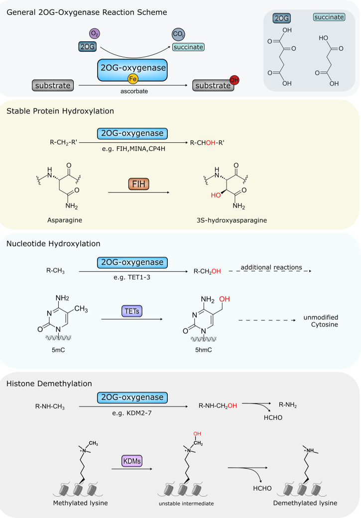
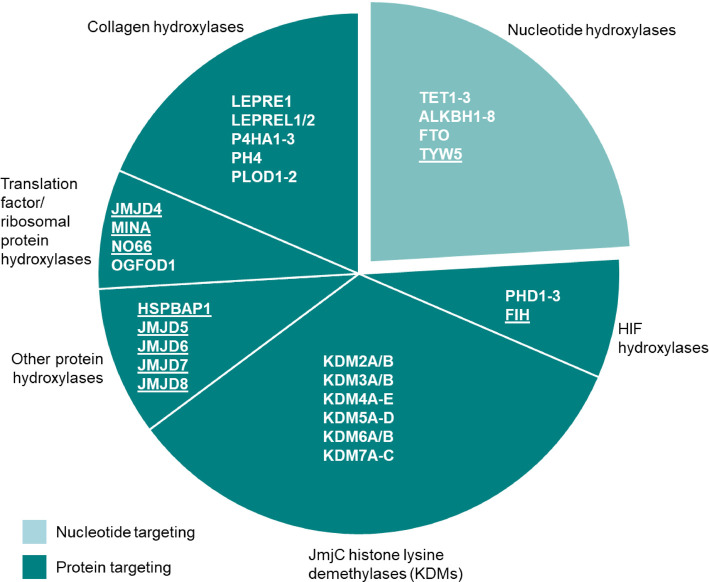
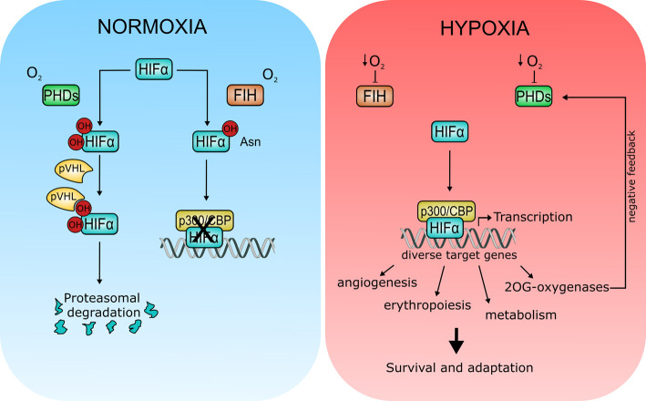
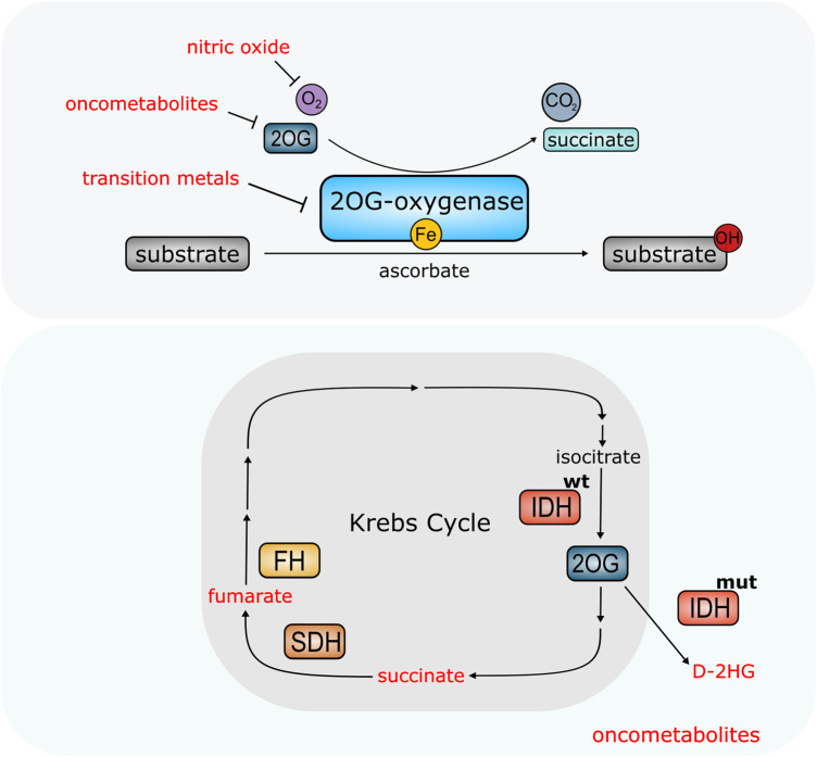
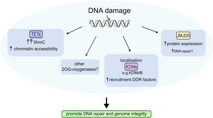
Fe(II)/2-oxoglutarate (2OG)-dependent oxygenases are a conserved enzyme class that catalyse diverse oxidative reactions across nature. In humans, these enzymes hydroxylate a broad range of biological substrates including DNA, RNA, proteins and some metabolic intermediates. Correspondingly, members of the 2OG-dependent oxygenase superfamily have been linked to fundamental biological processes, and found dysregulated in numerous human diseases. Such findings have stimulated efforts to understand both the biochemical activities and cellular functions of these enzymes, as many have been poorly studied. In this review, we focus on human 2OG-dependent oxygenases catalysing the hydroxylation of protein and polynucleotide substrates. We discuss their modulation by changes in the cellular microenvironment, particularly with respect to oxygen, iron, 2OG and the effects of oncometabolites. We also describe emerging evidence that these enzymes are responsive to cellular stresses including hypoxia and DNA damage. Moreover, we examine how dysregulation of 2OG-dependent oxygenases is associated with human disease, and the apparent paradoxical role for some of these enzymes during cancer development. Finally, we discuss some of the challenges associated with assigning biochemical activities and cellular functions to 2OG-dependent oxygenases.

Keywords: disease; hydroxylation; hypoxia; nutrient sensing; oxygenase; post translational modification.


Conflict of interest statement

The authors declare that there are no competing interests associated with the manuscript.

Figures

Figure 1.
Figure 1.. Major oxidative modifications catalysed by Human 2OG-oxygenases.
Top panel: Overview of 2OG-oxygenase reaction. Substrates are hydroxylated in a reaction requiring oxygen, 2OG and iron as cofactors. Succinate and CO2 are released as by-products in addition to the hydroxylated substrate. Catalytic centre of 2OG-oxygenases contains bound iron. Some 2OG-oxygenases also require ascorbate for optimal activity. Chemical structures of 2OG and succinate are shown inset. Second panel: Stable protein hydroxylation. For clarity only FIH-mediated asparagine hydroxylation is shown. However, hydroxylation of arginine, aspartate, histidine, lysine and proline residues is carried out by other 2OG-oxygenases. Third panel: nucleotide hydroxylation mediated by TET enzymes. TETs hydroxylate 5mC (5-methylcytosine) at the methyl group forming 5hmC (5-hydroxymethylcytosine). This is the first stage in a multi-step oxidation to facilitate removal of 5mC modification in DNA. Bottom panel: 2OG-oxygenase-mediated histone demethylation via hydroxylation creates an unstable intermediate that spontaneously decomposes into the unmethylated form, also releasing formaldehyde (HCHO). Only demethylation of a mono-methylated lysine residue is shown. However, 2OG oxygenase-mediated demethylation also occurs at di- and tri-methylated residues. The requirement for oxygen, 2OG and iron, as well as the CO2 and succinate by-products are omitted for simplicity in panels 2–4: see top panel for details.
Figure 2.
Figure 2.. Functional grouping of 2OG-oxygenases.
Nucleotide hydroxylases are shown in the light turquoise segment. These include enzymes targeting DNA (e.g. TET1–3) and RNA (e.g. TYW5). Protein-targeting enzymes are shown in the dark turquoise segments. Underlining indicates 2OG-oxygenase family members that can be phylogenetically classified as JmjC-only hydroxylases. NB: Not all members of the 2OG-oxygenase family are shown here (small molecule oxygenases have been omitted).
Figure 3.
Figure 3.. Regulation of hypoxia signalling by 2OG-oxygenases.
During normoxia (left panel), prolyl hydroxylases (PHD1–3) hydroxylate conserved proline residues in the HIFα subunit creating a recognition motif for the von Hippel–Lindau (pVHL) E3 ubiquitin ligase. Hydroxylation leads to HIFα degradation via the proteasome. HIFα is also hydroxylated at a conserved asparagine residue by FIH. Hydroxylation at this site impairs HIF transactivation activity by inhibiting binding to transcriptional coactivator p300/CBP. Note that these two hydroxylation pathways are not necessarily mutually exclusive. For clarity, the requirement for 2OG and iron, as well as the CO2 and succinate by-products are omitted in the PHD and FIH reaction scheme (See Figure 1, Panel 1 for general reaction scheme). Under hypoxia (right panel), inhibition of PHDs and FIH leads to HIFα stabilisation and activation. HIFα activation results in transcription of genes involved in a wide range of cellular processes to promote survival and adaptation under low oxygen conditions. Some 2OG-oxygenases are also HIFα transcriptional targets, including PHD2/3 which form part of a negative feedback loop to limit HIF activity.
Figure 4.
Figure 4.. 2OG-oxygenases are molecular sensors modified by the cellular environment.
Top panel: Availability of nutrients (ascorbate, iron, oxygen) and metabolite 2OG affect activity of 2OG-oxygenases. Other molecules can also interfere with oxygenase activity. Nitric oxide can compete with oxygen. Certain transition metals can also displace iron from the catalytic site, blocking enzymatic activity. High levels of oncometabolites can compete with 2OG and inhibit 2OG-oxygenase activity in cancer. Bottom panel: Elevated levels of oncometabolites succinate and fumarate result from mutations in Succinate Dehydrogenase (SH) and Fumarate Hydratase (FH) which form part of the Krebs Cycle. Mutations in Isocitrate Dehydrogenase (IDHmut) confer neomorphic enzymatic activity resulting in the conversion of 2OG into oncometabolite D-2HG.
Figure 5.
Figure 5.. 2OG-oxygenases in the DNA damage response (DDR).
2OG-oxygenases contribute to the cellular response to DNA damage through several mechanisms. Elevated 5hmC levels, produced by the TET enzymes are found at DNA damage sites and are thought to increase chromatin accessibility to repair complexes. Multiple KDMs localise to sites of DNA damage, which enhances recruitment of DDR proteins. JMJD5 protein levels are also increased by DNA damage, although its precise function in the DDR is unknown. Damage-dependent regulation of 2OG-oxygenases is thought to facilitate DNA repair thereby promoting genome integrity. Other 2OG-oxygenases may also be involved.

Similar articles

Cited by

References

    1. Herr C.Q. and Hausinger R.P. (2018) Amazing diversity in biochemical roles of Fe(II)/2-oxoglutarate oxygenases. Trends Biochem. Sci. 43, 517–532 10.1016/j.tibs.2018.04.002 - DOI - PMC - PubMed
    1. Martinez S. and Hausinger R.P. (2015) Catalytic mechanisms of Fe(II)- and 2-oxoglutarate-dependent oxygenases. J. Biol. Chem. 290, 20702–20711 10.1074/jbc.R115.648691 - DOI - PMC - PubMed
    1. Hyland J., Ala-Kokko L., Royce P., Steinmann B., Kivirikko K.I. and Myllyla R. (1992) A homozygous stop codon in the lysyl hydroxylase gene in two siblings with Ehlers-Danlos syndrome type VI. Nat. Genet. 2, 228–231 10.1038/ng1192-228 - DOI - PubMed
    1. Islam M.S., Leissing T.M., Chowdhury R., Hopkinson R.J. and Schofield C.J. (2018) 2-oxoglutarate-dependent oxygenases. Annu. Rev. Biochem. 87, 585–620 10.1146/annurev-biochem-061516-044724 - DOI - PubMed
    1. Ploumakis A. and Coleman M.L. (2015) OH, the places you'll go! Hydroxylation, gene expression, and cancer. Mol. Cell 58, 729–741 10.1016/j.molcel.2015.05.026 - DOI - PubMed

Publication types