. 2021 Jan 18;60(3):1423-1432.
doi: 10.1002/anie.202012513. Epub 2020 Nov 16.

A Gold Nanoparticle Nanonuclease Relying on a Zn(II) Mononuclear Complex

Affiliations

A Gold Nanoparticle Nanonuclease Relying on a Zn(II) Mononuclear Complex

Joanna Czescik et al. Angew Chem Int Ed Engl. .

Abstract

Similarly to enzymes, functionalized gold nanoparticles efficiently catalyze chemical reactions, hence the term nanozymes. Herein, we present our results showing how surface-passivated gold nanoparticles behave as synthetic nanonucleases, able to cleave pBR322 plasmid DNA with the highest efficiency reported so far for catalysts based on a single metal ion mechanism. Experimental and computational data indicate that we have been successful in creating a catalytic site precisely mimicking that suggested for natural metallonucleases relying on a single metal ion for their activity. It comprises one Zn(II) ion to which a phosphate diester of DNA is coordinated. Importantly, as in nucleic acids-processing enzymes, a positively charged arginine plays a key role by assisting with transition state stabilization and by reducing the pKa of the nucleophilic alcohol of a serine. Our results also show how designing a catalyst for a model substrate (bis-p-nitrophenylphosphate) may provide wrong indications as for its efficiency when it is tested against the real target (plasmid DNA).

Keywords: DNA cleavage; enzyme mimicry; nanonuclease; nanozymes; phosphate cleavage.


Conflict of interest statement

The authors declare no conflict of interest.

Figures

Figure 1
Figure 1
Chemical structure of the thiolated molecules 16 used for the passivation of AuNPs and ligands TACN, BAPA and BAPASH discussed in this work. All amino acids are the (L) enantiomers.
Figure 2
Figure 2
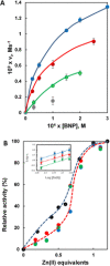
Cleavage of BNP by the different AuNPs. A) Initial rate constants obtained for the cleavage of BNP by AuNP1‐Zn(II) (blue symbols), AuNP2‐Zn(II) (green symbols), AuNP3‐Zn(II) (red symbols). The points in gray are for AuNP4‐Zn(II). The solid lines represent the fitting with the Michaelis–Menten equation. Conditions: [AuNP]=3×10⁻⁵ M, pH 8, 40 °C; 5 % DMSO added to the AuNP4 solution. Error bars in Panel A refer to data collected in three independent experiments. B) Dependence of the catalytic activity of AuNP13 (color codes are the same as in Panel A) and AuNPBAPASH (black symbols) on the number of equivalents of Zn(II) added. The dashed lines have been drawn to guide the eye. Conditions are the same as in Panel A. Inset: double log plots of vi versus [Zn(II)] for AuNP13. Color code as in Panel A.
Figure 3
Figure 3
Percentage of plasmid pBR322 cleaved by AuNP16. Data in the presence of 1 equivalent of Zn(II) (red bars) and without it (blue bars). Lighter symbols represent data collected after 1 h; darker ones after 24 h. Conditions: [AuNP]=45 μM; [pBR322]=19.3 μM (bp); pH 7.5; 37 °C; Original gel electrophoresis data are in Figures S8‐S10 and S16 of the SI.
Figure 4
Figure 4
Cleavage of plasmid pBR322 by AuNP4‐Zn(II). A) Dependence of the cleavage of BNP (red symbols, AuNP1‐Zn(II)) and pBR322 (blue symbols, AuNP4‐Zn(II)) on pH. All points have been normalized with respect to maximum efficiency (% cleavage for DNA, v i for BNP). The dashed line connecting the points for DNA was drawn to guide the eye; the solid one for BNP represents the best fitting for the two kinetically relevant pK a (7.6 and 9.1). Conditions: for DNA, 37 °C, [AuNP4‐Zn(II)]=35 μM, [pBR322]=19.3 μM bp−1, 3.5 h incubation time; conditions for BNP: [AuNP1‐Zn(II)]=3×10⁻⁵ M, [BNP]=1×10⁻⁴ M, 40 °C. B) Percentage of pBR322 cleaved by AuNP4 upon addition of increasing equivalents of Zn(II). Conditions: pH 7.5, 37 °C, [AuNP4]=35 μM, [pBR322]=19.3 μM bp−1, 3 h incubation time. Error bars refer to data collected in three independent experiments.
Figure 5
Figure 5
Molecular dynamic simulations for AuNP4. A) Top panel: Convergence of the RMSD value for the AuNP4‐Zn(II) MD simulations, calculated using as a reference the structure at time t=0 (i.e., the minimized model). Bottom panel: Number of pre‐catalytic binding sites on AuNP4‐Zn(II) over simulation time, calculated for different cut‐offs (0.8 nm in red, 0.7 nm in yellow, 0.6 nm in green, and 0.5 nm in blue). B) Fully equilibrated model of AuNP4‐Zn(II) (gold core in yellow and Zn Ions as gray spheres). C) The formation of pre‐catalytic binding sites is defined by a metric where at least two out of the following three distances are below a defined cut‐off distance: i) dist Zn/Arg, between the Zn ion and the C atom of arginine's guanidinium group; ii) dist Zn/Sera or dist Zn/Serb, between the Zn ion and the O atom of the hydroxyl group of the serine; iii) dist Arg/Sera or dist Arg/Serb, between C atom of arginine's guanidinium group and the alcoholic O atom of the serine side chain. In this way, the binding sites are defined so to contain one Zn atom, the guanidinium group of an arginine, and the side chain of at least one serine fragment (Sera or Serb, in the blue and orange binding site, respectively).
Figure 6
Figure 6
Pre‐catalytic binding complexes formed between AuNP4 and dsDNA during the MD simulations. A) Pre‐catalytic binding Complex1 formed by the coordination of the hydrolytic phosphate group (P) on top of the Zn ion (P‐O.Zn distance of 3.8 Å and via Zn‐bound water at the distance of 1.6 Å), while stabilized by one arginine Arg (2.9 Å) with nucleophilic serine (Ser) in close proximity (3.6 Å). The dsDNA is anchored to the monolayer by another phosphate‐Zn and phosphate‐arginine interactions along the backbone. B) Pre‐catalytic binding Complex2, where Zn atom coordinates one phosphate (P‐O.Zn distance of 1.8 Å), while the hydrolytic phosphate (P) is chelated by two arginine residues (at distances of 2.9 Å and 2.7 Å) with serine (Ser) in close proximity (4.5 Å) and water at the distance of 1.7 Å.
Figure 7
Figure 7
Suggested mechanism for the cleavage of a phosphate bond of plasmid DNA by AuNP4‐Zn(II) (A); and in the absence of the metal ion (B). Functions involved in acid catalysis (Lewis or proton) are in red; nucleophilic catalysis in blue.

References

    1. Wolfenden R., Annu. Rev. Biochem. 2011, 80, 645–667. - PubMed
    1. Yang W., Q. Rev. Biophys. 2011, 44, 1–93. - PMC - PubMed
    1. Diez-Castellnou M., Martinez A., Mancin F., Adv. Phys. Org. Chem. 2017, 51, 129–186.
    1. Dupureur C., Curr. Opin. Chem. Biol. 2008, 12, 250–255. - PubMed
    1. Palermo G., Cavalli A., Klein M., Alfonso-Prieto M., Peraro M., De Vivo M., Acc. Chem. Res. 2015, 48, 220–228. - PubMed

Publication types

LinkOut - more resources