

Systemic multipotent adult progenitor cells protect the cerebellum after asphyxia in fetal sheep
- PMID: 32985793
- PMCID: PMC7780812
- DOI: 10.1002/sctm.19-0157
Systemic multipotent adult progenitor cells protect the cerebellum after asphyxia in fetal sheep
Abstract
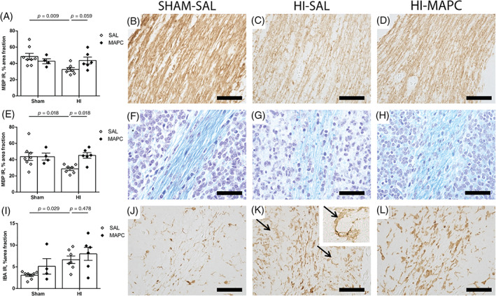
Involvement of the cerebellum in the pathophysiology of hypoxic-ischemic encephalopathy (HIE) in preterm infants is increasingly recognized. We aimed to assess the neuroprotective potential of intravenously administered multipotent adult progenitor cells (MAPCs) in the preterm cerebellum. Instrumented preterm ovine fetuses were subjected to transient global hypoxia-ischemia (HI) by 25 minutes of umbilical cord occlusion at 0.7 of gestation. After reperfusion, two doses of MAPCs were administered intravenously. MAPCs are a plastic adherent bone-marrow-derived population of adult progenitor cells with neuroprotective potency in experimental and clinical studies. Global HI caused marked cortical injury in the cerebellum, histologically indicated by disruption of cortical strata, impeded Purkinje cell development, and decreased dendritic arborization. Furthermore, global HI induced histopathological microgliosis, hypomyelination, and disruption of white matter organization. MAPC treatment significantly prevented cortical injury and region-specifically attenuated white matter injury in the cerebellum following global HI. Diffusion tensor imaging (DTI) detected HI-induced injury and MAPC neuroprotection in the preterm cerebellum. This study has demonstrated in a preclinical large animal model that early systemic MAPC therapy improved structural injury of the preterm cerebellum following global HI. Microstructural improvement was detectable with DTI. These findings support the potential of MAPC therapy for the treatment of HIE and the added clinical value of DTI for the detection of cerebellar injury and the evaluation of cell-based therapy.
Keywords: MAPC; asphyxia; cerebellum; hypoxic-ischemic encephalopathy; stem cells.
© 2020 The Authors. STEM CELLS TRANSLATIONAL MEDICINE published by Wiley Periodicals LLC on behalf of AlphaMed Press.
Conflict of interest statement
R.W.M. declared employment with Athersys, Inc., the company providing the cells in this study. The other authors declared no potential conflicts of interest.
Figures


Similar articles
-
Multipotent adult progenitor cells for hypoxic-ischemic injury in the preterm brain.J Neuroinflammation. 2015 Dec 23;12:241. doi: 10.1186/s12974-015-0459-5. J Neuroinflammation. 2015. PMID: 26700169 Free PMC article.
-
Systemic G-CSF attenuates cerebral inflammation and hypomyelination but does not reduce seizure burden in preterm sheep exposed to global hypoxia-ischemia.Exp Neurol. 2013 Dec;250:293-303. doi: 10.1016/j.expneurol.2013.09.026. Epub 2013 Oct 8. Exp Neurol. 2013. PMID: 24120465
-
Mesenchymal stem cells induce T-cell tolerance and protect the preterm brain after global hypoxia-ischemia.PLoS One. 2013 Aug 26;8(8):e73031. doi: 10.1371/journal.pone.0073031. eCollection 2013. PLoS One. 2013. PMID: 23991170 Free PMC article.
-
Perinatal hypoxic-ischemic brain injury in large animal models: Relevance to human neonatal encephalopathy.J Cereb Blood Flow Metab. 2018 Dec;38(12):2092-2111. doi: 10.1177/0271678X18797328. Epub 2018 Aug 28. J Cereb Blood Flow Metab. 2018. PMID: 30149778 Free PMC article. Review.
-
Role of instrumented fetal sheep preparations in defining the pathogenesis of human periventricular white-matter injury.J Child Neurol. 2006 Jul;21(7):582-9. doi: 10.1177/08830738060210070101. J Child Neurol. 2006. PMID: 16970848 Review.
Cited by
-
A preview of selected articles.Stem Cells Transl Med. 2021 Jan;10(1):1-4. doi: 10.1002/sctm.20-0519. Stem Cells Transl Med. 2021. PMID: 33373498 Free PMC article. No abstract available.
-
Key roles of glial cells in the encephalopathy of prematurity.Glia. 2024 Mar;72(3):475-503. doi: 10.1002/glia.24474. Epub 2023 Nov 1. Glia. 2024. PMID: 37909340 Free PMC article. Review.
-
Multi-layer perceptron classification & quantification of neuronal survival in hypoxic-ischemic brain image slices using a novel gradient direction, grey level co-occurrence matrix image training.PLoS One. 2022 Dec 13;17(12):e0278874. doi: 10.1371/journal.pone.0278874. eCollection 2022. PLoS One. 2022. PMID: 36512546 Free PMC article.
References
-
- Hammerl M, Zagler M, Griesmaier E, et al. Reduced cerebellar size at term‐equivalent age is related to a 17% lower mental developmental index in very preterm infants without brain injury. Neonatology. 2020;117:57‐64. - PubMed
-
- Hutton LC, Yan E, Yawno T, Castillo‐Melendez M, Hirst JJ, Walker DW. Injury of the developing cerebellum: a brief review of the effects of endotoxin and asphyxial challenges in the late gestation sheep fetus. Cerebellum. 2014;13:777‐786. - PubMed
