. 2020 Dec;133(1):e125.
doi: 10.1002/cpmb.125.

Optimized Transgene Delivery Using Third-Generation Lentiviruses

Affiliations

Optimized Transgene Delivery Using Third-Generation Lentiviruses

Katherine P Gill et al. Curr Protoc Mol Biol. 2020 Dec.

Abstract

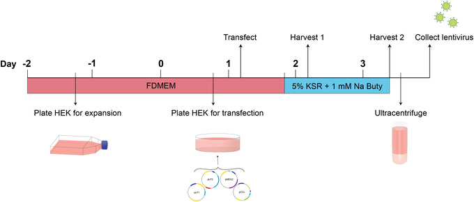
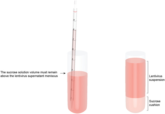
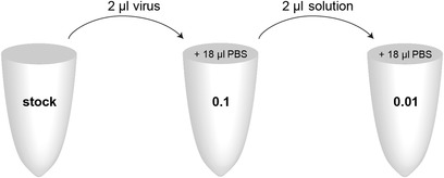
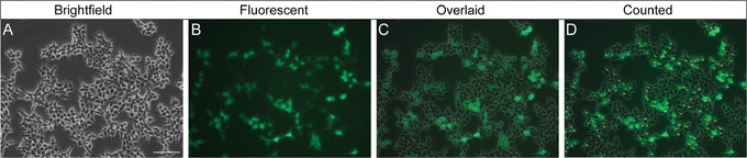
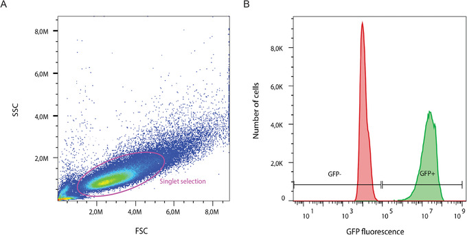
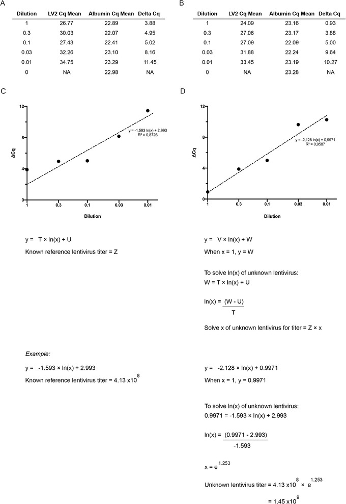
The lentivirus system enables efficient genetic modification of both dividing and non-dividing cells and therefore is a useful tool for elucidating developmental processes and disease pathogenesis. The development of third-generation lentiviruses has resulted in improved biosafety, low immunogenicity, and substantial packaging capabilities. However, because third-generation lentiviruses require successful co-transfection with four plasmids, this typically means that lower titers are attained. This is problematic, as it is often desirable to produce purified lentiviruses with high titers (>1 × 10⁸ TU/ml), especially for in vivo applications. The manufacturing process for lentiviruses involves several critical experimental factors that can influence titer, purity, and transduction efficiency. Here, we describe a straightforward, stepwise protocol for the reproducible manufacture of high-titer third-generation lentiviruses (1 × 10⁸ to 1 × 10⁹ TU/ml). This optimized protocol enhances transgene expression by use of Lipofectamine transfection and optimized serum replacement medium, a single ultracentrifugation step, use of a sucrose cushion, and addition of a histone deacetylation inhibitor. Furthermore, we provide alternate methods for titration analyses, including functional and genomic integration analyses, using common laboratory techniques such as FACS as well as genomic DNA extraction and qPCR. These optimized methods will be beneficial for investigating developmental processes and disease pathogenesis in vitro and in vivo. © 2020 The Authors. Basic Protocol 1: Lentivirus production Support Protocol: Lentivirus concentration Basic Protocol 2: Lentivirus titration Alternate Protocol 1: Determination of viral titration by FACS analysis Alternate Protocol 2: Determination of viral titration by genome integration analysis.

Keywords: high titer; lentiviral production; lipofection; third-generation; ultracentrifugation.


Figures

Figure 1
Figure 1
Experimental timeline for lentiviral production. HEK293T/17 cells are plated in the afternoon of day ‐2 for expansion. Approximately 48 hr later, the HEK cells are trypsinized and seeded into 10‐cm2 dishes for transfection. In the morning of day 1, the HEK cells are transfected with Opti‐MEM containing the Lipofectamine and DNA complex. Transfection media are removed 6 hr later and replaced with 5% KSR containing 1 mM sodium butyrate (denoted by the abbreviation “Na Buty”). The first lentivirus harvest is performed in the morning of day 2, 24 hr post‐transfection. The second lentivirus harvest is performed around midday of day 3, 52 hr post‐transfection. After ultracentrifuge concentration, the purified lentiviral particles can be aliquoted and stored.
Figure 2
Figure 2
Diagram illustrating the technique to create a sucrose cushion in the ultracentrifuge tubes. Using a long plastic pipet, slowly add 2 ml sucrose solution to the base of the tube, maintaining a volume in the pipet higher than the meniscus of the lentiviral supernatant.
Figure 3
Figure 3
Serial dilution series of lentivirus stock. Prepare a 0.1 dilution by transferring 2 μl of the undiluted virus stock into a single 1.5‐ml Eppendorf tube containing 18 μl PBS. Mix well. Using a new pipet tip, transfer 2 μl of this dilution into a new 1.5‐ml Eppendorf tube containing 18 μl PBS to prepare the 0.01 dilution.
Figure 4
Figure 4
Representative brightfield images (A) and fluorescent images (B) of HEK293T/17 cells transduced with a dilution of lentivirus with a GFP reporter. An overlaid image (C) can be generated to assist with identifying cell boundaries in the fluorescent channel. The labeling and measure functions in ImageJ are used to count cells (D). Scale bar: 100 µm.
Figure 5
Figure 5
Formula for determining lentiviral transducing units per milliliter (TU/ml) (A). An example of the pRRL‐PGK‐EGFP plasmid titration (B).
Figure 6
Figure 6
Representative flow cytometry analysis of a GFP reporter lentivirus. Singlet cells were isolated from the entire population by strict gating, in pink (A). The positive‐control (green) and negative‐control (red) samples were overlaid and used to set appropriate gates for GFP positivity (+) or negativity (–) (B). SSC, side scatter; FSC, forward scatter. GFP fluorescence intensity is shown in arbitrary units.
Figure 7
Figure 7
qPCR analysis for lentivirus titration. Representative triplicate Cq values for the LV2 and albumin genes are averaged to calculate the Cq mean for a serial dilution of lentivirus of known (A) or unknown (B) titration. A delta (Δ) Cq is calculated by subtracting the LV2 Cq mean from the albumin Cq mean. The Cq mean values for the lentivirus of known (C) and unknown (D) titration are plotted on an XY graph against dilution value. A natural log regression analysis is performed, and the unknown variables are used to calculate the unknown lentivirus titer. The letters (T‐W, Z) denote variables to be solved using the formula.

References

Literature Cited

    1. al Yacoub, N. , Romanowska, M. , Haritonova, N. , & Foerster, J. (2007). Optimized production and concentration of lentiviral vectors containing large inserts. Journal of Gene Medicine, 9(7), 579–584. doi: 10.1002/jgm.1052. - DOI - PubMed
    1. Boussif, O. , Lezoualc'h, F. , Zanta, M. A. , Mergny, M. D. , Scherman, D. , Demeneix, B. , & Behr, J. P. (1995). A versatile vector for gene and oligonucleotide transfer into cells in culture and in vivo: Polyethylenimine. Proceedings of the National Academy of Sciences of the United States of America, 92(16), 7297–7301. doi: 10.1073/pnas.92.16.7297. - DOI - PMC - PubMed
    1. Burns, J. C. , Friedmann, T. , Driever, W. , Burrascano, M. , & Yee, J. K. (1993). Vesicular stomatitis virus G glycoprotein pseudotyped retroviral vectors: Concentration to very high titer and efficient gene transfer into mammalian and nonmammalian cells. Proceedings of the National Academy of Sciences of the United States of America, 90(17), 8033–8037. doi: 10.1073/pnas.90.17.8033. - DOI - PMC - PubMed
    1. Butler, R. , & Bates, G. P. (2006). Histone deacetylase inhibitors as therapeutics for polyglutamine disorders. Nature Reviews Neuroscience, 7(10), 784–796. doi: 10.1038/nrn1989. - DOI - PubMed
    1. Chen, H. P. , Zhao, Y. T. , & Zhao, T. C. (2015). Histone deacetylases and mechanisms of regulation of gene expression (Histone deacetylases in cancer). Critical Reviews in Oncology/Hematology, 20(1–2), 35–47. doi: 10.1016/j.physbeh.2017.03.040. - DOI - PMC - PubMed

Publication types

LinkOut - more resources