Morphometric Study of the Cervical Spinal Canal Content and the Vertebral Artery
- PMID: 32986564
- PMCID: PMC7478068
- DOI: 10.14444/7060
Morphometric Study of the Cervical Spinal Canal Content and the Vertebral Artery
Abstract
Background: The morphological features of the cervical spinal nerves (C1-C8), their dimensions, and their anatomical relations with the vertebral artery are important for safe spinal surgery. The aim of the present study is to give detailed morphological data of the region to avoid complications.
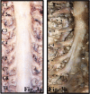
Methods: Five formalin-fixed adult cadavers were studied. The cervical spinal nerves and the vertebral artery were exposed via the posterior approach, and detailed anatomy and morphometric measurements were evaluated. The following measurements were documented: angles between the spinal nerve and the spinal cord of C1 to C8, width of the C1 to C8 spinal nerves at their origin, distance of the spinal cord to the vertebral artery, number of dorsal rootlets, length of the dorsal root entry zone of C1 to C8, and distance between respective spinal nerves. Further, the average length and width of the transverse foramen were measured.
Results: The average angle between the spinal cord and the spinal nerve within the vertebral canal ranged between 54 and 87 degrees and were most acute at C5 (54 degrees) compared to the rest of the cervical spinal nerves. The average width of the spinal nerves (mean ± SD), was thickest at C5 (5.7 ± 1.2 mm) and C6 (5.8 ± 0.7 mm). The average largest distance between the vertebral artery and the spinal cord was at C2 (14.3 ± 1.7 mm) and the smallest at C5 (7.3 ± 0.9 mm) and C6 (7.3 ± 2.2 mm) spinal levels. The number of dorsal rootlets was most numerous at C6 (8.25 ± 0.6) and C7 (7.25 ± 0.9). The dorsal root entry zone length was the largest at C5 (13.0 ± 1.6 mm) and C6 (13.75 ± 0.5 mm). The distance between respective spinal nerves was largest between C2 and C3 (11.8 ± 2.2) and C7 and C8 (11.5 ± 0.6).
Conclusion: The knowledge of detailed anatomy of the cervical spine (C1-C8) and its relations with the vertebral artery will reduce the unwanted damage to the vital structures of the region.
Keywords: dorsal cervical nerve roots; spinal cord; vertebral artery.
This manuscript is generously published free of charge by ISASS, the International Society for the Advancement of Spine Surgery. Copyright © 2020 ISASS.
Conflict of interest statement
Figures


Similar articles
-
Microsurgical anatomy of the dorsal cervical rootlets and dorsal root entry zones.Acta Neurochir (Wien). 2005 Feb;147(2):195-9; discussion 199. doi: 10.1007/s00701-004-0425-y. Acta Neurochir (Wien). 2005. PMID: 15565478
-
Surgical anatomy of the anterior cervical spine: the disc space, vertebral artery, and associated bony structures.Neurosurgery. 1996 Oct;39(4):769-76. doi: 10.1097/00006123-199610000-00026. Neurosurgery. 1996. PMID: 8880772
-
A cadaveric study of the cervical nerve roots and spinal segments.Eur Spine J. 2015 Dec;24(12):2828-31. doi: 10.1007/s00586-015-4070-3. Epub 2015 Jun 18. Eur Spine J. 2015. PMID: 26084787
-
Clinical anatomy of the C1 dorsal root, ganglion, and ramus: a review and anatomical study.Clin Anat. 2007 Aug;20(6):624-7. doi: 10.1002/ca.20472. Clin Anat. 2007. PMID: 17330847 Review.
-
Accessory Vertebral Artery: An Embryological Review With Translation from Adachi.Cureus. 2021 Feb 19;13(2):e13448. doi: 10.7759/cureus.13448. Cureus. 2021. PMID: 33767932 Free PMC article. Review.
Cited by
-
Anatomical description of the ventral and dorsal cervical rootlets in rats: A microsurgical study.Acta Cir Bras. 2022 Jun 1;37(3):e370307. doi: 10.1590/acb370307. eCollection 2022. Acta Cir Bras. 2022. PMID: 35674584 Free PMC article.
-
Anatomical Importance Between Neural Structure and Bony Landmark in Neuroventral Decompression for Posterior Endoscopic Cervical Discectomy.Neurospine. 2025 Mar;22(1):286-296. doi: 10.14245/ns.2448794.397. Epub 2025 Mar 31. Neurospine. 2025. PMID: 40211534 Free PMC article.
References
-
- Jeszenszky DJ, Haschtmann D, Pröbstl O, Kleinstück FS, Heyde CE, Fekete TF. Tumors and metastases of the upper cervical spine (C0-2). A special challenge. Orthopade. 2013;42(9):746–754. - PubMed
-
- Mattei TA, Mendel E. En bloc resection of primary malignant bone tumors of the cervical spine. Acta Neurochir (Wien) 2014;156(11):2159–2164. - PubMed
-
- Chibbaro S, Mirone G, Yasuda M, Marsella M, Di Emidio P, George B. Vertebral artery loop—a cause of cervical radiculopathy. World Neurosurg. 2012;78(3–4):375.e11–375.e13. - PubMed
-
- Yu YL, du Boulay GH, Stevens JM, Kendall BE. Morphology and measurements of the cervical spinal cord in computer-assisted myelography. Neuroradiology. 1985;27(5):399–402. - PubMed
LinkOut - more resources
Full Text Sources
Miscellaneous
