. 2020 Nov 4;48(19):11146-11161.
doi: 10.1093/nar/gkaa751.

Structural basis of DNA replication origin recognition by human Orc6 protein binding with DNA

Affiliations

Structural basis of DNA replication origin recognition by human Orc6 protein binding with DNA

Naining Xu et al. Nucleic Acids Res. .

Abstract

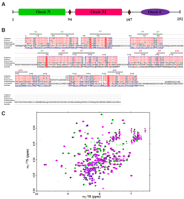
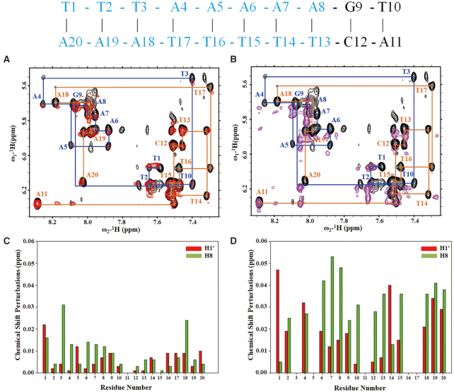
The six-subunit origin recognition complex (ORC), a DNA replication initiator, defines the localization of the origins of replication in eukaryotes. The Orc6 subunit is the smallest and the least conserved among ORC subunits. It is required for DNA replication and essential for viability in all species. Orc6 in metazoans carries a structural homology with transcription factor TFIIB and can bind DNA on its own. Here, we report a solution structure of the full-length human Orc6 (HsOrc6) alone and in a complex with DNA. We further showed that human Orc6 is composed of three independent domains: N-terminal, middle and C-terminal (HsOrc6-N, HsOrc6-M and HsOrc6-C). We also identified a distinct DNA-binding domain of human Orc6, named as HsOrc6-DBD. The detailed analysis of the structure revealed novel amino acid clusters important for the interaction with DNA. Alterations of these amino acids abolish DNA-binding ability of Orc6 and result in reduced levels of DNA replication. We propose that Orc6 is a DNA-binding subunit of human/metazoan ORC and may play roles in targeting, positioning and assembling the functional ORC at the origins.


Figures

Figure 1.
Figure 1.
(A) Schematic representation of the full-length human Orc6. (B) Sequence alignment of the Orc6 sequences from different species: Q9Y5N6, Homo sapiens; Q2HJF3, Bos taurus; Q9WUJ8 M. musculus; Q9Y1B2, Drosophila melanogaster; P38826, Saccharomyces cerevisiae; O74796, Schizosaccharomyces pombe. The dashes indicate the positions of gaps in eukaryotic sequences. The sequence alignment was produced with ClustalW2 (73) and plotted with ESPript 2.2 (74). The indicated secondary structure corresponds to the solution structure of full-length human Orc6 reported here. Residues are red-scaled based on percentage identity. (C) The superposition of 1H,15N HSQC NMR spectra of individual Orc6-N(green), Orc6-M (magenta), Orc6-C (purple) and full-length Orc6 (black) are shown.
Figure 2.
Figure 2.
(A)The ensemble of the lowest 20 energy structures of full-length Orc6 generated by superposition of backbone atoms of residues 1–252 (left) and the lowest structure of the ensemble shown in cartoon (right). Individual (B) Orc6-N (residues 1–94, green), (C) Orc6-M (residues 95–187, magenta) and (D) Orc6-C (residues 222–252, purple) domains are shown in superimposed ensemble and in a form of cartoon of the lowest structure. The secondary structure of each domain is colored as green in Orc6-N, magenta in Orc6-M and purple in Orc6-C. The loop in each domain is shown in gray. The region containing residues from 188 to 221 is flexible and is not shown in (D).
Figure 3.
Figure 3.
NMR study of the interaction between Orc6 and DNA. (A) The overlaid 1H,15N-HSQC spectra of full-length Orc6 in free form (red) titrated with 17b DNA at a molar ratio of 1:1 (green), 1:2 (purple) and 1:5 (blue). Residues that undergo significant changes in chemical shifts upon formation of the complex with DNA are indicated with arrows and labeled with peak assignments. (B) A zoomed part of overlaid 1H,15N-HSQC spectra shown in (A) with red square. (C) Weighted chemical shift perturbations for backbone 15N and 1HN resonances as calculated by the equation Δδ = [(ΔδHN)2+(ΔδN/5)2]0.5. The mean Δδ value (0.015 ppm) and the mean Δδ value plus 1 SD (0.038 ppm) of the chemical shift perturbations are plotted as solid lines. (D) Chemical shift perturbations in the presence of 17bp DNA are colored onto the structure of HsOrc6-DBD (resides 95–207) in ribbon representation. Residues with chemical shift perturbations ranging from 0.015 to 0.038 ppm are colored in blue, whereas residues with chemical shift perturbations larger than 0.038 ppm are shown in red. The residues, Lys158, Lys169, Arg174, Lys177, Lys181, Lys198, Arg199, Lys200 and Arg202, are shown in the stick model.
Figure 4.
Figure 4.
(A and B) Overlay of the fingerprint region showing intraresidue H1′–H6/H8 NOE peaks of 2D 13C/15N-filtered 1H-1H NOESY spectra of free (black) and Orc6-M bound with DNA (red) and HsOrc6-DBD (residues 95–207) bound with DNA (purple) at concentration ratio 1:2 (DNA, 0.5 mM: HsOrc6-DBD,1.0 mM). Intraresidue H1′–H6/H8 NOE peaks of free DNA are labeled by base type and number. The sequence the 10-mer DNA is shown above, with residues affected by Orc6 binding indicated in blue. (C and D) 1H chemical shift difference (Δδ) for H1′ (red) and H6/H8 (green) chemical shifts between free and Orc6-bound DNA corresponding to (A) and (B) are plotted against residue number.
Figure 5.
Figure 5.
DNA-binding ability of HsOrc6. (A) DNA binding of human Orc6 wild-type HsOrc6WT and mutants HsOrc6R198A/K199A/R200A/K201A, HsOrc6K169A, HsOrc6K168A/K169A/ D173A to radiolabeled origin fragment Lamin B2 in the presence of poly dGdC competitor DNA was monitored by EMSA. The amount of competitor was 50 and 100 ng. (B) Silver stained gel of purified wild-type human Orc6 (1) and mutants HsOrc6R198A/K199A/R200A/K201A (2), HsOrc6K169A (3), HsOrc6K168A/K169A/D173A (4). (C) Immunostaining of GFP- fused wild-type and mutant Orc6 proteins expressed in salivary glands of Drosophila third instar larvae. Orc6 was detected with anti-GFP antibodies. (D) The expression level of the GFP-tagged Orc6 proteins in salivary glands of fly strains used in (C). Salivary glands of Drosophila larvae expressing GFP-tagged Orc6 proteins were isolated and homogenized. The proteins in the extracts were separated by SDS-gel electrophoresis and analyzed by Western blotting using anti-GFP polyclonal antibodies. HsOrc6WT (lane 1), HsOrc6-R198A/K199A/R200A/K201A (lanes 2 and 3), HsOrc6-K169A (lanes 4 and 5), HsOrc6-K168A/K169A/D173A (lanes 6 and 7) are shown. Pnut protein was used as a loading control.
Figure 6.
Figure 6.
In vitro DNA replication in Orc6 depleted Xenopus extracts supplemented by the addition of increasing amounts of recombinant wild-type or mutant human Orc6 proteins. Xenopus sperm DNA was incubated for 30 min in Xenopus extract at a concentration of 2–5 ng/μl in a presence of [α32P]dCTP. Where indicated, extracts were depleted for Orc6 by using antibodies raised against human and Drosophila Orc6. Add back experiment was performed by the addition of 50, 100 or 200 ng of recombinant human Orc6 proteins to the depleted extracts; RE, non-depleted replication extract control (lane 1). HsOrc6WT (lanes 3–5), HsOrc6-R198A/K199A/R200A/K201A (lanes 6–8), HsOrc6-K169A (lanes 9–11) and HsOrc6-K168A/K169A/D173A (lanes 12–14) were used in rescue experiments. No recombinant protein was added to the Orc6 depleted extract in lane 2.
Figure 7.
Figure 7.
Structure model of Orc6/DNA complex generated by HADDOCK 2.2. (A) The sequence of 10 bp DNA used in structural model and the binding sequence with Orc6 is colored by blue. (B) Ribbon representation of Orc6/DNA (only residue 95–207 of Orc6 shown) complex in which Orc6 binds DNA like a clamp through the Orc6-M domain and the amino acid cluster, Arg198-Lys199-Arg200-Lys201, of Orc6-C domain.
Figure 8.
Figure 8.
The proposed model of HsORC/DNA complex based on the Saccharomycescerevisiae 3 Å cryo-EM structure ORC/DNA complex (PDB code: 5ZR1). The Orc1-5 is shown in gray. The domains of Orc6 are colored as green (Orc6-N), magenta (Orc6-M) and purple (Orc6-C).

References

    1. Yardimci H., Walter J.C.. Prereplication-complex formation: a molecular double take. Nat. Struct. Mol. Biol. 2014; 21:20–25. - PubMed
    1. O’Donnell M., Langston L., Stillman B.. Principles and concepts of DNA replication in bacteria, archaea, and eukarya. Cold Spring Harb. Perspect. Biol. 2013; 5:a010108. - PMC - PubMed
    1. Duderstadt K.E., Berger J.M.. A structural framework for replication origin opening by AAA+ initiation factors. Curr. Opin. Struct. Biol. 2013; 23:144–153. - PMC - PubMed
    1. Bell S.P., Labib K.. Chromosome duplication in saccharomyces cerevisiae. Genetics. 2016; 203:1027–1067. - PMC - PubMed
    1. Bell S.P., Stillman B.. ATP-dependent recognition of eukaryotic origins of DNA replication by a multiprotein complex. Nature. 1992; 357:128–134. - PubMed

Publication types