. 2021 Feb;162(2):194-207.
doi: 10.1111/imm.13273. Epub 2020 Oct 19.

A splice acceptor variant in HLA-DRA affects the conformation and cellular localization of the class II DR alpha-chain

Affiliations

A splice acceptor variant in HLA-DRA affects the conformation and cellular localization of the class II DR alpha-chain

Alessandro Didonna et al. Immunology. 2021 Feb.

Abstract

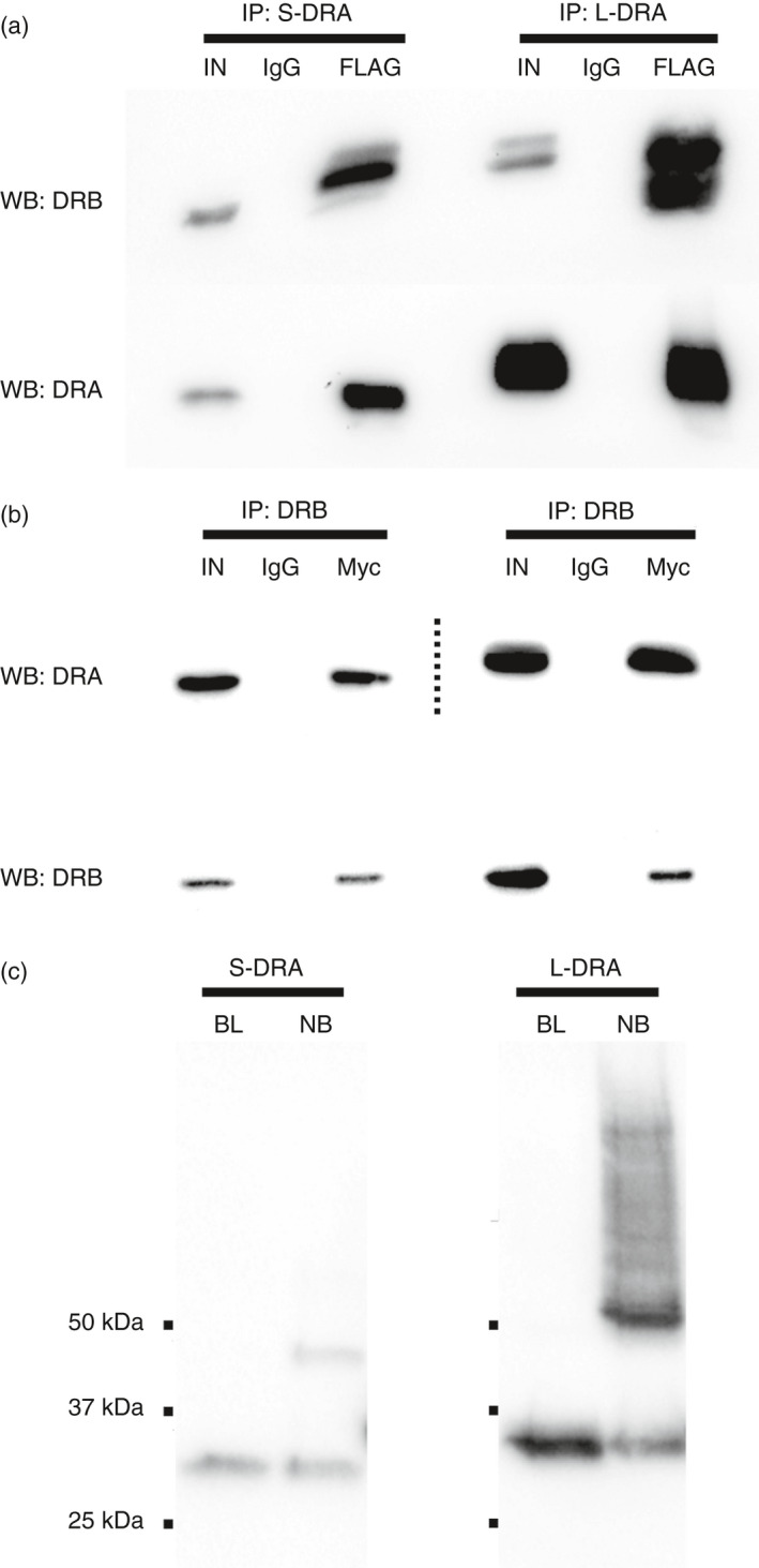
Class II human leucocyte antigen (HLA) proteins are involved in the immune response by presenting pathogen-derived peptides to CD4+ T lymphocytes. At the molecular level, they are constituted by α/β-heterodimers on the surface of professional antigen-presenting cells. Here, we report that the acceptor variant (rs8084) in the HLA-DRA gene mediates the transcription of an alternative version of the α-chain lacking 25 amino acids in its extracellular domain. Molecular dynamics simulations suggest this isoform undergoes structural refolding which in turn affects its stability and cellular trafficking. The short HLA-DRA isoform cannot reach the cell surface, although it is still able to bind the corresponding β-chain. Conversely, it remains entrapped within the endoplasmic reticulum where it is targeted for degradation. Furthermore, we demonstrate that the short isoform can be transported to the cell membrane via interactions with the peptide-binding site of canonical HLA heterodimers. Altogether, our findings indicate that short HLA-DRA functions as a novel intact antigen for class II HLA molecules.

Keywords: antigen presentation; human leucocyte antigen; immune response; protein folding.


Conflict of interest statement

The authors declare no competing interests.

Figures

Figure 1
Figure 1
SNP rs8084 drives HLADRA alternative splicing. (A) Constructs carrying the HLADRA minigenes with different alleles of rs8084 SNP (A, C or A converted to C by mutagenesis) or a control vector carrying the green fluorescent protein (GFP) coding sequence were transiently transfected in HeLa cells. After 24 h, their expression products were analysed by RT‐PCR using a forward primer designed on HLADRA exon 3 and a reverse primer on the vector backbone. This set of primers produces amplicons of 594 bp or 519 bp corresponding to the long and short isoforms, respectively. In the schematic are represented Exon 2, Exon 3 (the striped part represents the 75 bp portion only presents in the long transcript), and Exon 4. (B) Relative abundance of the alternative isoform of HLADRA in 1000 Genomes samples according to the genotypes of rs8084. Expression value ratio represents the expression value of the short transcript divided by the expression value of the long transcript. Reported P‐values are the results of Wilcoxon tests, one‐sided P‐value. (C,D) Expression analysis of long and short HLADRA isoforms in PBMCs from individuals carrying the following rs8084 genotypes: AA (n = 6), AC (n = 4), AA (n = 7). Cells were stimulated with 40 000 U/ml of human recombinant INF‐α for 24 h or left unstimulated. *P ≤ 0·05.
Figure 2
Figure 2
Short HLA‐DRA undergoes structural rearrangement. (A) The 25 missing amino acids of the short isoform map on two β strands in the α2 domain of the long isoform shown in magenta. This structural segment includes residue C132, which forms a disulphide bond with C188 in the long isoform. Two snapshots of the α2 domain shown at times 0 and 100 ns of the simulation are similar in shape indicating that the disulphide bond plays a critical role in stabilizing the structure. (B) In the short isoform, a linker region (orange) replaces the 25 amino acid stretch connecting α1 and α2 domains. Snapshots of the α2 domain of the short isoform clearly show misfolding of this domain due to the absence of the disulphide bond. (C) Minimal fluctuations in the SASA of the α2 domain throughout the simulation of the long isoform indicates no major conformational transitions, whereas significant structural rearrangements occurred at multiple time‐points of the short HLA‐DRA simulation (p < 2·2 × 10−16). (D) The RMSD distribution of the α1 domain measures deviation of the structure from the first frame, which shows no drastic difference between short and long isoforms. Particularly, the medians of distributions marked by the black line in the box plots, are very similar. (E, F) The PCA of the α2 domain confirmed the conformational transition of the short isoform. Each conformer is a point in the PC space. Specifically, there is no distinct grouping of the conformational states along PC1 to PC3 components (left panels), whereas there are two clear clusters recognizable in the short isoform in both PC1‐PC2 and PC1‐PC3 subspaces of the short isoform (right panels). The course of the trajectory is represented by the continuous colour gradient from blue to red. The percentage of the total mean square displacement of atom positional fluctuations along each eigenvector defined by the corresponding eigenvalue is shown in the bottom right panel in each case.
Figure 3
Figure 3
Short HLA‐DRA forms stable heterodimers with the β‐chain. (A) Co‐immunoprecipitation experiments of HLA‐DRB1 (encoded by HLADRB1*15:01 allele) with the short or long isoform of HLA‐DRA. HLA‐DRA and HLA‐DRB1 were FLAG‐ and Myc‐tagged respectively. Top band shows the detection of HLA‐DRB1 molecules after the co‐immunoprecipitation with either the short (left) or the long (right) HLA‐DRA isoform. Bottom bands confirm immunoprecipitation of HLA‐DRA molecules. Mock experiments using normal IgG were carried out in parallel. Input samples (IN) equal to 10% of total volume of lysates served as positive controls. (B) Reciprocal co‐immunoprecipitation experiments. Top band shows the detection of the HLA‐DRA molecules after co‐immunoprecipitation with HLA‐DRB1. Bottom bands show HLA‐DRB1 immunoprecipitation. Dotted lines separate contiguous lanes with two different exposure times. (C) HLA‐DRB1 was co‐expressed with short or long HLA‐DRA and heterodimers were immunoprecipitated using an anti‐FLAG antibody. Samples were then incubated in Laemmli buffer containing 2% SDS for 1 hour at RT or boiled for 10 minutes. Samples were then separated by SDS–PAGE and HLA‐DRA molecules were detected by Western blotting with an anti‐FLAG antibody. Signals around 25 kDa originate from monomeric α‐chains after denaturation while the bands of 50 kDa correspond to intact α/β heterodimers.
Figure 4
Figure 4
Short HLA‐DRA is retained within the cytosolic compartment. (A) HeLa cells expressing short or long HLA‐DRA isoforms, either alone or with HLA‐DRB1, were fixed and immunostained with an antibody recognizing the extracellular domain of HLA‐DRA (in red). Some cells were permeabilized with Triton X‐100 before staining, to detect the total amount of the protein. Other cells were left unpermeabilized, in order to label only HLA‐DRA molecules on the cell membrane. Nuclei were counterstained with DAPI (in blue). Only long HLA‐DRA was detected on the cell surface upon HLA‐DRB1 co‐expression. Scale bar=20 μm. (B) HeLa cell lysates expressing short or long HLA‐DRA, alone or in combination with HLA‐DRB1, were digested with EndoH enzyme for 1 h (+ lanes) or not (− lanes), before being separated by SDS–PAGE. HLA‐DRA molecules were then probed by Western blotting, using an anti‐FLAG antibody. Lower bands in +lanes correspond to immature HLA‐DRA molecules that are de‐glycosylated by the enzyme. Conversely, upper bands come from mature HLA‐DRA chains maintaining their glycosidic groups. Both isoforms are fully sensitive to EndoH activity when expressed alone, while long HLA‐DRA becomes partially resistant to de‐glycosylation upon co‐expression with HLA‐DRB1. (C) EndoH assay on cell lysates expressing HLA‐DRA isoforms in combination with HLA‐DRB1, HLA‐DM and invariant chain (Ii). Co‐expression of other factors involved in antigen presentation does not improve short HLA‐DRA maturation. (D, E) HeLa cells expressing short or long HLA‐DRA isoform, alone or together with HLA‐DRB1, were treated for increasing amount of time (4, 8 and 12 h) with cycloheximide (CHX). Lysates were then prepared and the levels of HLA‐DRA molecules were assessed at the different time‐points by Western blotting. Actin levels were used as internal controls. Both isoforms are rapidly degraded when expressed alone. On the contrary, long HLA‐DRA is stabilized by HLA‐DRB1 co‐expression. (F, G) CHX time‐course experiments in HeLa cells expressing both HLA‐DRA isoforms with or without HLA‐DRB1. Short HLA‐DRA becomes more stable when it is co‐expressed along the classic HLA heterodimer. Conversely, the short isoform is rapidly degraded when only long HLA‐DRA is co‐expressed.
Figure 5
Figure 5
Short HLA‐DRA interacts with canonical heterodimers. (A) HeLa cells expressing HLA‐DRA molecules in various combinations were seeded into 96‐well plates and probed for short HLA‐DRA cell membrane expression by in‐cell ELISA as described in the Methods section. The surface levels of the short isoform are significantly higher in cells co‐expressing also long HLA‐DRA and HLA‐DRB1. (B) Viability of cells expressing the different combinations of HLA‐DRA molecules as measured by XTT assay. No significant differences were found. (C) Co‐immunoprecipitation of long and short HLA‐DRA molecules. Top band shows the detection of long HLA‐DRA after pull‐down of the short HLA‐DRA isoform. Bottom bands confirm immunoprecipitation of short HLA‐DRA. The interaction of the two isoforms takes place only when HLA‐DRB1 is also co‐expressed. (D) In‐cell ELISA assays in HeLa demonstrating that short HLA‐DRA surface expression via HLA heterodimer is abolished when invariant chain (Ii) is also co‐expressed. **P ≤ 0·01, ***P ≤ 0·001.
Figure 6
Figure 6
Cell proliferation is lower in cells expressing short HLA‐DRA. PBMCs from individuals that are homozygous for rs8084, either CC (n = 22) or AA (n = 18), were cultured in serum‐free media. After 72 h, thymidine incorporation was assessed to test their intrinsic proliferation capacity. Cells carrying the CC genotype showed higher proliferation rates as compared to cells carrying the AA genotype. **P ≤ 0·01.

Similar articles

Cited by

References

    1. Kulski JK, Shiina T, Anzai T, Kohara S, Inoko H. Comparative genomic analysis of the MHC: the evolution of class I duplication blocks, diversity and complexity from shark to man. Immunol Rev. 2002; 190:95–122. - PubMed
    1. Stern LJ, Brown JH, Jardetzky TS, Gorga JC, Urban RG, Strominger JL, et al Crystal structure of the human class II MHC protein HLA‐DR1 complexed with an influenza virus peptide. Nature. 1994; 368:215–21. - PubMed
    1. Runnels HA, Weber DA, Moore JC, Westerman LE, Jensen PE. Intact proteins can bind to class II histocompatibility molecules with high affinity. Mol Immunol. 1997; 34:471–80. - PubMed
    1. Horton R, Wilming L, Rand V, Lovering RC, Bruford EA, Khodiyar VK, et al Gene map of the extended human MHC. Nat Rev Genet. 2004; 5:889–99. - PubMed
    1. Misra MK, Damotte V, Hollenbach JA. The immunogenetics of neurological disease. Immunology. 2018; 153:399–414. - PMC - PubMed

Publication types

MeSH terms