Review
. 2020 Nov 15;396(2):112300.
doi: 10.1016/j.yexcr.2020.112300. Epub 2020 Sep 26.

Control of craniofacial and brain development by Cullin3-RING ubiquitin ligases: Lessons from human disease genetics

Affiliations
Review

Control of craniofacial and brain development by Cullin3-RING ubiquitin ligases: Lessons from human disease genetics

Anthony J Asmar et al. Exp Cell Res. .

Abstract

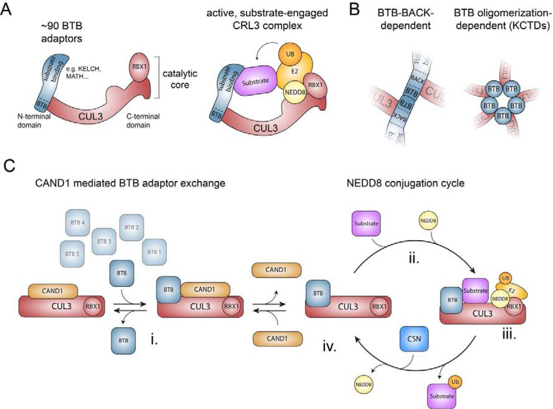
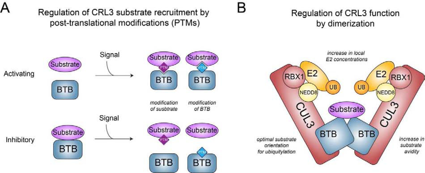
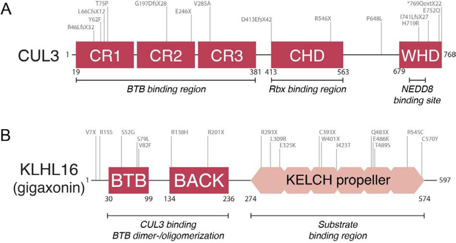
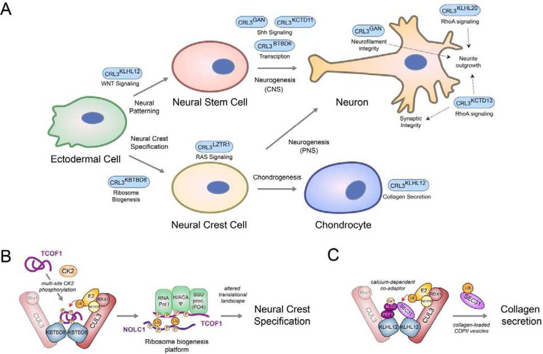
Metazoan development relies on intricate cell differentiation, communication, and migration pathways, which ensure proper formation of specialized cell types, tissues, and organs. These pathways are crucially controlled by ubiquitylation, a reversible post-translational modification that regulates the stability, activity, localization, or interaction landscape of substrate proteins. Specificity of ubiquitylation is ensured by E3 ligases, which bind substrates and co-operate with E1 and E2 enzymes to mediate ubiquitin transfer. Cullin3-RING ligases (CRL3s) are a large class of multi-subunit E3s that have emerged as important regulators of cell differentiation and development. In particular, recent evidence from human disease genetics, animal models, and mechanistic studies have established their involvement in the control of craniofacial and brain development. Here, we summarize regulatory principles of CRL3 assembly, substrate recruitment, and ubiquitylation that allow this class of E3s to fulfill their manifold functions in development. We further review our current mechanistic understanding of how specific CRL3 complexes orchestrate neuroectodermal differentiation and highlight diseases associated with their dysregulation. Based on evidence from human disease genetics, we propose that other unknown CRL3 complexes must help coordinate craniofacial and brain development and discuss how combining emerging strategies from the field of disease gene discovery with biochemical and human pluripotent stem cell approaches will likely facilitate their identification.

Keywords: BTB; Brain development; CRL; CRL3; Craniofacial development; Developmental diseases; Ubiquitin.


Conflict of interest statement

Conflict of Interest:

The authors declare no conflict of interest.

Figures

Figure 1:
Figure 1:. Structure and assembly of CRL3s
A) Schematic of the structure of a multi-subunit CRL3 complex. Left panel depicts a CRL3 that consists of CUL3 and the RING domain containing protein RBX1 (catalytic core) and one of ~90 interchangeable BTB proteins, which use their BTB domain to bind to the N-terminal domain of CUL3 and variable protein interaction domains to recruit specific sets of substrates. Right panel illustrates a model of an active, substrate-engaged CRL3 complex. Modification of CUL3 with NEDD8 at Lys-712 induces conformational changes and formation of ubiquitylation assemblies, in which RBX1 recruits an ubiquitin-charged E2 enzyme and the BTB protein position substrates for ubiquitin transfer. B) Schematic model of the two different modes how BTB proteins can interact with CUL3. C) Model of how CAND1-mediated BTB adaptor exchange and reversible NEDD8 modification are thought to regulate the cellular CRL3 repertoire (see text for details).
Figure 2:
Figure 2:. Regulatory principles underlying CRL3 function and substrate recruitment
A) Scheme depicting how signal-induced posttranslational modification (PTMs, e.g. phosphorylation, glycosylation, cysteine modifications) of BTB adaptors or substrates can promote or inhibit interaction and thus CRL3 function. B) Model of a homo-dimeric CRL3 complex highlighting the different mechanisms how dimerization is thought to regulate CRL3 function.
Figure 3:
Figure 3:. Mutations in CUL3 and BTB proteins lead to human diseases that affect the differentiation and maintenance of neuroectodermal tissues
A) Schematic overview of mutations in CUL3 (NM_003590.5) that cause / are associated with autism, schizophrenia, and neurodevelopmental disease. Adopted from [35], and only displaying probable pathogenic mutations and not deletions and non-coding variants. These mutations are generally considered to be loss-of-function, and frequently result in truncated protein versions, but can also inactivate CUL3 function through decreasing BTB binding (e.g. as experimentally demonstrated for V285A [35]) B) Schematic overview of mutations in KLHL16/GAN (NM_022041.4) that have been shown to cause giant axonal neuropathy (adopted from [82] and additional disease-causing variants added as described in [–171]). These mutations result in loss of protein function through generating truncated protein versions or variants that are deficient in homodimerization, CUL3 binding, and substrate recruitment. Mutations in other BTBs such as LZTR1 and KCTD7 have been shown to cause craniofacial and neurodevelopmental diseases in a similar manner.
Figure 4:
Figure 4:. CRL3s controlling neuroectodermal development
A) Schematic overview of various CRL3 complexes implicated in regulating aspects of neuroectodermal differentiation. B) Model of CRL3-KBTBD8-dependent neural crest specification. During embryonic development, multisite CK2-phopshorylation of the ribosome biogenesis factors TCOF1 and NOLC1 allows their recognition and monoubiquitylation by homo-dimeric CRL3-KBTBD8. This is thought to open the compact conformation of these ampholytic, intrinsically disordered proteins to promote recognition of ribosome biogenesis factors, including the RNA polymerase I, the pseudouridylation machinery (Ψ), and the SSU processome. These ubiquitin-dependent changes in ribosome biogenesis then trigger remodeling of translational networks required for neural crest specification, which likely occurs through newly synthesized, modified ribosomes C) Model of calcium-induced CRL3-KLHL12-dependent collagen secretion. During bone formation, homo-dimeric CRL3-KLHL12 complexes are thought to engage their substrate SEC31 using a co-adaptor complex consisting of PEF1 and ALG2. The ALG2 subunit is only able to engage SEC31 after calcium has been released from the endoplasmic reticulum, thus allowing CRL3-KLHL12 to mono-ubiquitylate and promote formation of large, collagen-loaded COPII vesicles in a calcium-dependent manner.

References

    1. Clague MJ, Urbe S, Komander D, Breaking the chains: deubiquitylating enzyme specificity begets function, Nat Rev Mol Cell Biol 20 (2019) 338–352. - PubMed
    1. Oh E, Akopian D, Rape M, Principles of Ubiquitin-Dependent Signaling, Annu Rev Cell Dev Biol 34 (2018) 137–162. - PubMed
    1. Komander D, Rape M, The ubiquitin code, Annu Rev Biochem 81 (2012) 203–229. - PubMed
    1. Yau R, Rape M, The increasing complexity of the ubiquitin code, Nat Cell Biol 18 (2016) 579–586. - PubMed
    1. Di Fiore PP, Polo S, Hofmann K, When ubiquitin meets ubiquitin receptors: a signalling connection, Nat Rev Mol Cell Biol 4 (2003) 491–497. - PubMed

Publication types