. 2020 Dec:42:101087.
doi: 10.1016/j.molmet.2020.101087. Epub 2020 Sep 26.

Reversal of diet-induced hepatic steatosis by peripheral CB1 receptor blockade in mice is p53/miRNA-22/SIRT1/PPARα dependent

Affiliations

Reversal of diet-induced hepatic steatosis by peripheral CB1 receptor blockade in mice is p53/miRNA-22/SIRT1/PPARα dependent

Shahar Azar et al. Mol Metab. 2020 Dec.

Abstract

Objective: The endocannabinoid (eCB) system is increasingly recognized as being crucially important in obesity-related hepatic steatosis. By activating the hepatic cannabinoid-1 receptor (CB1R), eCBs modulate lipogenesis and fatty acid oxidation. However, the underlying molecular mechanisms are largely unknown.

Methods: We combined unbiased bioinformatics techniques, mouse genetic manipulations, multiple pharmacological, molecular, and cellular biology approaches, and genomic sequencing to systematically decipher the role of the hepatic CB1R in modulating fat utilization in the liver and explored the downstream molecular mechanisms.

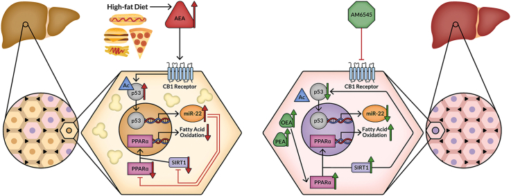
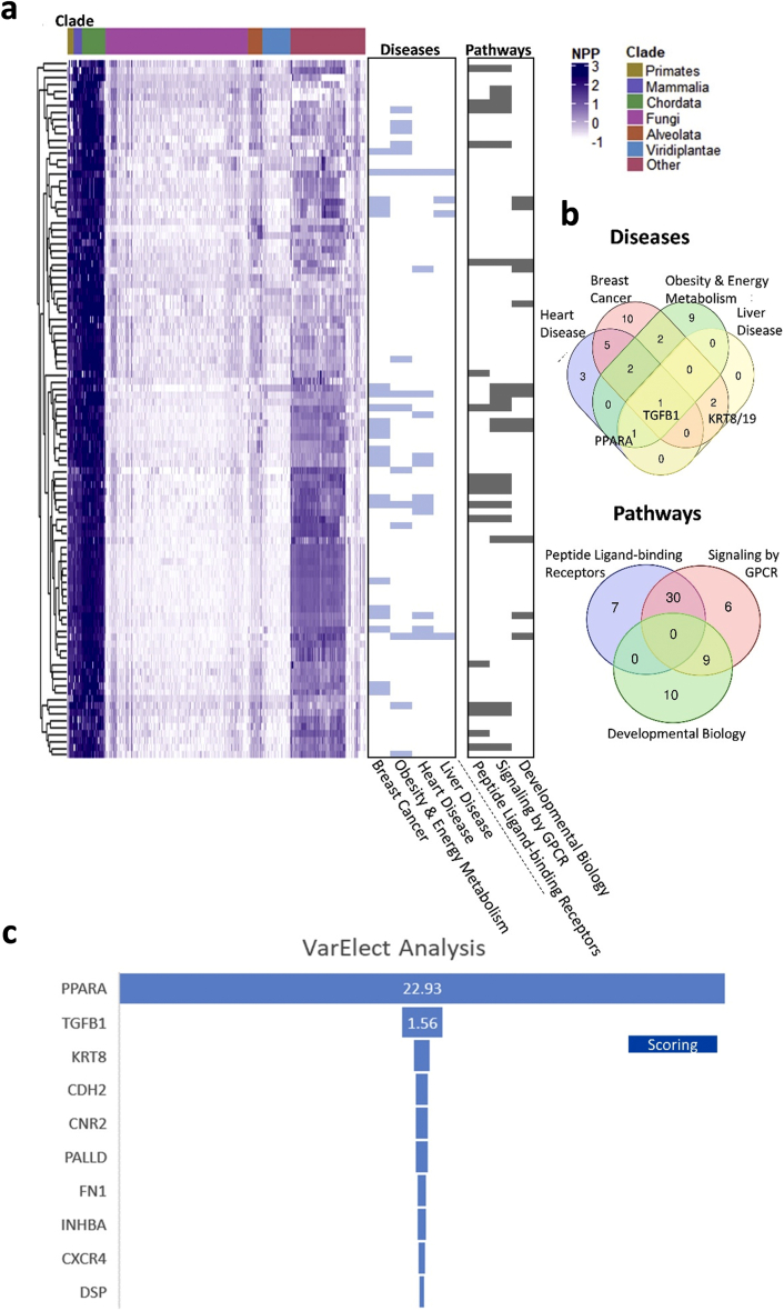
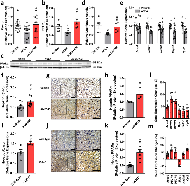
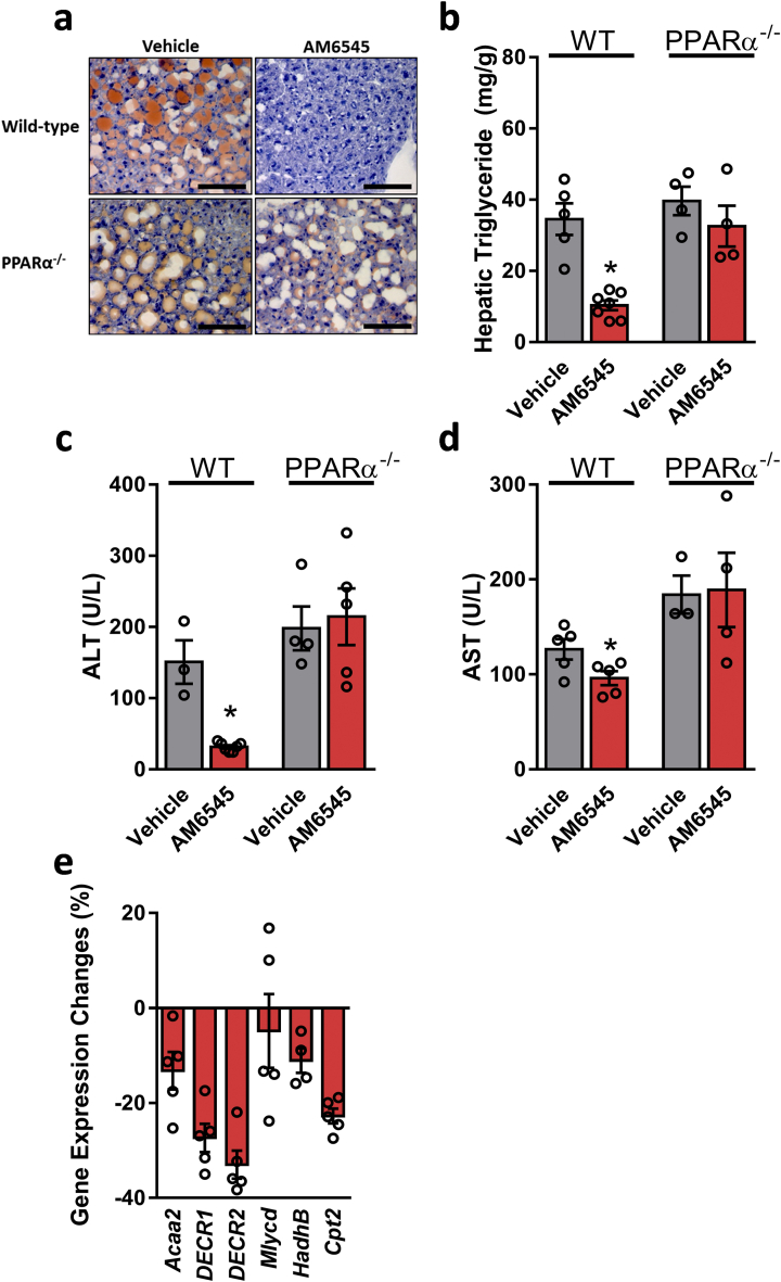
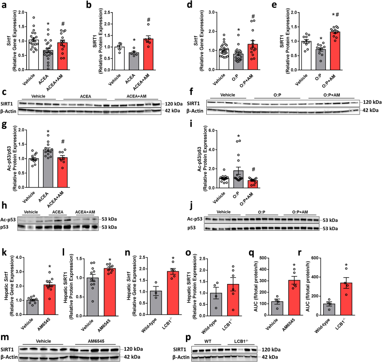
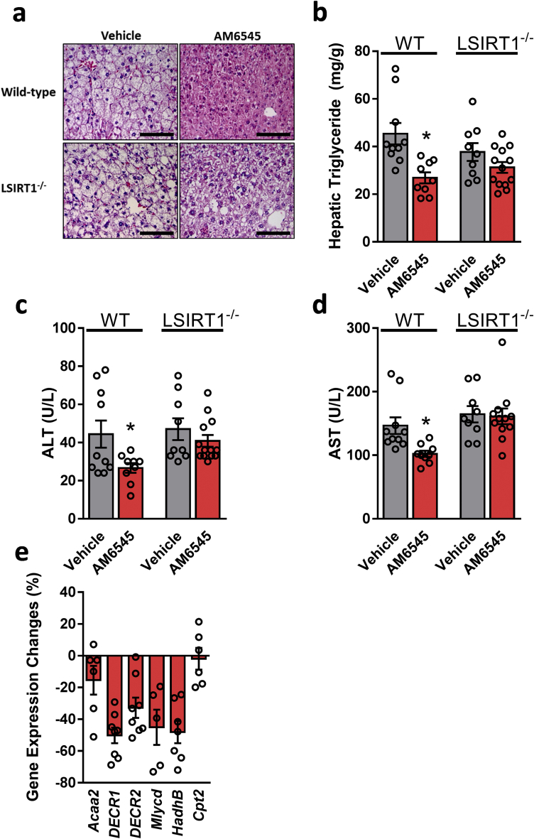
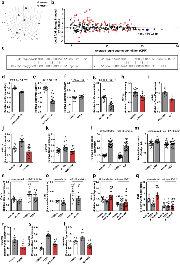
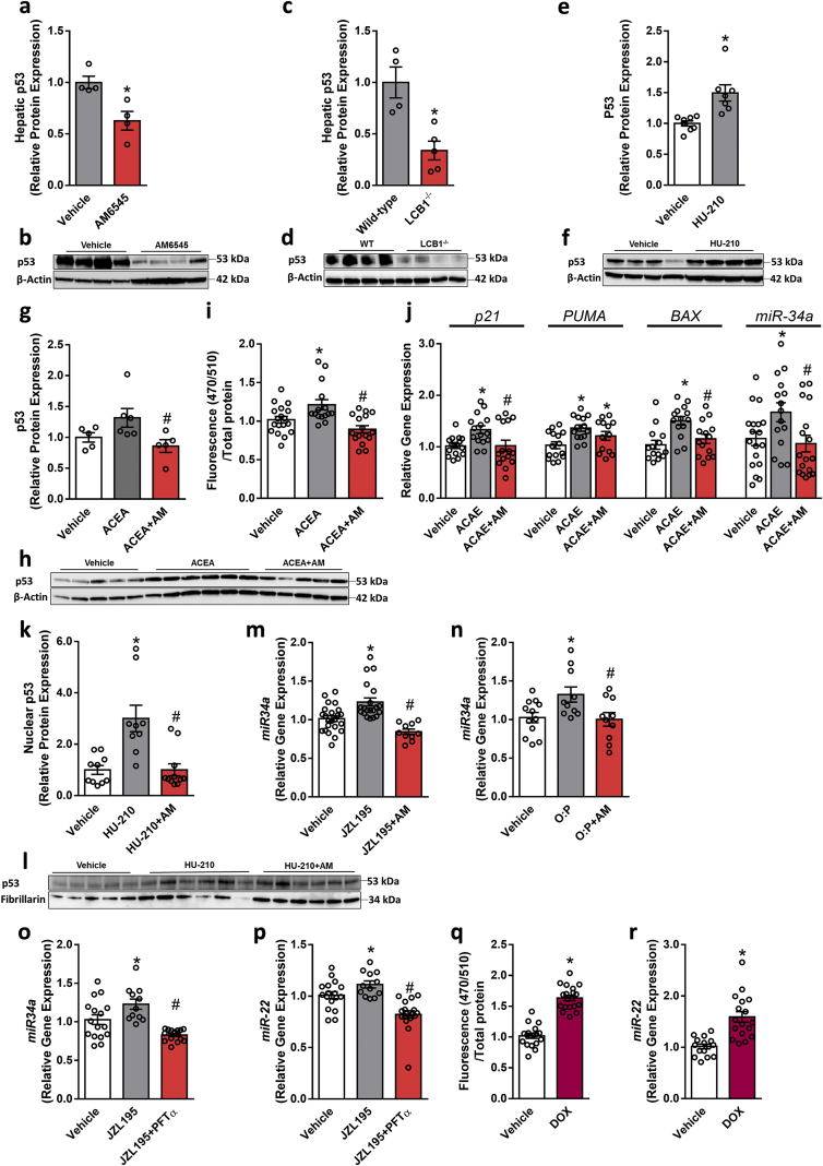
Results: Using an unbiased normalized phylogenetic profiling analysis, we found that the CB1R evolutionarily coevolves with peroxisome proliferator-activated receptor-alpha (PPARα), a key regulator of hepatic lipid metabolism. In diet-induced obese (DIO) mice, peripheral CB1R blockade (using AM6545) induced the reversal of hepatic steatosis and improved liver injury in WT, but not in PPARα-/- mice. The antisteatotic effect mediated by AM6545 in WT DIO mice was accompanied by increased hepatic expression and activity of PPARα as well as elevated hepatic levels of the PPARα-activating eCB-like molecules oleoylethanolamide and palmitoylethanolamide. Moreover, AM6545 was unable to rescue hepatic steatosis in DIO mice lacking liver sirtuin 1 (SIRT1), an upstream regulator of PPARα. Both of these signaling molecules were modulated by the CB1R as measured in hepatocytes exposed to lipotoxic conditions or treated with CB1R agonists in the absence/presence of AM6545. Furthermore, using microRNA transcriptomic profiling, we found that the CB1R regulated the hepatic expression, acetylation, and transcriptional activity of p53, resulting in the enhanced expression of miR-22, which was found to specifically target SIRT1 and PPARα.

Conclusions: We provide strong evidence for a functional role of the p53/miR-22/SIRT1/PPARα signaling pathway in potentially mediating the antisteatotic effect of peripherally restricted CB1R blockade.

Keywords: Endocannabinoids; Fatty liver; Nuclear receptor; Obesity; microRNAs.


Figures

Image 1
Graphical abstract
Figure 1
Figure 1
Analysis of phylogenetic profiling of the top 100 coevolved genes related to CNR1. (A) A normalized phylogenetic profile (NPP) gene by species; each row represents the NPP of a single gene across 1,028 species (columns) ordered by their phylogenetic distance from Homo sapiens. The colors in the heat map indicate the relative degree of conservation between human protein and its ortholog in a certain species (column). The bars on the right side represent genes enriched for (i) diseases: breast cancer, obesity and energy metabolism, heart disease and liver disease, or (ii) pathways: peptide ligand-binding receptors, signaling by GPCR, and developmental biology. (B) Venn diagrams representing the intersecting genes between the tested disease/pathway-associated genes analyzed with GeneAnalytics. TGFB1, PPARA, KRT8, and KRT19 are among the most representative genes within the tested diseases. (C) VarElect analysis ranking genes to phenotype/diseases terms. Applied on the top 100 coevolved genes with CNR1 annotated to NAFLD, revealing PPARA to be the top ranked gene.
Figure 2
Figure 2
CB1R regulates PPARα expression and activity. Changes in the mRNA (A) and protein (B and C) expression as well as activity (D) of PPARα in hepatocytes exposed to vehicle or ACEA (10 μM) in the absence/presence of AM6545 (1 μM) for 24 h. ACEA reduced the mRNA expression levels of PPARα-related target genes in hepatocytes (E). Male six-week-old C57Bl/6 and LCB1−/− mice were fed a HFD for 14 weeks. Then C57Bl/6 mice were treated with AM6545 (10 mg/kg, i.p.) for 7 days. Increased mRNA (F and I) and protein (G, H, J, and K) expression of hepatic PPARα was found in AM6545-treated and obese LCB1−/− mice. The relative expression levels of PPARα-related target genes in the livers of obese WT treated with AM6545 (L) or LCB1−/− (M). The values represent the percent fold change in expression relative to vehicle-treated animals (L) or WT littermate controls (M). In vitro data represent the mean ± SEM from 2 to 5 independent experiments. ∗P < 0.05 relative to vehicle-treated cells. #P < 0.05 relative to ACEA-treated cells. In vivo data represent the mean ± SEM from 5 to 11 mice per group. ∗P < 0.05 relative to vehicle-treated or wild-type groups. For panels G and J, original magnification of ×20 and × 40. Scale bars, 100 μm.
Figure 3
Figure 3
Reversal of HFD-induced hepatic steatosis and hepatic injury by peripheral CB1R blockade is PPARα dependent. Male six-week-old PPARα−/− and their littermate control mice were fed a HFD for 14 weeks and then treated with AM6545 (10 mg/kg, i.p.) for 7 days. AM6545 failed to reverse HFD-induced hepatic steatosis in PPARα−/− mice as documented by Oil Red O staining of liver sections (A), hepatic triglyceride quantification (B), and serum ALT (C) and AST (D) levels. The relative expression levels of PPARα-related target genes in the livers of PPARα−/− mice treated with AM6545 (E). The values represent the percent fold change in expression relative to the WT littermate controls. Data represent the mean ± SEM from 4 to 7 mice per group. ∗P < 0.05 relative to the vehicle-treated group from the same strain. For panel A, original magnification of × 40. Scale bars, 100 μm.
Figure 4
Figure 4
CB1R regulates SIRT1 expression and activity. Changes in the mRNA (A and D) and protein (B, C, E, and F) expression of SIRT1 in hepatocytes exposed to vehicle, ACEA (10 μM), or a 1 mM mixture of oleate and palmitate (O:P = 2:1, respectively) in the absence/presence of AM6545 (1 μM) for 24 h. In vitro SIRT1 activity (G–J) was assessed by monitoring the acetylation levels of p53, which was inhibited by ACEA and O:P and increased following pretreatment of the cells with AM6545. In vivo hepatic SIRT1 expression and activity were measured in male six-week-old C57Bl/6 and LCB1−/− mice fed a HFD for 14 weeks. Wild-type mice were treated with AM6545 (10 mg/kg, i.p.) for 7 days. Increased mRNA (K and N) and protein (L, M, O, and P) expression of hepatic SIRT1 was documented in AM6545-treated and LCB1−/− mice. AM6545 increased SIRT1 activity as measured using a SIRT1 activity assay in liver homogenates from WT DIO mice treated with AM6545 or obese LCB1−/− animals (Q, R). In vitro data represent the mean ± SEM from 3 to 5 independent experiments. ∗P < 0.05 relative to vehicle-treated cells. #P < 0.05 relative to ACEA- or O:P-treated cells. In vivo data represent the mean ± SEM from 5 to 12 mice per group. ∗P < 0.05 relative to the vehicle-treated or wild-type groups.
Figure 5
Figure 5
Reversal of HFD-induced hepatic steatosis and hepatic injury by peripheral CB1R blockade is SIRT1 dependent. Male six-week-old LSIRT1−/− and their littermate control mice were fed a HFD for 14 weeks and then treated with AM6545 (10 mg/kg, i.p.) for 7 days. AM6545 failed to reverse HFD-induced hepatic steatosis in LSIRT1−/− mice as manifested by fat accumulation in hepatocytes (A), hepatic triglyceride quantification (B), and elevated serum ALT (C) and AST (D) levels. The relative expression levels of PPARα-related target genes in the livers of LSIRT1−/− mice treated with AM6545 are shown (E). The values represent the percent fold change in expression relative to the WT littermate controls. Data represent the mean ± SEM from 4 to 15 mice per group. ∗P < 0.05 relative to the vehicle-treated group from the same strain. For panels a, original magnification of × 40. Scale bars, 100 μm.
Figure 6
Figure 6
Hepatic CB1R regulates PPARα and SIRT1 expression via miR-22-3-p. C57Bl/6 mice were fed a HFD for 14 weeks and then treated with AM6545 (10 mg/kg, i.p.) for 1 week. (A–B) miRNA profiles were obtained via deep sequencing of small RNA libraries prepared in house. Principal component analysis based on miR count profiles shows distinct discrimination by treatment (A). The results of miR sequence family differential expression analysis are displayed as an MA plot (B) in which red points denote P < 0.05. The mature sequences of miR-22 and base pairing alignments with the 3′ UTR of PPARα (left) and SIRT1 (right) are shown as predicted by miRanda (C). Co-transfection of hepatocytes with mimic-miR-22 and a luciferase reporter containing the 3′UTR sequence of the PPARα and SIRT1 genes that contained miR-22 recognition sequences reduced the luciferase activity of both PPARα (D) and SIRT1 (E). A reduction in luciferase activity in cells containing the 3′UTR sequence of SIRT1 (G) but not PPARα (F) was recorded when the cells were treated with ACEA. One-week treatment with AM6545 (H) or genetic ablation of hepatic CB1R (I) resulted in significant reductions in hepatic miR-22 levels. Similarly, AM6545 (1 μM) reduced the elevated expression levels of miR-22 in hepatocytes exposed to 0.6 mM oleate and palmitate (O:P = 2:1, respectively, J) or 1 μM ACEA (K) for 24 h. Transient transfection of hepatocytes with a miR-22 hairpin inhibitor reduced the ability of the cells to accumulate fat following exposure to O:P (L) or ACEA (M) treatment. ACEA was unable to reduce the expression of PPARα (N) and SIRT1 (O) in the transfected cells with a miR-22 hairpin inhibitor. AM6545 (1 μM) was unable to prevent the ACEA-induced reduction in PPARα (P) and SIRT1 (Q) in hepatocytes transiently transfected with mimic-miR-22. AM6545 reduced the hepatic expression of pri-mir-22 in DIO mice (R). Similarly, AM6545 (1 μM) reduced the elevated expression levels of pri-mir-22 in hepatocytes exposed to 1 μM ACEA (S) or 0.6 mM oleate and palmitate (O:P, 2:1, respectively, T) for 24 h. In vitro data represent the mean ± SEM from 2 to 3 independent experiments. ∗P < 0.05 relative to the vehicle-treated group. #P < 0.05 relative to the same treatment in the untransfected group. In vivo data represent the mean ± SEM from 5 to 14 mice per group. ∗P < 0.05 relative to the vehicle-treated or wild-type groups.
Figure 7
Figure 7
CB1R regulates miR-22 expression and activity via p53. Male six-week-old C57Bl/6 and LCB1−/− mice were fed a HFD for 14 weeks and then WT obese mice were treated with AM6545 (10 mg/kg, i.p.) for 7 days. Chronic CB1R blockade (A and B) or genetic ablation of hepatic CB1R (C and D) in obese mice resulted in reduction in the hepatic protein expression of p53. HU-210 (100 nM) increased the protein expression of p53 in HepG2 cells (E and F), whereas AM6545 (1 μM) prevented the elevated expression levels of p53 induced by ACEA (10 μM) in immortalized mouse primary hepatocytes (G and H). ACEA (10 μM) increased GFP fluorescence in hepatocytes transfected with a lentivector encoding GFP derived by p53 transcriptional response elements paired with a minimal CMV promotor, whereas AM6545 (1 μM) abolished this effect (I). AM6545 (1 μM) also reduced the elevated expression levels of p21, PUMA, BAX, and miR-34a induced by 1 μM ACEA in hepatocytes (J). HU-210 (100 nM) increased the nuclear localization of p53 in hepatocytes, an effect that was prevented by pretreating the cells with AM6545 (1 μM) (K and L). AM6545 (1 μM) prevented the increased expression of miR-34a induced by 250 nM JZL195 (M) or 0.6 mM O:P treatment (N) in hepatocytes. Exposing hepatocytes to PFT-α prevented the JZL195-induced elevation in the expression of miR-34a (O) and miR-22 (P). Doxorubicin (DOX, 1.9 μM) increased GFP fluorescence in hepatocytes transfected with a lentivector encoding GFP derived by p53 transcriptional response elements paired with a minimal CMV promotor (Q). DOX (1.9 μM) also induced elevation in miR-22 expression in hepatocytes (R). In vitro data represent the mean ± SEM from 2 to 5 independent experiments. ∗P < 0.05 relative to the vehicle-treated group. #P < 0.05 relative to the ACEA-, O:P-, or JZL195-treated groups. In vivo data represent the mean ± SEM from 4 to 9 mice per group. ∗P < 0.05 relative to the vehicle-treated or wild-type groups.

References

    1. Adinolfi L.E., Marrone A., Rinaldi L. Non-alcoholic fatty liver disease: beyond the liver is an emerging multifaceted systemic disease. Hepatobiliary Surgery and Nutrition. 2018;7:143–146. - PMC - PubMed
    1. Tam J., Liu J., Mukhopadhyay B., Cinar R., Godlewski G., Kunos G. Endocannabinoids in liver disease. Hepatology. 2011;53:346–355. - PMC - PubMed
    1. Osei-Hyiaman D., DePetrillo M., Pacher P., Liu J., Radaeva S., Batkai S. Endocannabinoid activation at hepatic CB1 receptors stimulates fatty acid synthesis and contributes to diet-induced obesity. Journal of Clinical Investigation. 2005;115:1298–1305. - PMC - PubMed
    1. Osei-Hyiaman D., Liu J., Zhou L., Godlewski G., Harvey-White J., Jeong W.I. Hepatic CB1 receptor is required for development of diet-induced steatosis, dyslipidemia, and insulin and leptin resistance in mice. Journal of Clinical Investigation. 2008;118:3160–3169. - PMC - PubMed
    1. Liu J., Zhou L., Xiong K., Godlewski G., Mukhopadhyay B., Tam J. Hepatic cannabinoid receptor-1 mediates diet-induced insulin resistance via inhibition of insulin signaling and clearance in mice. Gastroenterology. 2012;142:1218–1228 e1. - PMC - PubMed

Publication types

MeSH terms