. 2020 Dec:42:101089.
doi: 10.1016/j.molmet.2020.101089. Epub 2020 Sep 25.

P2Y2R contributes to the development of diabetic nephropathy by inhibiting autophagy response

Affiliations

P2Y2R contributes to the development of diabetic nephropathy by inhibiting autophagy response

Theodomir Dusabimana et al. Mol Metab. 2020 Dec.

Abstract

Objective: Diabetic nephropathy (DN) is one of the most common complications of diabetes and a critical risk factor for developing end-stage renal disease. Activation of purinergic receptors, including P2Y2R has been associated with the pathogenesis of renal diseases, such as polycystic kidney and glomerulonephritis. However, the role of P2Y2R and its precise mechanisms in DN remain unknown. We hypothesised that P2Y2R deficiency may play a protective role in DN by modulating the autophagy signalling pathway.

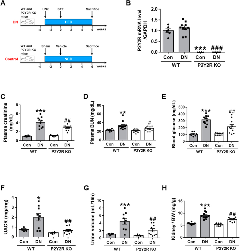
Methods: We used a mouse model of DN by combining a treatment of high-fat diet and streptozotocin after unilateral nephrectomy in wild-type or P2Y2R knockout mice. We measured renal functional parameter in plasma, examined renal histology, and analysed expression of autophagy regulatory proteins.

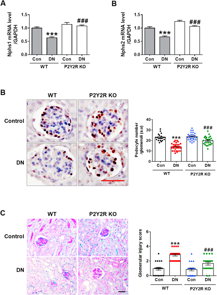
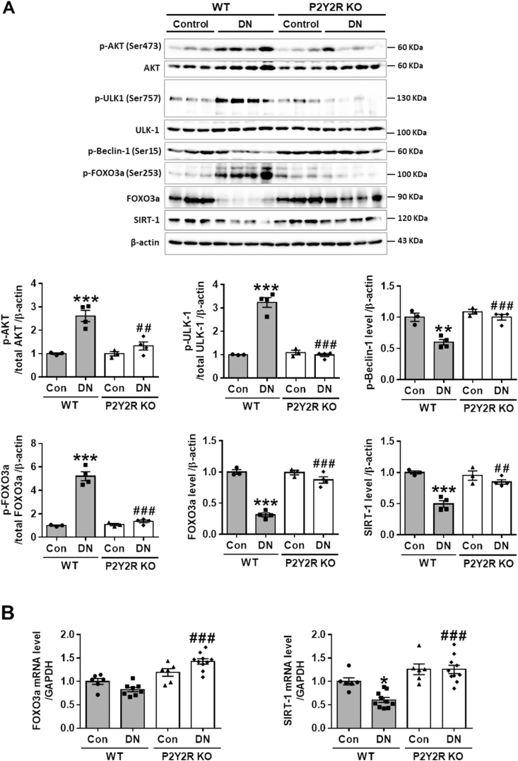
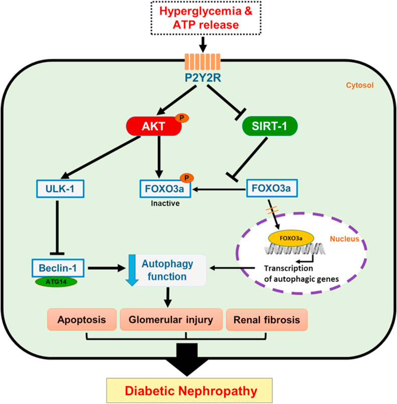
Results: Hyperglycaemia and ATP release were induced in wild type-DN mice and positively correlated with renal dysfunction. Conversely, P2Y2R knockout markedly attenuates albuminuria, podocyte loss, development of glomerulopathy, renal tubular injury, apoptosis and interstitial fibrosis induced by DN. These protective effects were associated with inhibition of AKT-mediated FOXO3a (forkhead box O3a) phosphorylation and induction of FOXO3a-induced autophagy gene transcription. Furthermore, inhibitory phosphorylation of ULK-1 was decreased, and the downstream Beclin-1 autophagy signalling was activated in P2Y2R deficiency. Increased SIRT-1 (sirtuin-1) and FOXO3a expression in P2Y2R deficiency also enhanced autophagy response, thereby ameliorating renal dysfunction in DN.

Conclusions: P2Y2R contributes to the pathogenesis of DN by impairing autophagy and serves as a therapeutic target for treating DN.

Keywords: Autophagy; Diabetic nephropathy; FOXO3a; Glomerulus; Kidney; P2Y2R.


Figures

Figure 1
Figure 1
P2Y2R KO mice decrease albuminuria and renal dysfunction induced by DN. DN animal model was generated by STZ injection (100 mg/kg) to the WT or P2Y2R KO mice after 3 weeks of UNx and HFD feeding and then sacrificed at 6 weeks post-STZ injection. The control mice were fed with a normal chow diet (NCD). Blood and urine samples were collected from WT and P2Y2R KO control (n = 8) or DN (n = 12–14). (A) Schematic representation of experimental design. (B) Expression of endogenous P2Y2R in kidney tissues from WT and KO mice (n = 3–5). (C, D, E) Plasma creatinine, blood urea nitrogen (BUN) and blood glucose levels were measured, respectively (control n = 8, DN n = 14). (F, G, H) Urine albumin/creatinine ratio (UACR), urine volume (mL/16 h), and kidney/body weight (BW) ratio at 6 weeks post-STZ injection (control n = 8, DN n = 14). Data are presented as mean ± SEM. One-way ANOVA was used for statistical analysis followed by Bonferroni's multiple comparisons test. ∗p < 0.05, ∗∗p < 0.01, ∗∗∗p < 0.001 vs WT control mice; and #p < 0.05, ##p < 0.01, ###p < 0.001 vs WT DN mice.
Figure 2
Figure 2
P2Y2R deficiency protects against podocyte loss and glomerular injury in DN. (A) Relative mRNA levels of Nphs1 (nephrin) and Nphs2 (podocin) were determined by real-time PCR analysis (n = 3–5). (B) Localisation of WT1, a podocyte marker, was analysed by immunohistochemistry, and representative images are shown. The number of stained podocytes per glomeruli was counted by using ImageJ software (n = 3). (C) Kidney sections were stained with PAS staining and glomerular morphological changes were scored as described in the method section (n = 3). Data are presented as mean ± SEM. One-way ANOVA was used for statistical analysis followed by Bonferroni's multiple comparisons test. ∗∗∗p < 0.001 vs WT control mice; and ###p < 0.001 vs WT DN mice. Scale bar, 50 μm.
Figure 3
Figure 3
P2Y2R KO mice reduce renal tubular injury and interstitial fibrosis in DN. (A) Kidney injury molecule-1 (KIM-1) and neutrophil gelatinase-associated lipocalin (NGAL), the specific biomarkers for renal tubular damage, were assessed by real-time PCR analysis (n = 3–5). (B) Renal tubular damage was analysed by PAS staining, the representative images were shown, and the tubular damage scores were evaluated (black arrows indicate damaged renal tubules), (n = 3). (C) Picro-Sirius Red staining was performed in kidney sections and fibrotic area was presented as percentages (%) by using ImageJ software (n = 3). Data are presented as mean ± SEM. One-way ANOVA was used for statistical analysis followed by Bonferroni's multiple comparisons test. ∗∗∗p < 0.001 vs WT control mice; and ###p < 0.001 vs WT DN mice. Scale bar, 50 μm.
Figure 4
Figure 4
P2Y2R deficiency attenuates renal apoptosis in DN. (A) Representative images of TUNEL staining that were processed in the kidney sections to determine DN-induced renal apoptosis. The number of TUNEL-positive cells/HPF was counted to present the severity of apoptosis (n = 3). (B) The extent of apoptosis was further determined by caspase-3 and PARP-1 cleavage in kidney tissue lysates using western blot analysis, and the quantitative values are shown (n = 3–4). Data are presented as mean ± SEM. One-way ANOVA was used for statistical analysis followed by Bonferroni's multiple comparisons test. ∗∗∗p < 0.001 vs WT control mice and ###p < 0.001 vs WT DN mice. Scale bar, 100 μm.
Figure 5
Figure 5
P2Y2R deficiency enhances autophagy signalling in DN. (A) Kidney tissues were lysed to perform western blot analysis, and the levels of autophagy proteins (ATG5, ATG12, Beclin-1, p62 and LC3B) and β-actin, as a loading control, were examined. Quantitative analysis of each protein was shown (n = 3–4). (B) Autophagy-related gene transcripts were determined by real-time PCR analysis. Relative mRNA expression was normalised to that of GAPDH (n = 3–5). (C) Representative images of immunofluorescence staining of p62 (red) were assessed by confocal microscopy (n = 3). Data are presented as mean ± SEM. One-way ANOVA was used for statistical analysis followed by Bonferroni's multiple comparisons test. ∗p < 0.05 vs WT control mice; and #p < 0.05 vs WT DN mice. Scale bar, 20 μm.
Figure 6
Figure 6
P2Y2R deficiency alters the protein levels of p-AKT, p-FOXO3a, p-ULK-1, p-Beclin-1 and SIRT-1 in DN. (A) The protein expression levels of p-AKT, p-FOXO3a, p-ULK-1, p-Beclin-1, SIRT-1 and β-actin, as a loading control, were examined by for western blot analysis in kidney tissue lysates. Quantitative analysis of each protein level is shown (n = 3–4). (B) Relative mRNA levels of FOXO3a and SIRT-1 were determined by real-time PCR analysis in the kidney tissues. Relative mRNA expression was normalised to that of GAPDH (n = 3–5). Data are presented as mean ± SEM. One-way ANOVA was used for statistical analysis followed by Bonferroni's multiple comparisons test. ∗p < 0.05, ∗∗p < 0.01, ∗∗∗p < 0.001 vs WT control mice; and ##p < 0.01, ###p < 0.001 vs WT DN mice.
Figure 7
Figure 7
Schematic molecular mechanism of P2Y2R in the development of DN. P2Y2R activation via hyperglycaemia and extracellular ATP increases AKT phosphorylation, and the increased AKT activity promotes an inhibitory ULK-1 phosphorylation that downregulates autophagy through inhibition of Beclin-1 and ATG14 complex required for phagophore formation, resulting in renal apoptosis, glomerular injury and interstitial fibrosis. In addition, the increased phosphorylation of FOXO3a by AKT activity and the inhibited SIRT-1 activity reduced autophagy response by downregulating transcription of autophagy genes. Conversely, P2Y2R knockout mice have reduced AKT phosphorylation and increased FOXO3a and SIRT-1 expression to rescue autophagy response and attenuate the progression of DN.

References

    1. Ding Y., Choi M.E. Autophagy in diabetic nephropathy. Journal of Endocrinology. 2015;224(1):R15–R30. - PMC - PubMed
    1. Jefferson J.A., Shankland S.J., Pichler R.H. Proteinuria in diabetic kidney disease: a mechanistic viewpoint. Kidney International. 2008;74(1):22–36. - PubMed
    1. Su J., Li S.J., Chen Z.H., Zeng C.H., Zhou H., Li L.S. Evaluation of podocyte lesion in patients with diabetic nephropathy: Wilms' tumor-1 protein used as a podocyte marker. Diabetes Research and Clinical Practice. 2010;87(2):167–175. - PubMed
    1. Collins A.J., Foley R.N., Chavers B., Gilbertson D., Herzog C., Ishani A. US renal data system 2013 annual data report. American Journal of Kidney Diseases. 2014;63(1 Suppl):A7. - PubMed
    1. Klionsky D.J., Emr S.D. Autophagy as a regulated pathway of cellular degradation. Science. 2000;290(5497):1717–1721. - PMC - PubMed

Publication types