Comparative Study
. 2020 Sep 24;12(10):610.
doi: 10.3390/toxins12100610.

Mycotoxin Deoxynivalenol Has Different Impacts on Intestinal Barrier and Stem Cells by Its Route of Exposure

Affiliations
Comparative Study

Mycotoxin Deoxynivalenol Has Different Impacts on Intestinal Barrier and Stem Cells by Its Route of Exposure

Hikaru Hanyu et al. Toxins (Basel). .

Abstract

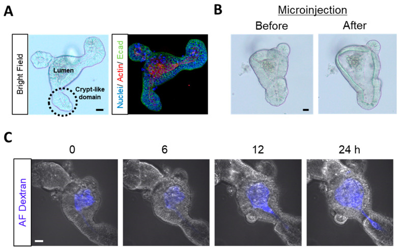
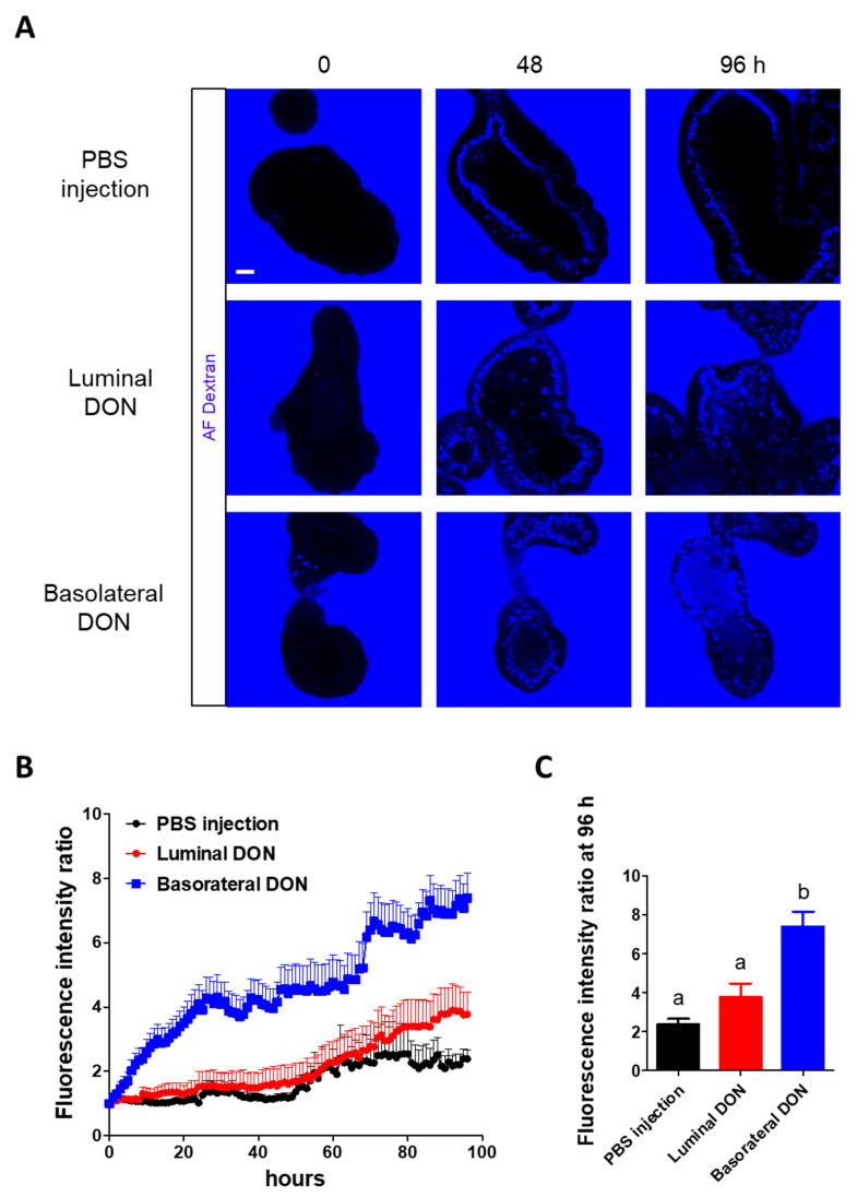
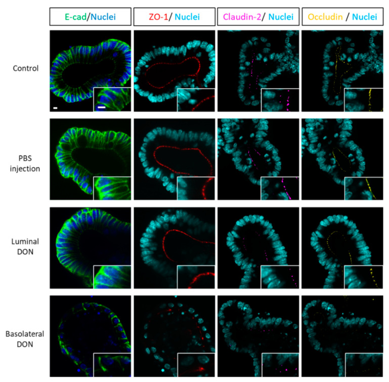
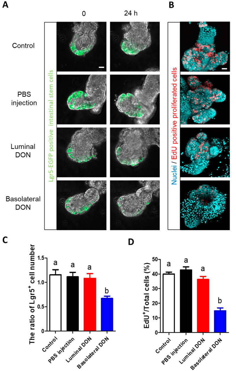
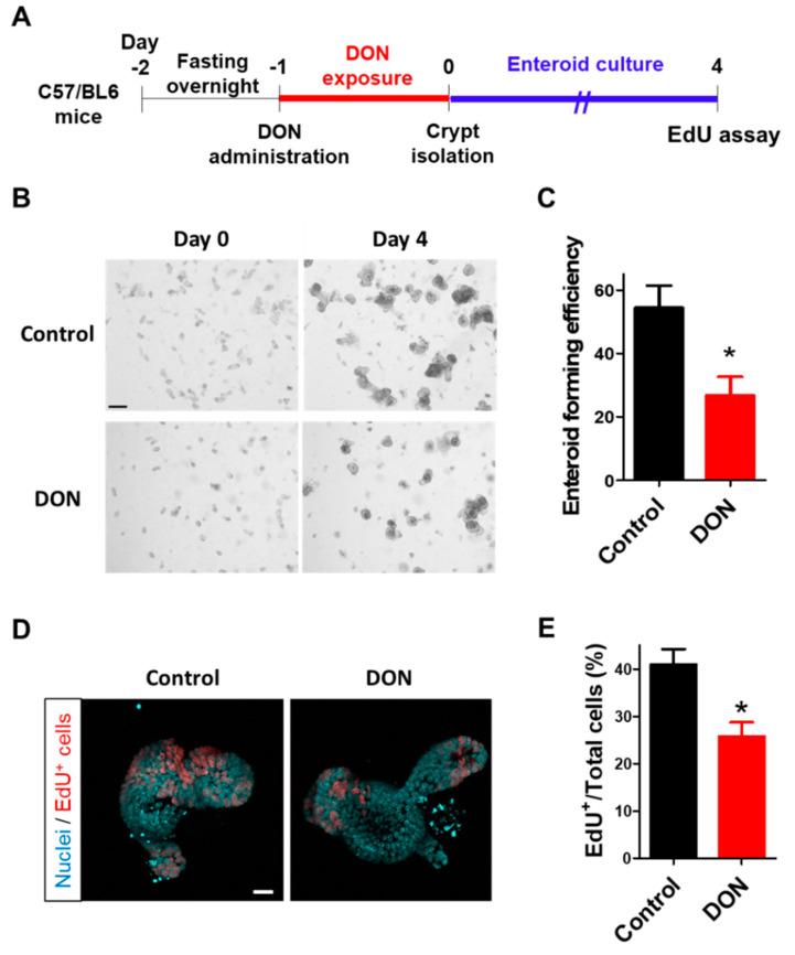
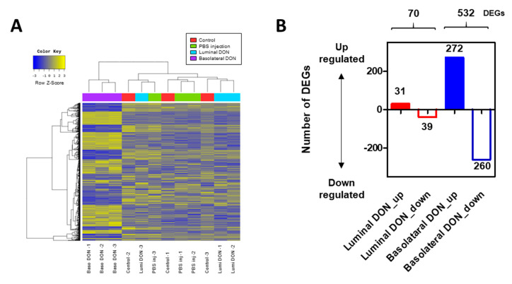
The different effects of deoxynivalenol (DON) on intestinal barrier and stem cells by its route of exposure remain less known. We explored the toxic effects of DON on intestinal barrier functions and stem cells after DON microinjection (luminal exposure) or addition to a culture medium (basolateral exposure) using three-dimensional mouse intestinal organoids (enteroids). The influx test using fluorescein-labeled dextran showed that basolateral DON exposure (1 micromolar (µM) disrupted intestinal barrier functions in enteroids compared with luminal DON exposure at the same concentration. Moreover, an immunofluorescence experiment of intestinal epithelial proteins, such as E-cadherin, claudin, zonula occludens-1 (ZO-1), and occludin, exhibited that only basolateral DON exposure broke down intestinal epithelial integrity. A time-lapse analysis using enteroids from leucine-rich repeat-containing G-protein-coupled receptor 5 (Lgr5)-enhanced green fluorescence protein (EGFP) transgenic mice and 5-ethynyl-2-deoxyuridine (EdU) assay indicated that only the basolateral DON exposure, but not luminal DON exposure, suppressed Lgr5+ stem cell count and proliferative cell ratio, respectively. These results revealed that basolateral DON exposure has larger impacts on intestinal barrier function and stem cells than luminal DON exposure. This is the first report that DON had different impacts on intestinal stem cells depending on the administration route. In addition, RNA sequencing analysis showed different expression of genes among enteroids after basolateral and luminal DON exposure.

Keywords: basolateral exposure; deoxynivalenol; enteroids; intestinal barrier; intestinal stem cells; luminal exposure; microinjection; mycotoxin; organoids.


Conflict of interest statement

The authors declare no conflict of interest.

Figures

Figure 1
Figure 1
Microinjection into enteroids as luminal exposure. (A) Representative images of mice enteroids in a bright field (left) and immunofluorescence (right; blue: Nuclei, red: Actin, and green: E-cadherin). (B) Representative images of enteroids before (left) and after (right) microinjection. (C) Representative confocal images of enteroids microinjected with Alexa Fluor® (AF) dextran (blue). Scale bars: 20 µm.
Figure 2
Figure 2
Effects of basolateral or luminal DON exposures on intestinal barrier functions. (A) Representative confocal images of enteroids with fluorescent dextran (blue) at 0, 48, and 96 h after treatments (phosphate-buffered saline (PBS) injection, 1-µM luminal DON exposure, or 1-µM basolateral DON exposure). Scale bar: 10 µm. (B) The time course of fluorescence intensity ratio of enteroids with fluorescent dextran. Mean ± standard error of the mean (SEM), n = 17–18. (C) Fluorescence intensity ratio of enteroids (black: Control, red: Luminal DON exposure, and blue: Basolateral DON exposure) at 96 h after treatments. Data were taken from Figure 2B. Mean ± SEM, n = 17–18. Different lowercase letters indicate significant differences (p < 0.05; Tukey′s post hoc test).
Figure 3
Figure 3
Effects of basolateral or luminal DON exposures on intestinal epithelial integrity. Representative confocal images of enteroids at 48 h after treatments (control, PBS injection, 1-µM luminal DON exposure, or 1-µM basolateral DON exposure). Immunofluorescence shows E-cadherin (green), ZO-1 (red), claudin-2 (pink), occludin (yellow), and nuclei (blue or sky blue). Scale bars: 10 µm.
Figure 4
Figure 4
Effects of basolateral or luminal exposures of DON on intestinal stem cells. (A) Representative confocal images of Lgr5-enhanced green fluorescence protein (EGFP) enteroids at 0 or 24 h after treatments (control, PBS injection, 1-µM luminal DON exposure, or 1-µM basolateral DON exposure). Lgr5-EGFP+ cells (green) shows Lgr5+ stem cells. (B) Representative confocal images of enteroids at 24 h after treatments. EdU+ cells (red) show proliferative cells and nuclei stained by Hoechst 33342 (sky blue). (C) The ratio of Lgr5+ cell numbers in enteroids at 24 h/0 h after treatments. Mean ± SEM, n = 8–16. (D) EdU+ cell quantification in enteroids at 24 h after treatments. The number of EdU+ cells was normalized with the number of total cells and expressed as EdU/total cells (%). Mean ± SEM, n = 16–22. Different lowercase letters indicate significant differences (p < 0.05; Tukey’s post hoc test). Scale bars: 20 µm.
Figure 5
Figure 5
Effects of oral administration of DON to mice on intestinal stem cells. (A) Scheme of the experimental design. After fasting overnight, Wild-type (WT) mice were orally administered with DON at a dose of 50 mg/kg body weight. After the crypts were isolated from the mice at 24 h after DON exposure, enteroids were prepared and cultured for four days. (B) Representative images of enteroids (at day 0 or 4 after crypt isolation) derived from mice with or without oral DON administration. Scale bar: 200 µm. (C) Enteroid-forming efficiency from mice with or without oral DON administration. Enteroid-forming efficiency was calculated from the ratio of the number of enteroids at day 4 to the number of crypts at day 0. Mean ± SEM, n = 8–12. Asterisk (*) indicates a significant difference. (p < 0.05; Student′s t-test.) (D) Representative confocal images of enteroids at day 4 after crypt isolation. EdU+ cells (red) show proliferative cells and nuclei stained by Hoechst 33342 (sky blue). Scale bar: 20 µm. (E) EdU+ cell quantification in enteroids at day 4 after crypt isolation. The number of EdU+ cells was normalized with the number of total cells and expressed as EdU/Total cells (%). Mean ± SEM, n = 12–18. Asterisk (*) indicates a significant difference (p < 0.05; Students t-test).
Figure 6
Figure 6
RNA sequencing (RNA-seq) analysis of enteroids after the basolateral or luminal DON exposure. (A) Heat map of 888 differentially expressed genes (DEGs) identified through comparison among control enteroids, PBS-injected enteroids, and enteroids after luminal or basolateral DON exposure. (B) Distribution of up- or downregulated DEGs in enteroids after luminal and basolateral DON treatment analyzed through RNA-seq analysis.
Figure 7
Figure 7
Kyoto Encyclopedia of Genes and Genomes (KEGG) pathway analysis of enteroids after basolateral or luminal DON exposure. Kyoto Encyclopedia of Genes and Genomes (KEGG) pathway of focal adhesion (mmu04510). Green- or red-colored gene name box indicates down- or upregulated genes in enteroids after luminal (left) or basolateral (right) DON exposures, compared with the control enteroids as log2 (fold change) of the expression value.

Similar articles

Cited by

References

    1. Marin S., Ramos A., Cano-Sancho G., Sanchis V. Mycotoxins: Occurrence, toxicology, and exposure assessment. Food Chem. Toxicol. 2013;60:218–237. doi: 10.1016/j.fct.2013.07.047. - DOI - PubMed
    1. Pestka J.J., Smolinski A.T. Deoxynivalenol: Toxicology and potential effects on humans. J. Toxicol. Env. Heal. Part B. 2005;8:39–69. doi: 10.1080/10937400590889458. - DOI - PubMed
    1. Sugita-Konishi Y., Park B.J., Kobayashi-Hattori K., Tanaka T., Chonan T., Yoshikawa K., Kumagai S. Effect of cooking process on the deoxynivalenol content and its subsequent cytotoxicity in wheat products. Biosci. Biotech. Biochem. 2006;70:1764–1768. doi: 10.1271/bbb.50571. - DOI - PubMed
    1. Bhat R., Ramakrishna Y., Beedu S., Munshi K. Outbreak of trichothecene mycotoxicosis associated with consumption of mould-damaged wheat products in Kashmir Valley, India. Lancet. 1989;333:35–37. doi: 10.1016/S0140-6736(89)91684-X. - DOI - PubMed
    1. Luo X. Fusarium toxins contamination of cereals in China. JSM Mycotoxins. 1988;1988:97–98. doi: 10.2520/myco1975.1988.1Supplement_97. - DOI

Publication types

MeSH terms