Review
. 2020 Sep 24;12(10):1069.
doi: 10.3390/v12101069.

Solid-State NMR for Studying the Structure and Dynamics of Viral Assemblies

Affiliations
Review

Solid-State NMR for Studying the Structure and Dynamics of Viral Assemblies

Lauriane Lecoq et al. Viruses. .

Abstract

Structural virology reveals the architecture underlying infection. While notably electron microscopy images have provided an atomic view on viruses which profoundly changed our understanding of these assemblies incapable of independent life, spectroscopic techniques like NMR enter the field with their strengths in detailed conformational analysis and investigation of dynamic behavior. Typically, the large assemblies represented by viral particles fall in the regime of biological high-resolution solid-state NMR, able to follow with high sensitivity the path of the viral proteins through their interactions and maturation steps during the viral life cycle. We here trace the way from first solid-state NMR investigations to the state-of-the-art approaches currently developing, including applications focused on HIV, HBV, HCV and influenza, and an outlook to the possibilities opening in the coming years.

Keywords: capsids; membrane proteins; solid-state NMR; structure; viral proteins.


Conflict of interest statement

The authors declare no conflict of interest.

Figures

Figure 1
Figure 1
NMR rotors, spectrometer and resonance signals: (a) NMR rotors for fast spinning: 110 kHz for 0.7 mm rotor, 150 kHz for 0.5 mm (picture courtesy of Susanne Penzel). (b) 1200 MHz magnet for solid-state NMR at ETH Zurich. (c) Schematic representation of anisotropic interactions arising from different orientation of the individual molecules with respect to the magnetic field. The superposition of all possible chemical shifts gives rise to broad peaks with a characteristic shape, the powder pattern. The anisotropic interactions can be averaged out using MAS, which results in a single resonance line centered at the isotropic chemical shift of the spin. In addition, spinning sidebands may appear. This spin can be correlated to a neighboring spin, in the example, the amide 1H to the amide 15N. Two-dimensional spectroscopy then shows peaks which represent, for the present example, the amide proton frequency in one dimension, and the nitrogen frequency in the other. Such a signal will be observed for every NH pair in the protein. The resonance-line position is given by the isotropic part of NMR chemical shift and is usually specified in ppm (parts per million) of the resonance frequency. To obtain high-resolution spectra, the linewidth should be as narrow as possible, as this allows to distinguish (resolve) a maximum of resonances. A narrowing of linewidths can be achieved by (i) using a spectrometer operating at a higher magnetic field, (ii) improving sample homogeneity and symmetry, and (iii) reducing the dipolar coupling interactions by decreasing the density of protons in the system (through protein deuteration, for example) and/or increasing the MAS frequency by using smaller diameter rotors.
Figure 2
Figure 2
Overview of human viruses where solid-state NMR contributed to the understanding of global organization, orientation in the membrane and protein structure and dynamics. Viral proteins investigated by solid-state NMR are labeled in red. (a) HIV-1 virion with envelope-embedded gp41 protein containing the fusion peptide and internal capsid. (b) Influenza virion with M2 ion channel. (c) Measles virion with nucleocapsid protein. (d) HBV virion with small surface protein S and internal capsid. (e) Membrane topology of HCV with p7 and NS4B membrane proteins. Panel (d) is reprinted from [86], Copyright (2014), with permission from Elsevier. Panel (e) is adapted from [87].
Figure 3
Figure 3
Solid-state NMR studies on the HIV and HBV capsids. (a) Illustration of the mature HIV-1 capsid (PDB: 3J3Y [99]); and the CA protein in (b), with a zoom on the backbone 15NH CSA tensor and its orientation in the protein molecular frame. (c) Transmission EM image of CA and (d) the corresponding 2D NCA spectrum recorded at 14 kHz MAS. (e) HBV capsid structure (PDB: 1QGT [7]) showing the pentamers formed by A subunits and hexamers formed by B, C and D subunits. (f) Negative-staining EM picture of HBV capsids and (g) corresponding 2D Dipolar Assisted Rotational Resonance (DARR) spectrum recorded at 17.5 kHz MAS. Residues with NMR peak splitting due to the asymmetric subunits are highlighted in yellow and are shown as spheres in (h) on the capsid structure. Pictures from panels (ad) were reprinted with permission from reference [94], Copyright (2016) American Chemical Society. Pictures from panels (eh) were taken with permissions from reference [55].
Figure 4
Figure 4
(a) 2D hNH spectrum of HBV capsids made of full-length core protein (Cp183) expressed and auto-assembled using WG-CFPS, recorded at 100 kHz. (b) Negative-staining EM pictures of the capsids prepared by WG-CFPS in the absence (top) and in the presence (bottom) of a JNJ-890 capsid assembly modulator, leading to opened objects. Figure adapted with permissions from reference [40].
Figure 5
Figure 5
(a) EM pictures of intact (grey) and cleaved (brown) measle nucleocapsids [111]. (b) 2D hNH spectra recorded at 60 kHz MAS of intact (blue) and cleaved (red) measle nucleocapsids and corresponding bulk 15N R1rho decays in (c), revealing longer life times and thus different dynamics for the cleaved form. Figure reprinted from [110], Copyright (2014), with permission from Elsevier.
Figure 6
Figure 6
(a) 2D hNH spectrum of DHBs small envelope protein produced using WG-CFPS, recorded at 110 kHz MAS. (b) Negative-staining EM pictures of the corresponding protein, which self-assembles into spherical particles of ~29 nm. Figures taken with permission from reference [41].
Figure 7
Figure 7
Solid-state NMR structures of (a) Vpu from HIV-1 (PDB: 2N28 [127]), and (b) closed and open influenza BM2 channels in lipid membranes (PDB: 6pvr (pH 7.5) and 6pvt (pH 4.5) [130]). Panel (a) reprinted from reference [127] Copyright (2015), with permission from Elsevier. Panel (b) reprinted from reference [130] Copyright (2020), with permission from Springer Nature.
Figure 8
Figure 8
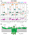
(a) 3D correlation spectra of deuterated NS4B from HCV with sequential connectivities. Selected strip plots representing the assignment of amino acid stretches of residues 119 to 125 and 237 to 241 using a set of 6 spectra recorded at 110 kHz MAS on two NS4B samples with different labeling schemes [60]. The orange spectrum was acquired at 60 kHz MAS. (b) A putative topology model of the NS4B protein adapted from [150], in which NS4B is proposed to contain four presumably amphipathic α-helices and four predicted transmembrane segments in the middle. The black boxes indicate the location of the two assigned regions shown in panel a. Figure reproduced with permission from reference [60].

References

    1. Hopper P., Harrison S.C., Sauer R.T. Structure of tomato bushy stunt virus. V. Coat protein sequence determination and its structural implications. J. Mol. Biol. 1984;177:701–713. doi: 10.1016/0022-2836(84)90045-7. - DOI - PubMed
    1. Silva A.M., Rossmann M.G. Refined structure of southern bean mosaic virus at 2.9 A resolution. J. Mol. Biol. 1987;197:69–87. doi: 10.1016/0022-2836(87)90610-3. - DOI - PubMed
    1. Krishnaswamy S., Rossmann M.G. Structural refinement and analysis of Mengo virus. J. Mol. Biol. 1990;211:803–844. doi: 10.1016/0022-2836(90)90077-Y. - DOI - PubMed
    1. Arnold E., Rossmann M.G. The use of molecular-replacement phases for the refinement of the human rhinovirus 14 structure. Acta Crystallogr. A Found. Crystallogr. 1988;44:270–282. doi: 10.1107/S0108767387011875. - DOI - PubMed
    1. Kim S.S., Smith T.J., Chapman M.S., Rossmann M.C., Pevear D.C., Dutko F.J., Felock P.J., Diana G.D., McKinlay M.A. Crystal structure of human rhinovirus serotype 1A (HRV1A) J. Mol. Biol. 1989;210:91–111. doi: 10.1016/0022-2836(89)90293-3. - DOI - PubMed

Publication types

MeSH terms

LinkOut - more resources