. 2020 Sep 24;12(10):614.
doi: 10.3390/toxins12100614.

Inhibitory Effects of a Reengineered Anthrax Toxin on Canine and Human Osteosarcoma Cells

Affiliations

Inhibitory Effects of a Reengineered Anthrax Toxin on Canine and Human Osteosarcoma Cells

Jonathan Mackowiak da Fonseca et al. Toxins (Basel). .

Abstract

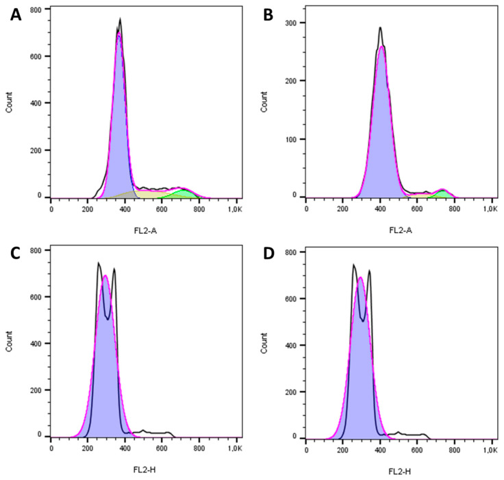
Canine and human osteosarcomas (OSA) share similarities. Novel therapies are necessary for these tumours. The Bacillus anthracis toxin was reengineered to target and kill cells with high expressions of matrix metalloproteinases (MMPs) and urokinase plasminogen activator (uPA). Since canine OSA express MMPs and uPA, we assessed whether the reengineered toxin could show efficacy against these tumours. Two OSA cell lines (canine D17 and human MG63) and a non-neoplastic canine osteoblastic cell line (COBS) were used. Cells were treated with different concentrations of the reengineered anthrax toxin and cell viability was quantified using MTT assay. The cell cycle, apoptosis, and necrosis were analysed by flow cytometry. The wound-healing assay was performed to quantify the migration capacity of treated cells. D17 and MG63 cells had significantly decreased viability after 24 h of treatment. Cell cycle analysis revealed that OSA cells underwent apoptosis when treated with the toxin, whereas COBS cells arrested in the G1 phase. The wound-healing assay showed that D17 and MG63 cells had a significantly reduced migration capacity after treatment. These results point for the first time towards the in vitro inhibitory effects of the reengineered anthrax toxin on OSA cells; this reengineered toxin could be further tested as a new therapy for OSA.

Keywords: Bacillus anthracis; anthrax; apoptosis; canine osteosarcoma; toxin.


Conflict of interest statement

The authors declare that they have no conflict of interest.

Figures

Figure 1
Figure 1
Photomicrograph representation of the expression of different markers in canine (D17) and human (MG63) osteosarcoma cell lines and canine osteoblastic (COBS) cell line, as tested by immunofluorescence. (A) Metalloproteinase 2 (MMP2) expression in canine (D17) and human (MG63) osteosarcoma cell lines. (B) The urokinase plasminogen activator (uPA)expression in canine (D17) and human (MG63) osteosarcoma cell lines. (C) Membrane-type 1 matrix metalloproteinase MT1-MMP expression in canine (D17) and human (MG63) osteosarcoma cell lines. (D) Membrane-type 1 matrix metalloproteinase (MT1-MMP) and Metalloproteinase 2 (MMP2) expression in Canine Osteoblast Spitz (COBS) cell line. Note: Alexa Fluor 488: green; DAPI: blue. Original magnification 40×.
Figure 2
Figure 2
Response of the canine osteosarcoma cell line (D17) to the reengineered anthrax toxin. (A) D17 cells treated with LF + PA at differing PA concentrations; (B) D17 cells treated with LF + PAL1/PAU2 at differing PA concentrations. (C) D17 cells treated with FP59 + PA at differing PA concentrations. (D) D17 cells treated with FP59 + PAL1/PAU2 at differing PA concentrations. Optical density was compared to that of untreated cells (only LF or FP59). Note: * p < 0.05; *** p < 0.001. LF: lethal factor; PA: protective antigen; PAL1: PA-L1-l210; PAU2: PA-U2-R200A; FP59: the A exotoxin of Pseudomonas aeruginosa combined with the LF.
Figure 3
Figure 3
Response of the human osteosarcoma cell line (MG63) to the reengineered anthrax toxin. (A) MG63 cells treated with LF + PA at differing PA concentrations. (B) MG63 cells treated with LF + PAL1/PAU2 at differing PA concentrations. (C) MG63 cells treated with FP59 + PA at differing PA concentrations. (D) MG63 cells treated with FP59 + PAL1/PAU2 at differing PA concentrations. Optical density was compared to that of untreated cells (only LF or FP59). Note: ** p < 0.01; *** p < 0.001. LF: lethal factor; PA: protective antigen; PAL1: PA-L1-l210; PAU2: PA-U2-R200A; FP59: the A exotoxin of Pseudomonas aeruginosa combined with the LF.
Figure 4
Figure 4
Response of the non-neoplastic canine osteoblastic cell line (COBS) to the reengineered anthrax toxin. (A) COBS cells treated with LF + PA at differing PA concentrations. (B) COBS cells treated with LF + PAL1/PAU2 at differing PA concentrations. (C) COBS cells treated with FP59 + PA at differing PA concentrations. (D) COBS cells treated with FP59 + PAL1/PAU2 at differing PA concentrations. Optical density was compared to that of untreated cells (only LF or FP59). Note: * p < 0.05; ** p < 0.01; *** p < 0.001. LF: lethal factor; PA: protective antigen; PAL1: PA-L1-l210; PAU2: PA-U2-R200A; FP59: the A exotoxin of Pseudomonas aeruginosa combined with the LF.
Figure 5
Figure 5
Graphs showing the cell viability of canine and human osteosarcoma cell lines (D17 and MG63, respectively) and canine osteoblast cell line (COBS) after treatment with reengineered anthrax toxins: (A) LF + PA; (B) LF + PAL1/PAU2; (C) FP + PA; (D) FP + PAL1/PAU2. The IC50 values are shown in Table 1.
Figure 6
Figure 6
Cell cycle histograms of the canine osteosarcoma cell line D17 after 24 h of treatment with the reengineered anthrax toxin from Bacillus anthracis: (A) LF only; (B) LF + PAL1/PAU2 50 ng/mL; (C) LF + PAL1/PAU2 500 ng/mL; (D) LF + PAL1/PAU2 5000 ng/mL. LF: lethal factor; PAL1: PA-L1-l210; PAU2: PA-U2-R200A. The black line represents a cell population histogram in respect to iodide propide fluorescence, while the pink line represents the univariate cell cycle model created by FlowJo software to de-convolute the populations in order to assign percentage values to each population, since adjacent populations overlap each other, resulting in the purple, yellow, and green areas, which represent G0/G1, S, and G2/M phases, respectively.
Figure 7
Figure 7
Cell cycle histograms of the human osteosarcoma cell line MG63 after 24 h of treatment with the reengineered anthrax toxin from Bacillus anthracis: (A) LF only; (B) LF + PAL1/PAU2 50 ng/mL; (C) LF + PAL1/PAU2 500 ng/mL; (D) LF + PAL1/PAU2 5000 ng/mL. LF: lethal factor; PAL1: PA-L1-l210; PAU2: PA-U2-R200A.
Figure 8
Figure 8
Cell cycle histograms of the non-neoplastic COBS cell line after 24 h of treatment with the reengineered anthrax toxin from Bacillus anthracis: (A) LF only; (B) LF + PAL1/PAU2 50 ng/mL: (C) LF + PAL1/PAU2 500 ng/mL; (D) LF + PAL1/PAU2 5000 ng/mL. LF: lethal factor; PAL1: PA-L1-l210; PAU2: PA-U2-R200A.
Figure 9
Figure 9
Relative width of the lesion in the wound-healing assay in the canine osteosarcoma cell line D17 with and without treatment of LF + PAL1/PAU2 at a concentration of 5000 ng/mL PA. (A) D17 cells treated with LF + PAL1/PAU2 at 5000 ng/mL PA. (B) Representative images of the wound-healing assay in the canine osteosarcoma cell line D17. Note: *** p < 0.001 in relation to the control at 24 h. LF: lethal factor; PAL1: PA-L1-l210; PAU2: PA-U2-R200A.
Figure 10
Figure 10
Relative area of the lesion in the wound-healing assay in the human osteosarcoma cell line MG63 with and without LF + PAL1/PAU2 at 5000 ng/mL PA. (A) MG63 cells treated with LF + PAL1/PAU2 at 5000 ng/mL PA. (B) Representative images of the wound-healing assay in the human osteosarcoma cell line MG63. Note: ** p < 0.01 in relation to the control at 24 h. LF: lethal factor; PAL1: PA-L1-l210; PAU2: PA-U2-R200A.
Figure 11
Figure 11
Photomicrograph representative of canine (D17) and human (MG63) osteosarcoma cell lines and canine osteoblastic cell line (COBS) in culture: (A) canine osteosarcoma cell line (D17); (B) human osteosarcoma cell line (MG63); (C) canine osteoblastic cell line (COBS). Original magnification 40×.

Similar articles

References

    1. Morello E., Martano M., Buracco P. Biology, diagnosis and treatment of canine appendicular osteosarcoma: Similarities and differences with human osteosarcoma. Vet. J. 2011;189:268–277. doi: 10.1016/j.tvjl.2010.08.014. - DOI - PubMed
    1. Withrow S.J., Powers B.E., Straw R.C., Wilkins R.M. Comparative aspects of osteosarcoma. Dog versus man. Clin. Orthop. Relat. Res. 1991:159–168. - PubMed
    1. Misdorp W., Van der Heul R.O. Tumours of bones and joints. Bull. World Health Organ. 1976;53:265–282. - PMC - PubMed
    1. Covey J.L., Farese J.P., Bacon N.J., Schallberger S.P., Amsellem P., Cavanaugh R.P., Milner R.J. Stereotactic radiosurgery and fracture fixation in 6 dogs with appendicular osteosarcoma. Vet. Surg. 2014;43:174–181. doi: 10.1111/j.1532-950X.2014.12082.x. - DOI - PubMed
    1. Selvarajah G.T., Bonestroo F.A., Kirpensteijn J., Kik M.J., van der Zee R., van Eden W., Timmermans-Sprang E.P., Slob A., Mol J.A. Heat shock protein expression analysis in canine osteosarcoma reveals HSP60 as a potentially relevant therapeutic target. Cell Stress Chaperones. 2013;18:607–622. doi: 10.1007/s12192-013-0414-2. - DOI - PMC - PubMed

Publication types

MeSH terms