. 2020 Sep 25;18(1):158.
doi: 10.1186/s12964-020-00630-w.

Inhibition of αvβ3 integrin impairs adhesion and uptake of tumor-derived small extracellular vesicles

Affiliations

Inhibition of αvβ3 integrin impairs adhesion and uptake of tumor-derived small extracellular vesicles

Wanessa F Altei et al. Cell Commun Signal. .

Abstract

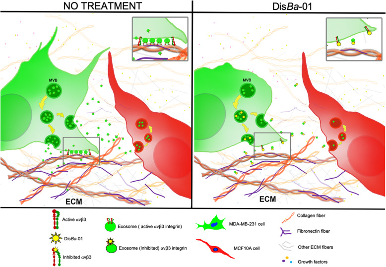
Background: Extracellular vesicles (EVs) are lipid-bound particles that are naturally released from cells and mediate cell-cell communication. Integrin adhesion receptors are enriched in small EVs (SEVs) and SEV-carried integrins have been shown to promote cancer cell migration and to mediate organ-specific metastasis; however, how integrins mediate these effects is not entirely clear and could represent a combination of EV binding to extracellular matrix and cells.

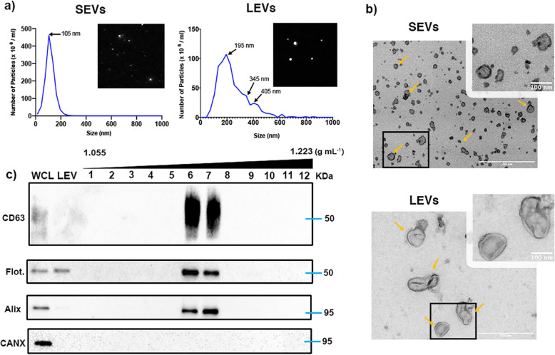
Methods: To probe integrin role in EVs binding and uptake, we employed a disintegrin inhibitor (DisBa-01) of integrin binding with specificity for αvβ3 integrin. EVs were purified from MDA-MB-231 cells conditioned media by serial centrifugation method. Isolated EVs were characterized by different techniques and further employed in adhesion, uptake and co-culture experiments.

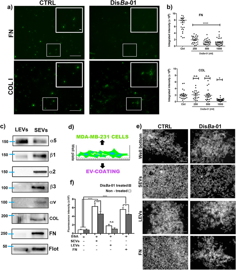
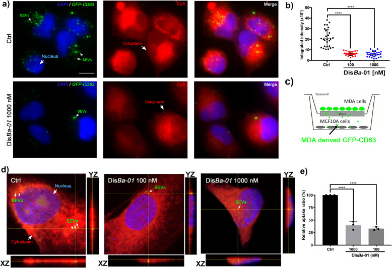
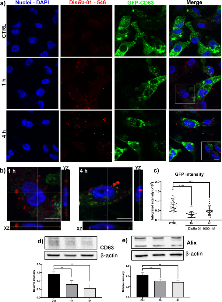
Results: We find that SEVs secreted from MDA-MB-231 breast cancer cells carry αvβ3 integrin and bind directly to fibronectin-coated plates, which is inhibited by DisBa-01. SEV coating on tissue culture plates also induces adhesion of MDA-MB-231 cells, which is inhibited by DisBa-01 treatment. Analysis of EV uptake and interchange between cells reveals that the amount of CD63-positive EVs delivered from malignant MDA-MB-231 breast cells to non-malignant MCF10A breast epithelial cells is reduced by DisBa-01 treatment. Inhibition of αvβ3 integrin decreases CD63 expression in cancer cells suggesting an effect on SEV content.

Conclusion: In summary, our findings demonstrate for the first time a key role of αvβ3 integrin in cell-cell communication through SEVs. Video Abstract.

Keywords: Adhesion; Breast cancer; Small extracellular vesicles; Uptake; αvβ3 integrin.


Conflict of interest statement

The authors declare that they have no competing interests. The work was funded in part by National Institute of Health [NIH grants: 1R01GM117916 and 1R01CA206458 to AMW].

Figures

Fig. 1
Fig. 1
Validation of cancer cell-derived EV isolation and characterization. a Representative trace and video acquisition snapshot from nanoparticle tracking analysis of small and large EVs. Both traces show vesicles within a typical size profile. b Transmission electron microscopy of density gradient-purified SEVs and LEVs. Yellow arrows point to representative EVs. Scale bar: 500 nm (large image) and 100 nm (zoomed images). c Western blotting for the EV markers CD63, Flotillin and Alix across 12 iodixanol density gradient fractions, large vesicles (LEV) and whole cell lysate (WCL) of the EV donor cells. Calnexin (CANX) antibody was used as a negative control
Fig. 2
Fig. 2
SEVs adhere to ECM components and support MDA-MB-231 cell adhesion. a Representative epifluorescence images of CFSE-labeled-SEV adhesion to fibronectin and collagen coating. Treated (DisBa-01500 nM) and untreated (CTRL) groups are compared. Scale bar: 100 μm (whole images); 10 μm (zoomed images). b Quantitation of SEV adhesion to fibronectin and collagen. **** p < 0.0001; * p < 0.05. c Western blot analysis of integrin subunits, α5, β1, α2, β3, and αv and ECM proteins, FN and COL on isolated SEVs and LEVs. d Schematic cartoon of cell adhesion assay to EV coating. e Representative images of Calcein AM-labeled MDA cell adhesion to well bottom, SEVs, LEV and FN coating comparing cell adhesion in control (CTRL) and DisBa-01 (100 nM) treated conditions. f Calcein AM fluorescence intensity of adherent cells. Comparison of adhesion in different coating and effect of DisBa-01 at 100 nM. * p < 0.05; *** p < 0.001; **** p < 0.0001
Fig. 3
Fig. 3
Inhibition of SEV uptake by αvβ3 integrin blocking. a Representative images of SEV uptake in MCF 10A cells labeled with cell tracker red. Arrows indicate internalized SEVs. Ctrl, control. Scale bar, 50 μm. b Integrated intensity of CFSE-labeled SEV internalized in MCF 10A cells; ****p < 0.0001. c Schematic view of transwell system. d Orthogonal view of MCF 10A co-cultured with MDA-MB-GFP-CD63 cells showing cells-derived GFP-CD63 uptake in control (Ctrl) and DisBa-01 (100 and 1000 nM) treated cells. Arrows indicate internalized GFP-CD63. Scale bar, 10 μm. e Quantitation of relative uptake of vesicles; ****p < 0.0001
Fig. 4
Fig. 4
DisBa-01 inhibits SEV uptake in a co-culture system. a Scanning electron microscopy of MDA-MB-231 and MCF 10A cells in single and co-culture systems. Ctrl, control. DisBa-01, 1000 nM. Scale bar, 10 μm. b Effect of integrin inhibition on MDA-MB-231 cell morphology; **p < 0.01. c Representative epifluorescence images highlighting the distribution of GFP-CD63-enriched vesicles between MDA-MB-231 expressing GFP-CD63 (green) and MCF 10A (red) cells. Scale bar, 50 μm. d Effect of integrin inhibition on MDA-MB-231-GFP-CD63 cell morphology; ***p < 0.001. e Extracellular GFP-CD63-EVs from tumor to non-malignant cells. Z-stack slices from confocal acquisition show reduction of internalized and extracellular SEVs in DisBa-01 treated cells. f Orthogonal view of MCF 10A cells showing internalized GFP-CD63-SEVs and their reduction upon DisBa-01 treatment. g Effect of integrin inhibition on SEV uptake; ***p < 0.001. h Effect of integrin inhibition on extracellular GFP-CD63; ****p < 0.001
Fig. 5
Fig. 5
DisBa-01 decreases GFP-CD63 content. (a) Stacks from confocal images showing reduction of GFP-CD63 intensity 1 h and 4 h after DisBa-01 treatment. CTRL, control. (b) Orthogonal view of DisBa-01-Alexa Fluor-546 internalized on MDA-MB-231-GFP-CD63 cells, evidencing an increased level of red signal in 4 h condition. (c) Quantitation of green fluorescent signal of GFP-CD63. Reduced expression level of CD63 (d) and Alix (e) after treatment with DisBa-01 in western blotting analysis. **p < 0.01; ***p < 0.001; ****p < 0.0001. Scale bar, 10 μm
Fig. 6
Fig. 6
Proposed model for inhibition of SEV adhesion and cellular uptake by recipient cells upon blockage of αvβ3 integrin. Left panel: in the absence of DisBa-01, tumor cells secrete SEVs that bind to ECM and are taken by non-malignant cells. Right panel: blockage of αvβ3 integrin by DisBa-01 decreases SEV secretion, adhesion to ECM proteins and uptake by recipient cells

Similar articles

Cited by

References

    1. Wong MS, Sidik SM, Mahmud R, Stanslas J. Molecular targets in the discovery and development of novel antimetastatic agents: current progress and future prospects. Clin Exp Pharmacol Physiol. 2013;40(5):307–319. doi: 10.1111/1440-1681.12083. - DOI - PubMed
    1. Crotti S, Piccoli M, Rizzolio F, Giordano A, Nitti D, Agostini M. Extracellular matrix and colorectal Cancer: how surrounding microenvironment affects Cancer cell behavior? J Cell Physiol. 2017;232(5):967–975. doi: 10.1002/jcp.25658. - DOI - PubMed
    1. Lu P, Weaver VM, Werb Z. The extracellular matrix: A dynamic niche in cancer progression. J Cell Biol. 2012;196(4):395–406. - PMC - PubMed
    1. Minciacchi VR, Freeman MR, Di Vizio D. Extracellular vesicles in Cancer: Exosomes, microvesicles and the emerging role of large Oncosomes. Semin Cell Dev Biol. 2015;40:41–51. doi: 10.1016/j.semcdb.2015.02.010. - DOI - PMC - PubMed
    1. Maia J, Caja S, Strano Moraes MC, Couto N, Costa-Silva B. Exosome-based cell-cell communication in the tumor microenvironment. Front Cell Dev Biol. 2018;6:1–19. doi: 10.3389/fcell.2018.00018. - DOI - PMC - PubMed

Publication types

Substances