. 2020 Oct 15;7(5):ENEURO.0189-20.2020.
doi: 10.1523/ENEURO.0189-20.2020. Print 2020 Sep/Oct.

Upregulation of nAChRs and Changes in Excitability on VTA Dopamine and GABA Neurons Correlates to Changes in Nicotine-Reward-Related Behavior

Affiliations

Upregulation of nAChRs and Changes in Excitability on VTA Dopamine and GABA Neurons Correlates to Changes in Nicotine-Reward-Related Behavior

Austin T Akers et al. eNeuro. .

Abstract

Previous reports indicate that nicotine reward is mediated through α4β2*, α6β2*, and α4α6β2* nicotinic acetylcholine receptors (nAChRs; * indicates that additional nAChR subunits may be present). Little is known about α4α6β2* nAChR involvement in reward and reinforcement because of a lack of methods that allow the direct investigation of this particular nAChR subtype. Here, we use male and female mice that contain α4-mCherry and α6-GFP nAChR subunits to show that concentrations of nicotine sufficient to evoke reward-related behavior robustly upregulate α4* and α4α6* nAChRs on midbrain dopamine (DA) and GABA neurons. Furthermore, the extent of α4α6* nAChR upregulation on ventral tegmental area (VTA) DA neurons aligns with the magnitude of nicotine reward-related behavior. We also show that the upregulation of nAChRs is accompanied by a functional change in firing frequency of both DA and GABA neurons in the VTA that is directly linked to nicotine reward-related behavior.

Keywords: excitability; nicotine; nicotinic receptor; reward; upregulation.


Figures

Figure 1.
Figure 1.
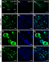
Presence of α6-GFP overlaps with presence of TH. A1–A3, 10× images of an α6-GFP mouse coronal brain slice (bregma, −3.1) immunostained with anti-GFP/Alexa Fluor 488 and anti-TH/Alexa Fluor 647. B, 20× image of neurons in the LVTA displaying overlap of α6-GFP and TH presence. C, 20× (with 5× digital zoom) images of LVTA. D, 20× Images of SNc neurons. Scale bars: 100 μm (A1–A3), 50 μm (B1–B3, D1–D3), and 10 μm (C1–C3). Figure Contributions: Brandon J. Henderson performed the experiments.
Figure 2.
Figure 2.
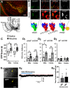
α4-mCherryα6-GFP mice reveal multiple subtypes of nAChRs on VTA pDA neurons. A, Schematic of target mouse brain region (bottom) and sample 10× image of a mouse coronal brain slice at approximately bregma −3.1 mm (top, no immunofluorescence was used here). Scale bar: 250 μm. B, Sample images of control and nicotine-treated VTA DA neurons, set to the same intensity scale, from α4-mCherryα6-GFP mice used in a CPP assay. Scale bar: 10 μm. C, Male and female mice were administered intraperitoneal injections of saline or 0.5 mg/kg nicotine in a CPP assay [n = 14 (8 males and 6 females) and 25 (14 males and 11 females) for saline and nicotine, respectively]. D1–D3, RID of α4α6*, α4*, and α6* nAChRs in saline-treated and nicotine-treated mice (from CPP assays). Individual dots represent the RID of individual mice [n = 11 (6 males and 5 females) and 19 (12 males and 7 females) for saline and nicotine, respectively]. For each mouse, 41–71 putative LVTA DA neurons were imaged. E1, Representative image of a putative VTA DA neuron in a brain slice (bregma, −3.1) from an α6-GFP mouse (scale bars: 20 μm). E2, Representative voltage-clamp recording of putative LVTA DA neurons during a 10-s puff of 300 and 500 nm nicotine. Blue bar indicates duration of nicotine puff and red dotted line represents baseline before nicotine puff. All data are mean ± SEM; *p < 0.05, **p < 0.05, ***p < 0.005; unpaired, two-tailed t test. Exact p values are given in Results. Figure Contributions: Austin T. Akers, Zachary J. Baumgard, Skylar Y. Cooper, Gabriella P. Casinelli, Alicia J. Avelar, and Brandon J. Henderson performed the experiments and analyzed the data.
Figure 3.
Figure 3.
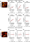
Upregulation of α4α6* and α4* nAChRs in VTA pDA neurons correlates with nicotine reward-related behavior. A1, C1, Representative merged images of LVTA DA neurons in a α4-mCherryα6-GFP brain slice. Scale bar, 10 μm. In nicotine-treated mice, changes in nAChR RID was correlated to CPP score for α4α6* nAChRs, α4* nAChRs, and α6* nAChRs for male (A2, A3, A4) and female (B1, B2, B3) mice. Linear fits (red line) with 95% confidence intervals (dotted red lines). In saline-treated mice, changes in nAChR RID was correlated to CPP score for α4α6* nAChRs (C2), α4* nAChRs (C3) and α6* nAChRs (C4). Linear fits (red line) with 95% confidence intervals (dotted red lines) were applied using Graphpad Prism software. Nicotine correlations used 7 male and 8 female α4-mCherryα6-GFP mice and the saline correlations used 5 α4-mCherryα6-GFP mice (3 males and 2 females). For each mouse, 36–71 neurons were imaged.Figure Contributions: Austin T. Akers, Zachary J. Baumgard, and Brandon J. Henderson performed the experiments and analyzed the data.
Figure 4.
Figure 4.
Upregulation of α4* nAChRs on SNr and VTA putative GABAergic neurons may not correlate with nicotine reward-related behavior. A1, B1, and C1, Representative images of neurons in the LVTA (A1), SNr (B1), or dentate gyrus (C) in a α4-mCherryα6-GFP brain slice. Scale bar, 10 μm (A1 and B1), 250 μm (C1), and 25 μm (C2, C3). A2, B2, RID of saline- and nicotine-treated mice from CPP assays for SNr GABA neurons or VTA GABA neurons. In A2 and B2 the n of individual male and female mice are indicated by individual dots. In nicotine-treated mice, changes in nAChR RID was correlated to CPP score for α4* nAChRs in SNr GABA neurons or VTA GABA neurons. In A3, A4, B3, and B4 the n of individual male and female mice are indicated by individual dots. D1, RID/area of saline- and nicotine-treated mice from CPP assays for dentate gyrus (n = 6, 3 male and 3 female). D2, Changes in nAChR RID/area was correlated to CPP score for α4* nAChRs in the dentate gyrus. Linear fits (red line) with 95% confidence intervals (dotted red lines) were applied using Graphpad Prism software. Data is Mean ± SEM; *p = 0.05, **p = 0.01; unpaired, two-tailed t test. Figure Contribution: Austin T. Akers, Zachary J. Baumgard, Skylar Y. Cooper, Alicia J. Avelar, and Brandon J. Henderson performed the experiments and analyzed the data.
Figure 5.
Figure 5.
Decreases in VTA pDA neuron firing frequency correlates with reward-related behavior. A, Schematic of target neurons within the LVTA (target bregma, −3.1). B1, B2, Representative images of VTA pDA neurons in DIC (B1) and GFP (B2) imaging modes. Scale bar: 20 μm. Mice used in CPP assays (C) were used to measure firing frequency of pDA neurons in the VTA (D, E). D, Representative cell-attached recordings of VTA pDA neuron baseline firing frequency. E, Mean VTA pDA neuron firing frequency in mice treated with saline or nicotine in CPP assays. Data are mean ± SEM, dots represent data from individual mice (n = 8–9 per condition). F, Reward-related behavior (CPP Score) was correlated to baseline firing frequency of LVTA pDA neurons; *p <0.05, **p <0.01; unpaired t test. Exact p values are given in text. Figure Contributions: Brandon J. Henderson performed the experiments and analyzed the data.
Figure 6.
Figure 6.
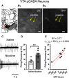
Increase in VTA pGABA neuron firing frequency correlates with reward-related behavior. A, Schematic of target pGABA neurons within the LVTA (target bregma, −3.1). B1, B2, Representative images of VTA pGABA neurons in DIC (B1) and GFP (B2) imaging modes. Scale bar: 20 μm. C, Representative cell-attached recordings of VTA pGABA neuron baseline firing frequency from mice assigned to saline or 0.5 mg/kg nicotine CPP cohorts. D, Mean VTA pGABA neuron firing frequency in mice treated with saline or nicotine in CPP assays. Data are mean ± SEM E, Reward-related behavior (CPP Score) were correlated to baseline firing frequency of VTA pGABA neurons; **p < 0.01 with; unpaired t test. Exact p values are given in text. Figure Contributions: Brandon J. Henderson performed the experiments and analyzed the data.

References

    1. Avelar AJ, Akers AT, Baumgard ZJ, Cooper SY, Casinelli GP, Henderson BJ (2019) Why flavored vape products may be attractive: green apple tobacco flavor elicits reward-related behavior, upregulates nAChRs on VTA dopamine neurons, and alters midbrain dopamine and GABA neuron function. Neuropharmacology 158:107729 10.1016/j.neuropharm.2019.107729 - DOI - PMC - PubMed
    1. Cooper SY, Akers AT, Henderson BJ (2020) Green apple e-cigarette flavorant farnesene triggers reward-related behavior by promoting high-sensitivity nAChRs in the ventral tegmental area. eNeuro 7:ENEURO.0172-20.2020 10.1523/ENEURO.0172-20.2020 - DOI - PMC - PubMed
    1. Engle SE, Shih PY, McIntosh JM, Drenan RM (2013) α4α6β2* nicotinic acetylcholine receptor activation on ventral tegmental area dopamine neurons is sufficient to stimulate a depolarizing conductance and enhance surface AMPA receptor function. Mol Pharmacol 84:393–406. 10.1124/mol.113.087346 - DOI - PMC - PubMed
    1. Fowler CD, Kenny PJ (2012) Habenular signaling in nicotine reinforcement. Neuropsychopharmacology 37:306–307. 10.1038/npp.2011.197 - DOI - PMC - PubMed
    1. Fowler CD, Lu Q, Johnson PM, Marks MJ, Kenny PJ (2011) Habenular α5 nicotinic receptor subunit signalling controls nicotine intake. Nature 471:597–601. 10.1038/nature09797 - DOI - PMC - PubMed

LinkOut - more resources