. 2020 Nov 19;202(24):e00363-20.
doi: 10.1128/JB.00363-20. Print 2020 Nov 19.

Influence of Shigella flexneri 2a O Antigen Acetylation on Its Bacteriophage Sf6 Receptor Activity and Bacterial Interaction with Human Cells

Affiliations

Influence of Shigella flexneri 2a O Antigen Acetylation on Its Bacteriophage Sf6 Receptor Activity and Bacterial Interaction with Human Cells

Min Yan Teh et al. J Bacteriol. .

Abstract

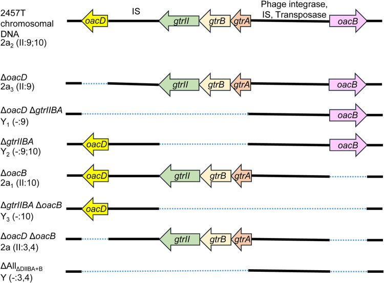
Shigella flexneri is a major causative agent of bacillary dysentery in developing countries, where serotype 2a2 is the prevalent strain. To date, approximately 30 serotypes have been identified for S. flexneri, and the major contribution to the emergence of new serotypes is chemical modifications of the lipopolysaccharide (LPS) component O antigen (Oag). Glucosylation, O-acetylation, and phosphoethanolamine (PEtN) modifications increase the Oag diversity, providing benefits to S. flexneri LPS Oag acts as a primary receptor for bacteriophage Sf6, which infects only a limited range of S. flexneri serotypes (Y and X). It uses its tailspike protein (Sf6TSP) to establish initial interaction with LPS Oags that it then hydrolyzes. Currently, there is a lack of comprehensive study on the parent and serotype variant strains from the same genetic background and an understanding of the importance of LPS Oag O-acetylations. Therefore, a set of isogenic strains (based on S. flexneri 2457T [2a2]) with deletions of different Oag modification genes (oacB, oacD, and gtrII) that resemble different naturally occurring serotype Y and 2a strains was created. The impacts of these Oag modifications on S. flexneri sensitivity to Sf6 and the pathogenesis-related properties were then compared. We found that Sf6TSP can hydrolyze serotype 2a LPS Oag, identified that 3/4-O-acetylation is essential for resistance of serotype 2a strains to Sf6, and showed that serotype 2a strains have better invasion ability. Lastly, we revealed two new serotype conversions for S. flexneri, thereby contributing to understanding the evolution of this important human pathogen.IMPORTANCE The emergence of antibiotic-resistant strains and lack of efficient vaccines have made Shigella a priority organism for the World Health Organization (1). Therefore, bacteriophage therapy has received increasing attention as an alternative therapeutic approach. LPS Oag is the most variable part of LPS due to chemical modifications and is the target of bacteriophage Sf6 (S. flexneri specific). We dissected the evolution of S. flexneri serotype Y to 2a2, which revealed a new role for a gene acquired during serotype conversion and furthermore identified new specific forms of LPS receptor for Sf6. Collectively, these results unfold the importance of the acquisition of those Oag modification genes and further our understanding of the relationship between Sf6 and S. flexneri.

Keywords: O antigen; O-acetylation; Sf6; Shigella flexneri; bacteriophages; glucosylation; lipopolysaccharide; serotypes.


Figures

FIG 1
FIG 1
Chemical structures of S. flexneri serotypes 1 through 5, 7, X, and Y. The basic O-polysaccharide backbone (serotype Y, group O-factor 3,4) is comprised of repeat units of three l-rhamnose residues (RhaIII-RhaII-RhaI) and one N-acetylglucosamine (GlcNAc). Each serotype differs by the addition of a glucosyl group, O-acetyl group, or phosphoethanolamine (PEtN) on different sugars within the repeat unit via the linkages indicated. Specific O antigen modification genes are indicated in parentheses. Each serotype has one type-specific (Roman numeral) and one or more group-specific (Arabic numeral) antigenic determinants. Serotypes 2a, 3b, 4a, 5a, and Y possess group O-factor 3,4, which is associated with the O-polysaccharide backbone but is omitted from the antigenic formula when other group O-factors are present.
FIG 2
FIG 2
Schematic diagram of various S. flexneri isogenic strains. Serotypes shown are both internationally approved and provisional, based on O antigen modifications such as O-acetylation (by oacD or oacB) and glucosylation (by gtrII). A dotted line represents the extent of the deleted region. IS, insertion sequence.
FIG 3
FIG 3
LPS profiles of various S. flexneri isogenic strains. S. flexneri strains were grown for 4 h at 37°C with aeration, followed by LPS sample preparation and SDS-PAGE and LPS silver staining as described in Materials and Methods. The open arrowheads indicate the higher-molecular-weight Oag repeat unit due to glucosylation on RhaI, while the filled arrowheads indicate the Oag repeat unit of serotype Y strains that does not have modification on RhaI. Serotypes are indicated in parentheses.
FIG 4
FIG 4
Sensitivity of various S. flexneri mutants to Sf6c phage. For bacteriophage Sf6c plaque assay, 100 μl of overnight cultures of various S. flexneri isogenic strains was incubated with 100 μl Sf6c phage (10−8), followed by the addition of 3 ml of soft LB agar, and overlaid on 25-ml LB agar plates. The plates were incubated at 37°C overnight. Serotypes are indicated in parentheses. Shigella strains were classified into three classes according to three phenotypes: class A, clear plaque with halo; class B, clear plaque without halo; class C, no plaque. Scale bars, 10 mm.
FIG 5
FIG 5
Complementation of the gtrIIBA operon in S. flexneri isogenic strains. LPS profiles of S. flexneri isogenic strains (ΔoacD ΔgtrIIBA, ΔgtrIIBA ΔoacB, ΔgtrIIBA, and ΔAllΔDIIBA+B) complemented with the gtrIIBA operon encoded on pRMM264 or pBCKS+ empty plasmid are shown. S. flexneri strains were grown for 4 h at 37°C with aeration, followed by LPS sample preparation and SDS-PAGE and LPS silver staining as described in Materials and Methods. The open arrowheads indicate the shift in Oag repeat unit size due to glucosylation on residue RhaI.
FIG 6
FIG 6
LPS profiles of various S. flexneri isogenic strains after hydrolysis with different concentrations of purified Sf6TSP. Overnight cultures of various S. flexneri strains were formalin fixed for 45 min at RT, washed twice with PBS, and resuspended in 40 μl of Sf6TSP (22 ng/ml and 44 ng/ml) in PBS or in PBS alone (no-Sf6TSP control). The bacterial and Sf6TSP mixtures were incubated at 37°C for 1 h, washed twice with Milli-Q water, and resuspended in 50 μl lysis buffer. LPS samples were subsequently prepared and subjected to SDS-PAGE and LPS silver staining as described in Materials and Methods. RU, repeat unit.
FIG 7
FIG 7
Serotype conversion of 2a1 and 2a strains into 3b1 and 3b. Serotype conversion of S. flexneri as a result of incorporation of the Sf6 genome is shown. OacA adds an acetyl group at position 2 of RhaI, which competes with GtrII that adds a glucosyl group at position 4 of RhaI.
FIG 8
FIG 8
Characterization of the invasion ability of various S. flexneri isogenic strains. HeLa cell plaque (a) and invasion (b) assays were performed using virulent S. flexneri isogenic strains. (a) Confluent HeLa cell monolayers were infected with mid-log-phase S. flexneri strains for 2 h at 37°C with 5% CO2, and plaques were observed at 48 h postinfection. The plaque size of each strain was normalized to that of 2457T. (b) Semiconfluent HeLa cell monolayers were infected with mid-log-phase S. flexneri strains at an MOI of 300, spun at 2,000 rpm for 7 min, and incubated at 37°C with 5% CO2 for 60 min. Infected monolayers were washed thrice with Dulbecco’s PBS, followed by the addition of MEM supplemented with 5% (vol/vol) FCS and 40 μg/ml gentamicin and incubation at 37°C with 5% CO2 for 90 min. Infected HeLa cells were washed thrice with PBS and lysed with 0.1% (vol/vol) Triton X-100 in PBS. Viable counts were performed by plating onto LB agar plates, and results are presented as percentage of invaded bacteria normalized to 2457T. Data are presented as mean ± standard error of the mean (SEM) from three experiments. One-way analysis of variance (ANOVA) was used to perform statistical analysis. **, P ≤ 0.01; ****, P ≤ 0.0001.
FIG 9
FIG 9
Serotype conversion pathways of S. flexneri. Serotype conversions mediated by bacteriophages SfII and Sf6 are highlighted in yellow and pink boxes, respectively. Serotype conversion mediated by the oacB transposon-like mobile element is highlighted in green boxes. Gene mutations are marked with asterisks and highlighted in gray boxes. The figure is based on this study and a study by Knirel et al. (5). Serotypes that have not been identified naturally are within dashed boxes. The order of serotype conversion from Y to 2a2 is not known; hence, two separate pathways are illustrated.

Similar articles

Cited by

References

    1. Tacconelli E, Carrara E, Savoldi A, Harbarth S, Mendelson M, Monnet DL, Pulcini C, Kahlmeter G, Kluytmans J, Carmeli Y, Ouellette M, Outterson K, Patel J, Cavaleri M, Cox EM, Houchens CR, Grayson ML, Hansen P, Singh N, Theuretzbacher U, Magrini N, WHO Pathogens Priority List Working Group. 2018. Discovery, research, and development of new antibiotics: the WHO priority list of antibiotic-resistant bacteria and tuberculosis. Lancet Infect Dis 18:318–327. doi:10.1016/S1473-3099(17)30753-3. - DOI - PubMed
    1. Khalil IA, Troeger C, Blacker BF, Rao PC, Brown A, Atherly DE, Brewer TG, Engmann CM, Houpt ER, Kang G, Kotloff KL, Levine MM, Luby SP, MacLennan CA, Pan WK, Pavlinac PB, Platts-Mills JA, Qadri F, Riddle MS, Ryan ET, Shoultz DA, Steele AD, Walson JL, Sanders JW, Mokdad AH, Murray CJL, Hay SI, Reiner RC Jr. 2018. Morbidity and mortality due to Shigella and enterotoxigenic Escherichia coli diarrhoea: the Global Burden of Disease Study 1990–2016. Lancet Infect Dis 18:1229–1240. doi:10.1016/S1473-3099(18)30475-4. - DOI - PMC - PubMed
    1. DuPont HL, Levine MM, Hornick RB, Formal SB. 1989. Inoculum size in shigellosis and implications for expected mode of transmission. J Infect Dis 159:1126–1128. doi:10.1093/infdis/159.6.1126. - DOI - PubMed
    1. Kotloff KL, Riddle MS, Platts-Mills JA, Pavlinac P, Zaidi AKM. 2018. Shigellosis. Lancet 391:801–812. doi:10.1016/S0140-6736(17)33296-8. - DOI - PubMed
    1. Knirel YA, Sun Q, Senchenkova SN, Perepelov AV, Shashkov AS, Xu J. 2015. O-antigen modifications providing antigenic diversity of Shigella flexneri and underlying genetic mechanisms. Biochemistry (Mosc) 80:901–914. doi:10.1134/S0006297915070093. - DOI - PubMed

Publication types

LinkOut - more resources