

RIPK3 Promotes Mefv Expression and Pyrin Inflammasome Activation via Modulation of mTOR Signaling
- PMID: 32989095
- PMCID: PMC9447007
- DOI: 10.4049/jimmunol.2000244
RIPK3 Promotes Mefv Expression and Pyrin Inflammasome Activation via Modulation of mTOR Signaling
Abstract
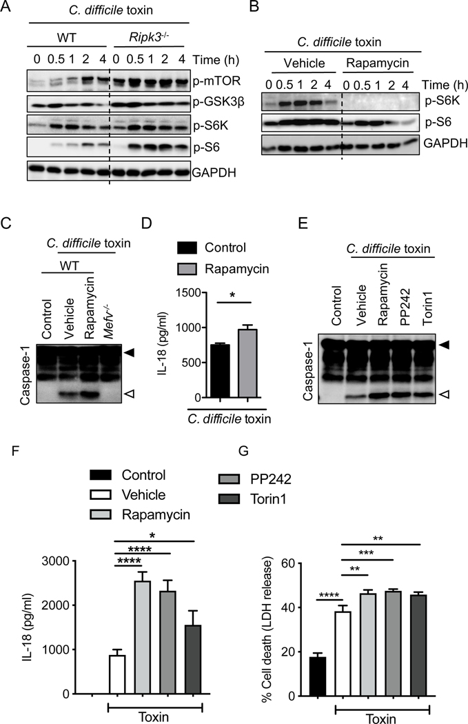
Mutations in MEFV, the gene encoding pyrin in humans, are associated with the autoinflammatory disorder familial Mediterranean fever. Pyrin is an innate sensor that assembles into an inflammasome complex in response to Rho-modifying toxins, including Clostridium difficile toxins A and B. Cell death pathways have been shown to intersect with and modulate inflammasome activation, thereby affecting host defense. Using bone marrow-derived macrophages and a murine model of peritonitis, we show in this study that receptor-interacting protein kinase (RIPK) 3 impacts pyrin inflammasome activation independent of its role in necroptosis. RIPK3 was instead required for transcriptional upregulation of Mefv through negative control of the mechanistic target of rapamycin (mTOR) pathway and independent of alterations in MAPK and NF-κB signaling. RIPK3 did not affect pyrin dephosphorylation associated with inflammasome activation. We further demonstrate that inhibition of mTOR was sufficient to promote Mefv expression and pyrin inflammasome activation, highlighting the cross-talk between the mTOR pathway and regulation of the pyrin inflammasome. Our study reveals a novel interaction between molecules involved in cell death and the mTOR pathway to regulate the pyrin inflammasome, which can be harnessed for therapeutic interventions.
Copyright © 2020 by The American Association of Immunologists, Inc.
Conflict of interest statement
Figures


Similar articles
-
Familial Mediterranean fever mutations are hypermorphic mutations that specifically decrease the activation threshold of the Pyrin inflammasome.Rheumatology (Oxford). 2018 Jan 1;57(1):100-111. doi: 10.1093/rheumatology/kex373. Rheumatology (Oxford). 2018. PMID: 29040788
-
Familial Mediterranean fever mutations lift the obligatory requirement for microtubules in Pyrin inflammasome activation.Proc Natl Acad Sci U S A. 2016 Dec 13;113(50):14384-14389. doi: 10.1073/pnas.1613156113. Epub 2016 Nov 22. Proc Natl Acad Sci U S A. 2016. PMID: 27911804 Free PMC article.
-
Transcriptional licensing is required for Pyrin inflammasome activation in human macrophages and bypassed by mutations causing familial Mediterranean fever.PLoS Biol. 2022 Nov 7;20(11):e3001351. doi: 10.1371/journal.pbio.3001351. eCollection 2022 Nov. PLoS Biol. 2022. PMID: 36342970 Free PMC article.
-
The Pyrin Inflammasome in Health and Disease.Front Immunol. 2019 Aug 7;10:1745. doi: 10.3389/fimmu.2019.01745. eCollection 2019. Front Immunol. 2019. PMID: 31456795 Free PMC article. Review.
-
The pyrin inflammasome in host-microbe interactions.Curr Opin Microbiol. 2020 Apr;54:77-86. doi: 10.1016/j.mib.2020.01.005. Epub 2020 Feb 28. Curr Opin Microbiol. 2020. PMID: 32120337 Free PMC article. Review.
Cited by
-
mTORC1 beyond anabolic metabolism: Regulation of cell death.J Cell Biol. 2022 Dec 5;221(12):e202208103. doi: 10.1083/jcb.202208103. Epub 2022 Oct 25. J Cell Biol. 2022. PMID: 36282248 Free PMC article. Review.
-
The unsolved mystery of MEFV variants variable expressivity in Familial Mediterranean Fever.Intern Emerg Med. 2022 Aug;17(5):1255-1259. doi: 10.1007/s11739-022-03027-4. Epub 2022 Jul 9. Intern Emerg Med. 2022. PMID: 35809153 No abstract available.
-
Inflammasomes and their role in PANoptosomes.Curr Opin Immunol. 2024 Dec;91:102489. doi: 10.1016/j.coi.2024.102489. Epub 2024 Sep 27. Curr Opin Immunol. 2024. PMID: 39340880 Review.
-
Inflammasome activation by Gram-positive bacteria: Mechanisms of activation and regulation.Front Immunol. 2023 Jan 24;14:1075834. doi: 10.3389/fimmu.2023.1075834. eCollection 2023. Front Immunol. 2023. PMID: 36761775 Free PMC article. Review.
-
Inflammasome activation: from molecular mechanisms to autoinflammation.Clin Transl Immunology. 2022 Jul 7;11(7):e1404. doi: 10.1002/cti2.1404. eCollection 2022. Clin Transl Immunology. 2022. PMID: 35832835 Free PMC article. Review.
References
-
- Bernot A CC, Dasilva C, Devaud C, Petit JL, Caloustian C, Cruaud C, Samson D, Pulcini F, Weissenbach J,R, Notanicola C, Domingo C, Rozenbaum M, Benchetrit E, Topaloglu R, Dewalle M, Dross C, Hadjari P, Dupont M, Demaille J, Touitou I, Smaoui N, Nedelec B, Méry JP, Chaabouni H, Delpech M, Grateau G. 1997. A candidate gene for familial Mediterranean fever. Nat Genet 17: 25–31. - PubMed
-
- Consortium*, T. I. F. 1997. Ancient missense mutations in a new member of the RoRet gene family are likely to cause familial Mediterranean fever. The International FMF Consortium. Cell 90: 797–807. - PubMed
-
- Xu H, Yang J, Gao W, Li L, Li P, Zhang L, Gong Y-N, Peng X, Xi JJ, Chen S, Wang F, and Shao F. 2014. Innate immune sensing of bacterial modifications of Rho GTPases by the Pyrin inflammasome. Nature 513: 237–241. - PubMed
Publication types
MeSH terms
Substances
Grants and funding
LinkOut - more resources
Full Text Sources
Molecular Biology Databases
Miscellaneous
