. 2020 Oct 21;40(43):8306-8328.
doi: 10.1523/JNEUROSCI.0860-20.2020. Epub 2020 Sep 28.

Serial Prefrontal Pathways Are Positioned to Balance Cognition and Emotion in Primates

Affiliations

Serial Prefrontal Pathways Are Positioned to Balance Cognition and Emotion in Primates

Mary Kate P Joyce et al. J Neurosci. .

Abstract

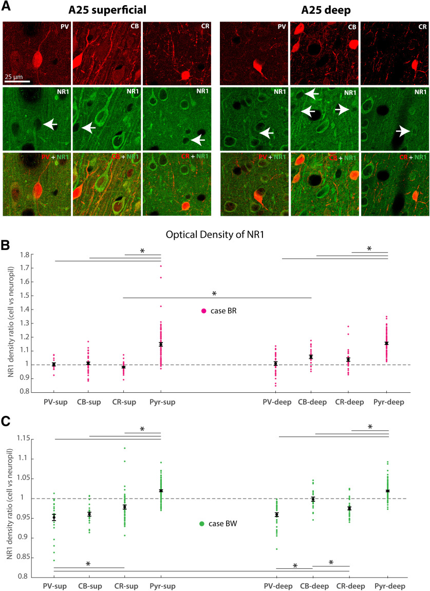
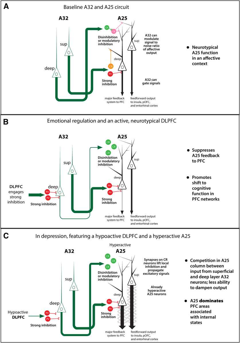
The delicate balance among primate prefrontal networks is necessary for homeostasis and behavioral flexibility. Dorsolateral prefrontal cortex (dlPFC) is associated with cognition, while the most ventromedial subgenual cingulate area 25 (A25) is associated with emotion and emotional expression. Yet A25 is weakly connected with dlPFC, and it is unknown how the two regions communicate. In rhesus monkeys of both sexes, we investigated how these functionally distinct areas may interact through pregenual anterior cingulate area 32 (A32), which is strongly connected with both. We found that dlPFC innervated the deep layers of A32, while A32 innervated all layers of A25, mostly targeting spines of excitatory neurons. Approximately 20% of A32 terminations formed synapses on inhibitory neurons in A25, notably the powerful parvalbumin inhibitory neurons in the deep layers, and the disinhibitory calretinin neurons in the superficial layers. By innervating distinct inhibitory microenvironments in laminar compartments, A32 is positioned to tune activity in columns of A25. The circuitry of the sequential pathway indicates that when dlPFC is engaged, A32 can dampen A25 output through the parvalbumin inhibitory microsystem in the deep layers of A25. A32 thus may flexibly recruit or reduce activity in A25 to maintain emotional equilibrium, a process that is disrupted in depression. Moreover, pyramidal neurons in A25 had a heightened density of NMDARs, which are the targets of novel rapid-acting antidepressants. Pharmacologic antagonism of NMDARs in patients with depression may reduce excitability in A25, mimicking the effects of the neurotypical serial pathway identified here.SIGNIFICANCE STATEMENT The anterior cingulate is a critical hub in prefrontal networks through connections with functionally distinct areas. Dorsolateral and polar prefrontal areas that are associated with complex cognition are connected with the anterior cingulate in a pattern that allows them to indirectly control downstream activity from the anterior cingulate to the subgenual cingulate, which is associated with heightened activity and negative affect in depression. This set of pathways provides a circuit mechanism for emotional regulation, with the anterior cingulate playing a balancing role for integration of cognitive and emotional processes. Disruption of these pathways may perturb network function and the ability to regulate cognitive and affective processes based on context.

Keywords: anterior cingulate; depression; dlPFC; inhibitory neurons; laminar connections; subgenual cingulate.


Figures

Figure 1.
Figure 1.
Experimental design. A, Schematic represents the serial pathway under study on the lateral (left) and medial (right) surfaces of the rhesus monkey brain. A bidirectional tracer injected into A32 retrogradely labeled neurons in the dlPFC/A10 and anterogradely labeled axonal terminations in A25 to produce a sequential set of labeled pathways. B, Example plot of a prefrontal coronal section exhaustively sampled to map retrogradely labeled neurons (green). C, Schematic represents the sampling of axonal terminations for density computation and quantification of bouton size. Darkfield photomicrograph (left) of a coronal slice through A25. BDA from an A32 injection appears bright, forming a columnar patch (white arrowhead) just above the rostral sulcus. Dotted lines indicate the bottom of layer I and the top of layer V. Middle, A simplified version of the optical fractionator method of stereological sampling, where a grid is placed over the ROI and fields are sampled at a rate that minimizes a coefficient of error. Brightfield photomicrographs (right) were taken at different focal planes through the depth of tissue, and the boutons were measured for major diameter. D, Series of photomicrographs represent the analysis of the postsynaptic targets of A32 terminations in A25. Left, Coronal section through A25 at the rostral sulcus. BDA (green) forms a columnar patch near the top of the image. CB neurons (blue) are more prevalent in the superficial layers, and PV neurons (red) are most prevalent in the middle to deep layers. Middle, Maximum projection of high magnification z stack (CM) in the deep layers of A25 represents A32 (green) axonal terminations in some cases closely apposing PV neuronal processes (red). Right, Electron photomicrograph represents the end of an A32 bouton labeled with DAB (dark, black precipitate) forming a synapse on a dendrite that has abundant gold labeling for PV (deeply black clustered quanta, white arrowheads). Numbers indicate cortical areas according to Barbas and Pandya (1989). ac, Anterior commissure; All, allocortex; AON, anterior olfactory nucleus; cc, corpus callosum; cd, caudate; LOT, lateral olfactory tract; MPAll, medial periallocortex; OPAll, orbital periallocortex; ROI, region of interest; thal, thalamus.
Figure 2.
Figure 2.
Injection of bidirectional tracers into A32. A, Injection sites in A32 on the medial surface of the rhesus monkey brain. Six injection sites are represented in a normalized brain space through the anterior to posterior aspects of A32. Numbers indicate cortical areas according to Barbas and Pandya (1989). B, Injection site of BDA in A32 in all layers (Case BS). Black line indicates boundary between layer VI and the superficial white matter. ac, anterior commissure; cc, corpus callosum; cg, cingulate sulcus; MPAll, medial periallocortex; wm, white matter.
Figure 3.
Figure 3.
System-level mapping of serial pathway from dlPFC and A10 to A25 through A32. A, Coronal sections show plots of neurons (green) projecting to A32 injection site. Projection neurons are distributed mostly in the superficial layers. Areas marked by hatching were not mapped in this study. B, Distribution of retrogradely labeled neurons directed to A32 injection site from areas in dlPFC and A10, expressed as percent of neurons found in the superficial layers. C, Sampling of coronal sections for stereological computation of A32 termination density in A25. Sections were sampled at 1 mm through the extent of A25. Laminar categories of A25 were outlined, and stereological sampling was conducted in small fields of 25 μm2. Markers used to count A32 terminations were enlarged so the fields are visible. D, Distribution of A32 terminations in subregions of A25, shown in color by each injection site. Stronger color represents denser distribution of A32 axonal terminations. E, Laminar distribution of A32 terminations in A25, expressed as the ratio of bouton density in a laminar category to the bouton density in all layers. F, Color-coded summary schematic of laminar pattern of connections in the serial pathway. Connections between cortical areas are in accordance with the Structural Model (Barbas and Rempel-Clower, 1997). Connections from the six-layered dlPFC/A10 flow to A32 in a feedforward pattern (red), from the superficial layers of dlPFC/A10 to the deep layers of A32. A32 and A25 both contain fewer than six layers, and have a lateral pattern of connections (yellow), meaning that the superficial and deep layers of both areas engage in connections between them. G, Photomicrographs of coronal sections in A46, A32, and A25 stained for Nissl. A46 has six distinct layers, while A32 and A25 do not. Color-coded arrows indicate the laminar connections in the serial pathway, and follow the color key represented in F. Roman numerals indicate cortical layers. Numbers indicate cortical areas according to Barbas and Pandya (1989). aon, anterior olfactory nucleus; cc, corpus callosum; cg, cingulate sulcus; lo, lateral orbital sulcus; mo, medial orbital sulcus; p, principal sulcus; rh, rhinal sulcus; ro, rostral sulcus; str, striatum; sup, superficial; v, ventricle; WM, white matter.
Figure 4.
Figure 4.
CB-labeled neurons in A25. A, Photomicrograph of coronal section in A46 immunolabeled for CB (brown) and counterstained with Nissl. B, Photomicrograph of coronal section in A32 immunolabeled for CB and counterstained with Nissl. C, Photomicrograph of coronal section in A25 immunolabeled for CB and counterstained with Nissl. D, Inset, Pyramidal-shaped (excitatory) neurons lightly labeled with CB (green arrows) and multipolar-shaped presumed inhibitory neurons that have a uniform dark CB label (red arrows) in the superficial layers of A25. E, Higher-magnification inset, Pyramidal-shaped neurons lightly labeled with CB (green arrows) and multipolar-appearing neuron with a uniform dark CB label (red arrow) in the superficial layers of A25. F, Photomicrograph of the superficial layers of A25 immunolabeled with GAD-67, a marker of inhibitory neurons. Red arrows indicate neurons with cytoplasm strongly labeled by GAD-67. Green asterisks indicate morphologically distinct pyramidal shaped (excitatory) neurons that show GAD-67 puncta targeting their cell bodies but an otherwise absence of GAD-67 labeling. G, The superficial layers of A25 immunolabeled for GABA show inhibitory neurons labeled by green fluorophores (red arrow, red arrowheads). Empty spaces represent the cell bodies of non-GABA-labeled neurons (green arrow, green asterisk). H, The same field of view (FOV) in the superficial layers of A25 immunolabeled with CB shows a darkly labeled CB neuron (red arrow), a lightly labeled neuron above it (green arrow), and empty spaces representing cell bodies of non-CB-labeled neurons (red arrowhead, green asterisk). I, Merged GABA and CB. Red arrow indicates an inhibitory neuron labeled for both GABA and CB. Red arrowhead indicates a GABA-positive neuron that is CB-negative, presumably a PV or CR inhibitory neuron. Green arrow indicates a lightly labeled CB neuron that is GABA-negative, presumably a CB-positive pyramidal neuron. Green asterisk indicates the shadow of a cell body that is both GABA- and CB-negative, presumably a CB-negative excitatory neuron. Roman numerals indicate cortical layers. J, Average neuronal density of CB+ presumed excitatory neurons in superficial layers of A46, A32, and A25 based on computations performed from stereological sampling. Circles represent values from individual cases. Data are mean ± SE. K, Average neuronal density of CB+ presumed excitatory neurons in deep layers of A46, A32, and A25 based on computations performed from stereological sampling. Circles represent values from individual cases. Data are mean ± SE. L, Average neuronal density of CB+ presumed excitatory neurons in all layers of A46, A32, and A25 based on computations performed from stereological sampling. Circles represent values from individual cases. Data are mean ± SE. WM, White matter.
Figure 5.
Figure 5.
The distribution of PV, CB, and CR neurons in A46, A32, and A25. A, Cortical columns photographed from coronal sections of A46, A32, and A25 after immunofluorescence labeling for PV (red), CB (cyan), and CR (green). Note the increase in CB labeling in the superficial layers from A46 to A25, which is indicative of the increase in CB pyramidal neuron labeling. B, Higher-magnification inset of the superficial layers of A25 shows a dominance of CB and CR neurons. Layer I contains only CR neurons. C, Higher-magnification inset of the mid-to deep layers of A25 shows the preponderance of PV neurons. D, Higher-magnification inset of the middle layers of A46. Note the dominance of PV neurons. E, Quantification of PV, CB, and CR neuron density in layers I-VI of each area using unbiased stereological sampling. F, Quantification of CR neuron density in layer I of each area using unbiased stereological sampling. G, Quantification of PV, CB, and CR density in layers II-III of each area using unbiased stereological sampling. H, Quantification of PV, CB, and CR density in layer IV of A46 using unbiased stereological sampling. I, Quantification of PV, CB, and CR density in layers V-VI of each area using unbiased stereological sampling. Roman numerals indicate cortical layers. Colored rectangles represent averages. Black circles represent individual cases. *p < 0.05. WM, White matter.
Figure 6.
Figure 6.
Appositions between A32 axons and CBP+ structures in A25 as obtained via CM. A, Normalized brain space depicting the regions sampled for appositional analysis (Cases BI, BS). Left, The medial surface of the rhesus monkey brain. Right, Vertical lines indicate the sampling plane for the coronal sections sampled. Temporal pole not shown in coronal sections. Scale bar, 2 mm. B, Photomicrographs of z stack maximum projections taken through coronal sections of A25 laminar regions represent A32 terminations (green) and CBP+-labeled neurons (red). A maximum projection is all the images in a z stack collapsed together. Left, A32 terminations interact with CR+ neuronal processes in the superficial layers. Insets, The selection adjusted for brightness and contrast, a single focal plane depicting an apposition between the A32 bouton and a CR+ dendrite, and a rotated 3D rendering of the selection depicting that the presynaptic and postsynaptic sites are apposed in all dimensions. Middle, A32 terminations interact with CB+ neuronal processes in the superficial layers. Insets, A higher magnification of the selection adjusted for brightness and contrast and a 3D-rendered rotation to show that the A32 termination apposes the CB+ dendrite. Right, A32 terminations interact with PV+ neuronal processes in the deep layers. Insets, 3D-rendered rotations to show that the A32 termination apposes the PV+ dendrite. C, Average proportion (colored boxes) of A32 boutons apposing CBP+ neuronal processes in the superficial and deep layers across 2 cases (BI and BS). Colored circles represent individual cases. Data are mean ± SD. *p = 0.05. D, Summary schematic of the inhibitory microcircuitry in the cortical column of A25. a, arcuate sulcus; aon, anterior olfactory nucleus; cg, cingulate sulcus; lo, lateral orbital sulcus; mo, medial orbital sulcus; p, principal sulcus; rh, rhinal sulcus; ro, rostral sulcus; str, striatum.
Figure 7.
Figure 7.
Postsynaptic targets of A32 terminations in A25 obtained using synaptic analysis via EM. A, Normalized brain space depicting the regions sampled for synaptic analysis (Cases BI, BS). Left, The medial surface of the rhesus monkey brain. Right, Vertical lines indicate the sampling plane for the coronal sections sampled. Scale bar, 2 mm. B, Photomicrograph of an A32 axon terminal labeled with DAB (uniform dark precipitate, white arrow) that forms a synapse (white arrowhead) on a CBP+ dendritic shaft in A25 that is positive for PV, which was labeled with gold (small black quanta, sometimes clustered, black arrowheads). C, Pattern of A32 postsynaptic targets in the superficial layers of A25. Left, Average across 2 cases (BI, BS). Right, Breakdown in individual cases. If an A32 bouton formed a synapse on the shaft or spine of a CBP+ sparsely spiny dendrite, these interactions were presumed to be with an inhibitory postsynaptic structure. If spines could not be traced back to their dendrite for classification as sparsely spiny or densely spiny, and thus presumed inhibitory or excitatory respectively, we grouped them in a separate “indeterminate” category, which was very low for both PV and CR. Synaptic interactions between A32 and CBP+ spines on densely spiny dendrites, which were common for CB but very rare for PV or CR, were grouped in their own category. D, Pattern of A32 postsynaptic targets in the deep layers of A25. Left, Average across 2 cases (BI, BS). Right, Breakdown in individual cases. *p < 0.05. a, arcuate sulcus; aon, anterior olfactory nucleus; cg, cingulate sulcus; lo, lateral orbital sulcus; mo, medial orbital sulcus; mt, mitochondria; o, olfactory tubercule; p, principal sulcus; rh, rhinal sulcus; ro, rostral sulcus; str, striatum.
Figure 8.
Figure 8.
3D reconstruction of an A32 terminal field in A25 from serial EM. 3D reconstruction of an ROI (200 serial sections spanning 10 µm) in the deep layers of A25 that contained 22 individual A32 terminations, demonstrating the extraordinary density of this pathway, even using the highest resolution methods. This ROI was double-labeled using immunohistochemistry for EM. A32 terminations were labeled with DAB, and CR was labeled with gold. A, An A32 axon terminal (blue, white triangle) forming a synapse on a CR+ soma (pink). B, The same FOV but with the addition of another A32 axon terminal (blue) forming a synapse on an inhibitory dendrite (red). This dendrite was not CR+, and was likely a PV+ dendrite. The dendrite is aspiny, or smooth, which is typical of the dendrites of inhibitory neurons. C, The full ROI containing all reconstructed structures. All axons from A32, including their terminal boutons, are labeled in blue. All postsynaptic structures receiving synapses from A32 terminations were reconstructed to the extent possible. Light green structures are spiny, presumed excitatory neuronal elements in A25. Green and blue represent excitatory neuronal processes. Red or pink represents inhibitory neuronal processes. Scale box, 1 µm3.
Figure 9.
Figure 9.
Structural characteristics of A32 terminals and their postsynaptic targets in A25 using EM. A, The superficial layers of A25, double-immunolabeled for A32 axonal processes (DAB, uniform dark precipitate), and PV (gold, black quanta, sometimes clustered). An A32 axonal termination (brown) forms synapses with three spines (sp), shaded in red, none of which contains PV+ gold labeling. The A32 axonal termination formed a perforated synapse (ps) with one of these spines. Structures shaded in blue represent unlabeled axon terminations in the surrounding ROI that form asymmetric synapses, which are presumed excitatory and were used for neuropil analysis. Green shading represents postsynaptic structures of presumed excitatory boutons in the neuropil. B, Bouton diameters of labeled A32 terminations, obtained using BM and EM, and of terminations forming asymmetric synapses in the surrounding neuropil (dashed lines) in the superficial and deep layers of A25. C, Comparison of the average bouton diameter of A32 terminations and asymmetric synapses in the surrounding neuropil, in the superficial and deep layers of A25. D, Comparison of the proportion of A32 axonal terminations containing mitochondria with the proportion of terminations forming asymmetric synapses in the neuropil that contained mitochondria, in the superficial and deep layers of A25. E, The proportion of A32 axonal terminations that formed perforated asymmetric synapses in comparison with synapses in the surrounding neuropil, in the superficial and deep layers of A25. F, Proportion of A32 axonal terminations forming synapses on spines that contained a spine apparatus in comparison with the surrounding neuropil, in the superficial and deep layers of A25. G, Distribution of A32 bouton volumes in the superficial and deep layers of A25 for all 3D reconstructed A32 terminations as measured in serial sections. H, Average bouton volume of A32 terminations in the superficial and deep layers of A25. I, PSD plotted against bouton volume for all 3D reconstructed A32 terminations. J, PSD plotted against bouton diameter for all 3D reconstructed A32 terminations. Data are mean ± SD. Black circles represent individual values. *p = 0.05. b, Bouton; d, dendrite; m, mitochondria; Neur, neuropil; ps, perforated synapse; sa, spine apparatus; sp, spine.
Figure 10.
Figure 10.
NR1 colocalization with PV, CB, and CR neurons in A25. A, Single focal planes obtained by CM show FOVs in superficial (left) and deep (right) layers of A25. FOVs are double-labeled using immunofluorescence for PV, CB, or CR (top row, red), and NR1 (middle row, green), the obligatory NMDAR subunit. Bottom row, Merged channels for CBP+ and NR1 labeling. White arrows point to CBP+-labeled inhibitory cell bodies. Surrounding pyramidal neurons are rich in NMDARs. B, C, Quantification of NR1 labeling across presumed pyramidal (excitatory) and presumed inhibitory CBP+ cell bodies in 2 cases (B, Case BR; C, Case BW). Colored dots represent NR1 density ratio using all cells (of respective type) in an individual image. Black dots represent average ± SEM over all images. Segmentation masks were prepared to outline CBP+ cell bodies and pyramidal-shaped, presumed excitatory cell bodies. For CB neurons, only those that were morphologically consistent with inhibitory neurons and that featured dark CB+ labeling were used. Segmentation masks were also prepared for sections of the neuropil, as a control for normalization within each image. This produced three masks for each image: one for pyramidal cell bodies, one for CBP+ cell bodies (PV, CB, or CR), and one for neuropil regions. For each image, NR1 labeling intensity was computed as a density (over area) within each mask. The ratio of NR1 density in cells versus the neuropil in each image was then compared across CBP+ neurons and pyramidal neurons in superficial and deep layers of A25. *p < 0.05.
Figure 11.
Figure 11.
Summary of findings. Superficial layer neurons from the dlPFC send feedforward projections to the deep layers of A32. A32 sends projections to A25 originating in superficial and deep layers of A32. In the superficial layers of A25, A32 axonal terminations form synapses with CR (green) followed by CB (pink) neurons in the superficial layers. In the deep layers of A25, A32 axon terminals form synapses with PV (red) neurons, followed by CR (orange) and CB (pink) neurons. CR neurons (green) are thought to be largely disinhibitory in primate superficial cortical layers through synapses on surrounding inhibitory neurons. By predominantly targeting disinhibitory neurons in the superficial layers, pathways from A32 to the superficial layers of A25 may allow excitatory signals to propagate through the local circuitry. In the deep layers of A25, CR neurons are thought to have a net inhibitory effect. By predominantly targeting PV neurons in the deep layers, A32 engages a stronger inhibitory system and likely has a stronger ability to dampen activity in the local circuitry.
Figure 12.
Figure 12.
Summary circuit schematic of serial pathway from dlPFC to A32 to A25 for various functional states. A, A circuit model of the synaptic interactions from A32 to A25 based on the present findings. The model leaves out other contributions to cortical microcircuitry (e.g., neuromodulatory and thalamocortical input), is unidirectional according to the serial pathway under study, and thus does not reflect reciprocal connections. Green represents neurons with excitatory effects. Shades of red represent neurons with inhibitory effects on the circuit. In the superficial layers of A25, terminations from A32 modulate the signal-to-noise ratio through synapses with the disinhibitory CR (light green) and modulatory CB (pink) inhibitory microsystems. In the deep layers of A25, terminations from A32 can gate signals through the powerful PV (red) and inhibitory CR (orange) microsystem. B, A putative model of neurotypical emotional regulation, based on the patterns of projections from the dlPFC to A32 to A25 and physiologic studies. For simplicity, the inhibitory microsystems illustrated in A have been collapsed to CR in the superficial layers and PV in the deep layers because they are the primary inhibitory targets of A32 in A25. The dlPFC and A32 are necessary for emotional regulation as summarized in functional studies. dlPFC terminations in A32 may suppress the deep layers of A32 through the strong PV inhibitory microsystem, reducing output to the superficial layers of A25. This leaves the superficial layers of A32 active, which engage the strong PV inhibitory microsystem in the deep layers of A25, dampening the contribution of A25 to ongoing processes in the PFC. Deep layer neurons of A25 project to many PFC areas that contribute to internal state (Joyce and Barbas, 2018); and when that output is suppressed, resources can be focused elsewhere as needed. C, A putative model in depression. Metabolic A25 activity is heightened, according to reported functional pathology in depression. Here, those findings have been interpreted to mean that individual neurons are in a more active state than in a neurotypical A25 (indicated by sinusoidal waves around axons). Functional pathology in depression also involves a hypoactive dlPFC. An attenuated influence of the dlPFC on A32 predicts that both the superficial and deep layers of A32 exert influence on A25. In the superficial layers of A25, lifted inhibition by A32 terminations on CR neurons allows local excitatory signals to propagate unfettered and potentially exacerbates the already hyperactive A25 neurons. In the deep layers, terminations from A32 may not exert enough inhibition to dampen A25 output. In short, A25 neurons are hyperactive, and A32 cannot suppress the abnormally high activity. A25 output to other PFC areas that are associated with internal states is exaggerated (thick arrows). pOFC, Posterior orbitofrontal cortex; sup, superficial.

Similar articles

Cited by

References

    1. Alexander L, Clarke HF, Roberts AC (2019a) A focus on the functions of area 25. Brain Sci 9:129 10.3390/brainsci9060129 - DOI - PMC - PubMed
    1. Alexander L, Gaskin PL, Sawiak SJ, Fryer TD, Hong YT, Cockcroft GJ, Clarke HF, Roberts AC (2019b) Fractionating blunted reward processing characteristic of anhedonia by over-activating primate subgenual anterior cingulate cortex. Neuron 101:307–320.e306. 10.1016/j.neuron.2018.11.021 - DOI - PMC - PubMed
    1. Arnsten AF. (2013) The neurobiology of thought: the groundbreaking discoveries of Patricia Goldman-Rakic 1937-2003. Cereb Cortex 23:2269–2281. 10.1093/cercor/bht195 - DOI - PMC - PubMed
    1. Anderson MC, Bunce JG, Barbas H (2015) Prefrontal-hippocampal pathways underlying inhibitory control over memory. Neurobiol Learn Mem, 134 Pt A, 145–161. 10.1016/j.nlm.2015.11.008 - DOI - PMC - PubMed
    1. Barbas H. (1993) Organization of cortical afferent input to orbitofrontal areas in the rhesus monkey. Neuroscience 56:841–864. 10.1016/0306-4522(93)90132-Y 10.1016/0306-4522(93)90132-y - DOI - DOI - PubMed

Publication types

LinkOut - more resources