. 2020 Oct 21;40(43):8355-8366.
doi: 10.1523/JNEUROSCI.1503-20.2020. Epub 2020 Sep 28.

Prefrontal Neural Ensembles Develop Selective Code for Stimulus Associations within Minutes of Novel Experiences

Affiliations

Prefrontal Neural Ensembles Develop Selective Code for Stimulus Associations within Minutes of Novel Experiences

Kaori Takehara-Nishiuchi et al. J Neurosci. .

Abstract

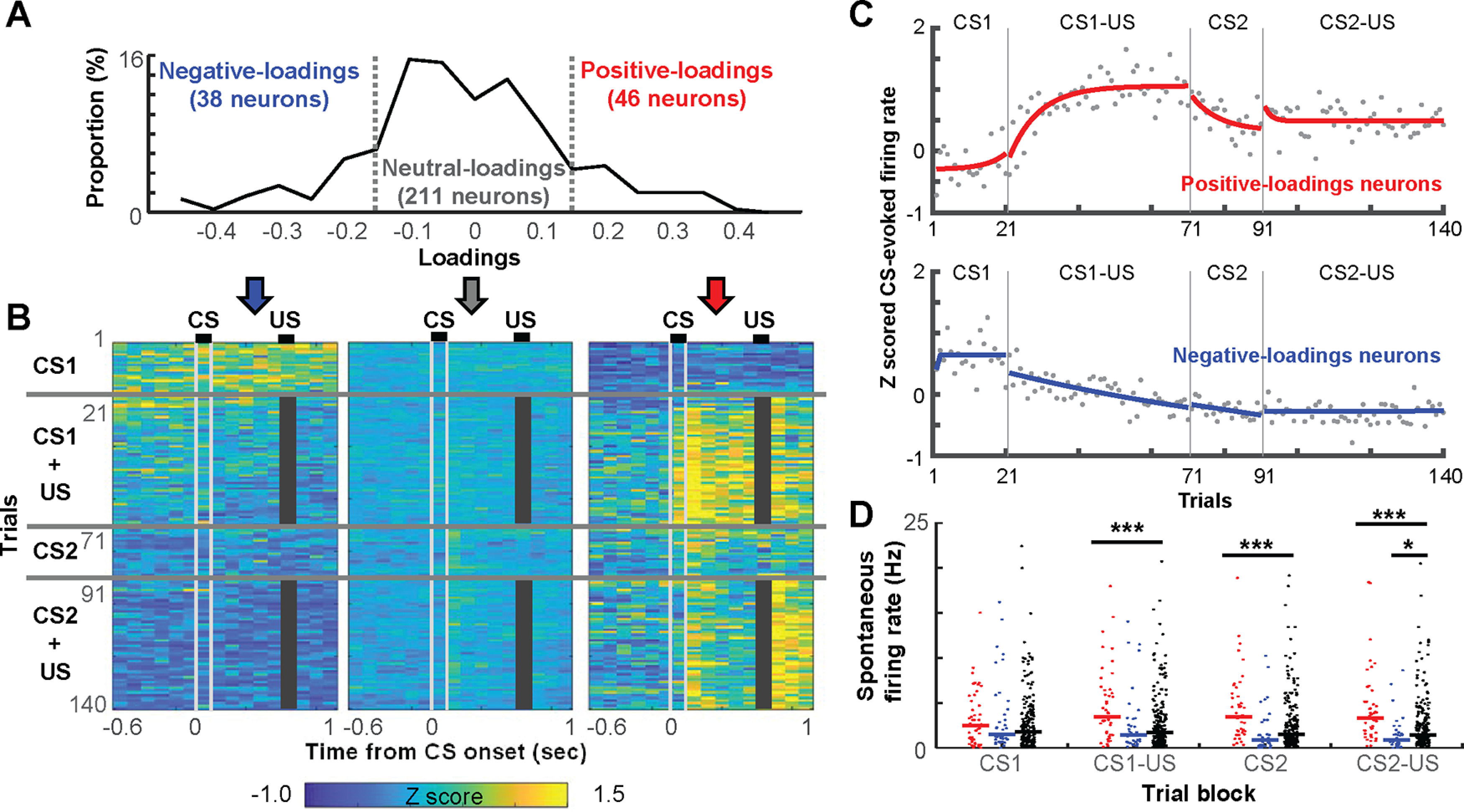
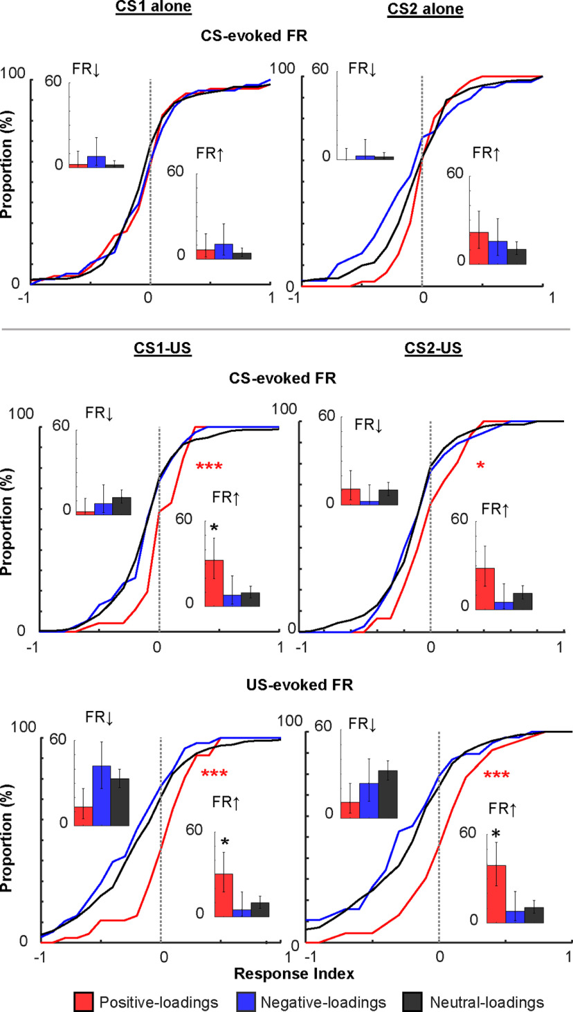
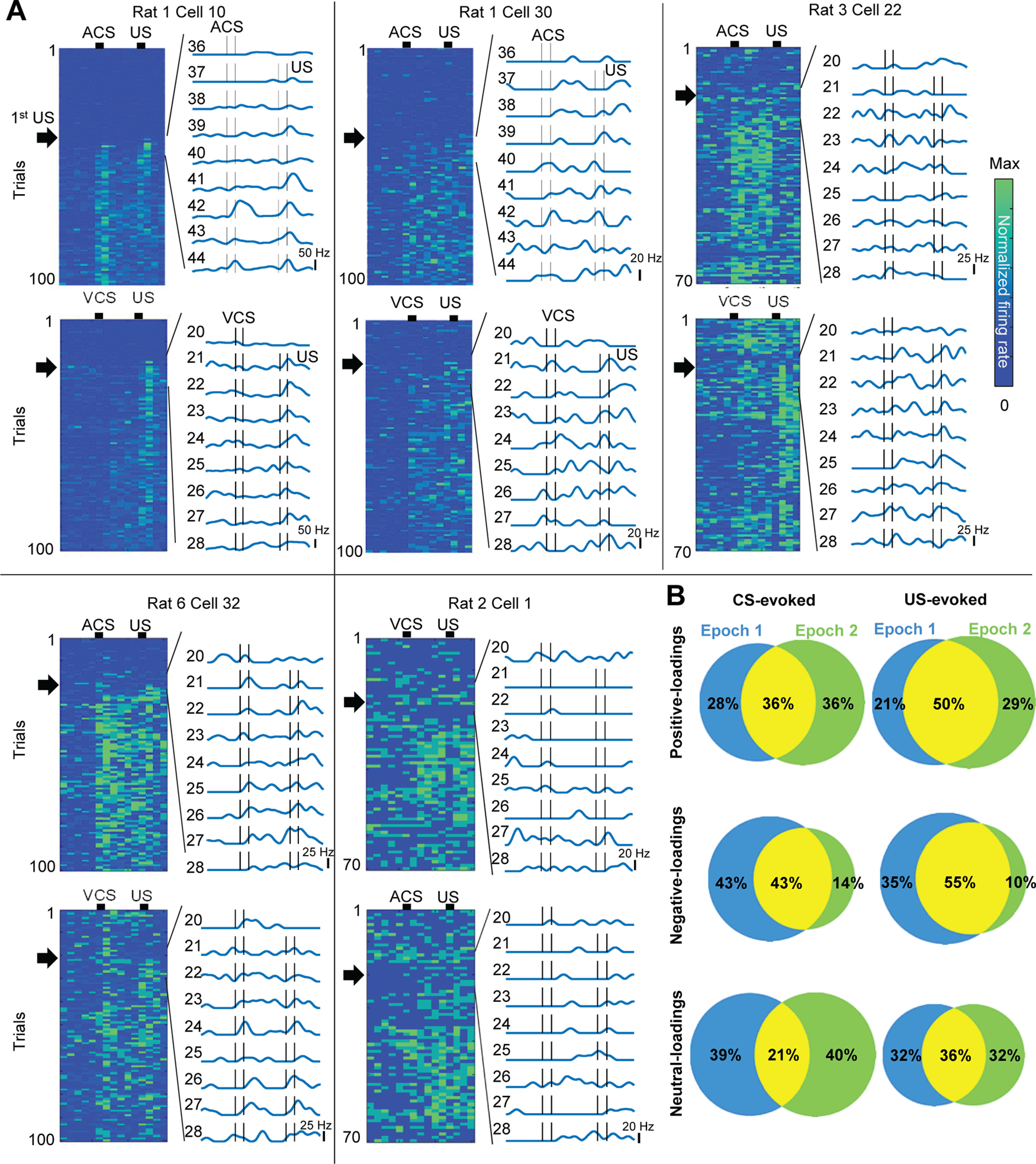
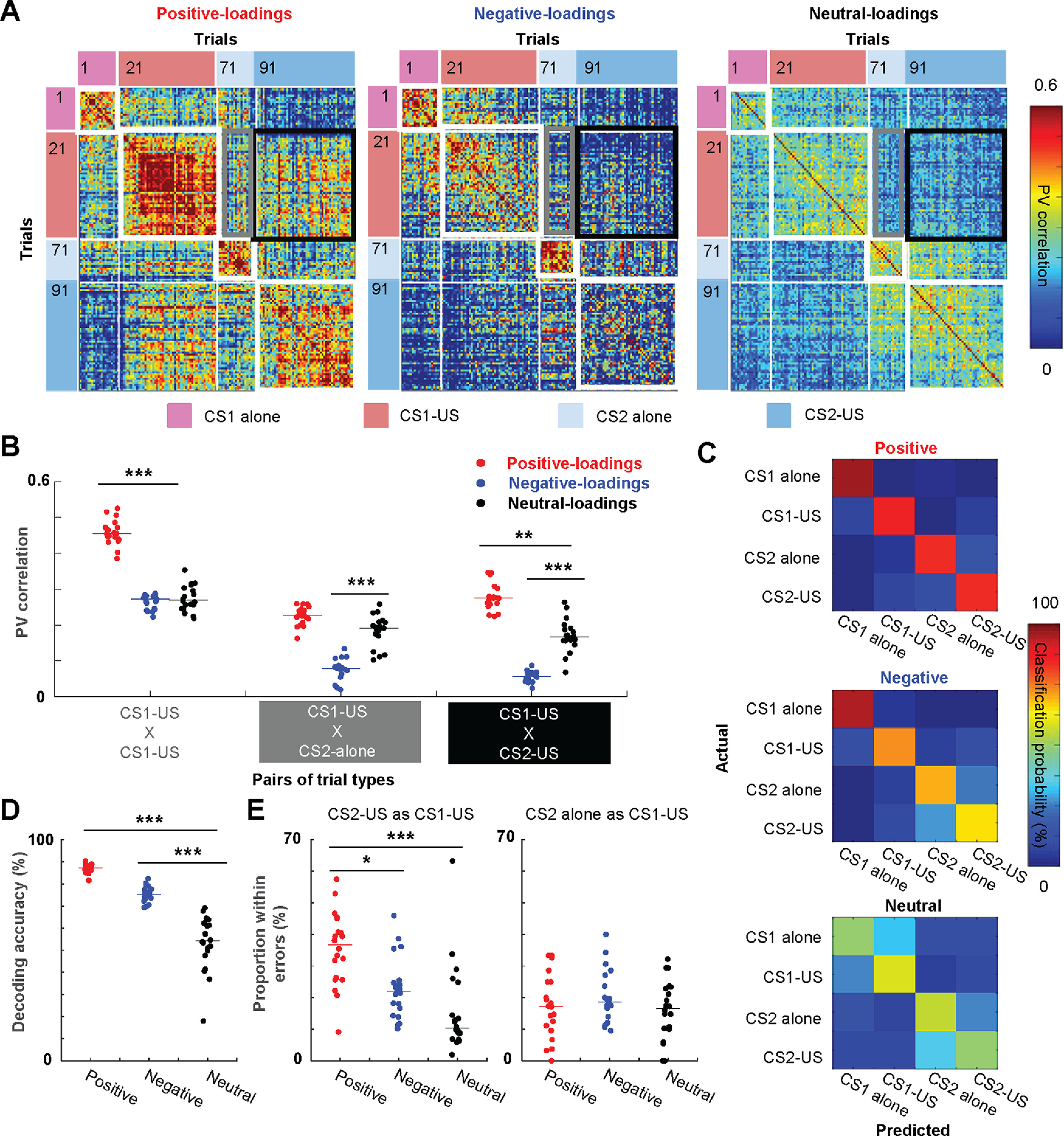
Prevailing theories posit that the hippocampus rapidly learns stimulus conjunctions during novel experiences, whereas the neocortex learns slowly through subsequent, off-line interaction with the hippocampus. Parallel evidence, however, shows that the medial prefrontal cortex (mPFC; a critical node of the neocortical network supporting long-term memory storage) undergoes rapid modifications of gene expression, synaptic structure, and physiology at the time of encoding. These observations, along with impaired learning with disrupted mPFC, suggest that mPFC neurons may exhibit rapid neural plasticity during novel experiences; however, direct empirical evidence is lacking. We extracellularly recorded action potentials of cells in the prelimbic region of the mPFC, while male rats received a sequence of stimulus presentations for the first time in life. Moment-to-moment tracking of neural ensemble firing patterns revealed that the prelimbic network activity exhibited an abrupt transition within 1 min after the first encounter of an aversive but not neutral stimulus. This network-level change was driven by ∼15% of neurons that immediately elevated their spontaneous firing rates (FRs) and developed firing responses to a neutral stimulus preceding the aversive stimulus within a few instances of their pairings. When a new sensory stimulus was paired with the same aversive stimulus, about half of these neurons generalized firing responses to the new stimulus association. Thus, prelimbic neurons are capable of rapidly forming ensemble codes for novel stimulus associations within minutes. This circuit property may enable the mPFC to rapidly detect and selectively encode the central content of novel experiences.SIGNIFICANCE STATEMENT During a new experience, a region of the brain, called the hippocampus, rapidly forms its memory and later instructs another region, called the neocortex, that stores its content. Consistent with this dominant view, cells in the neocortex gradually strengthen the selectivity for the memory content over weeks after novel experiences. However, we still do not know precisely when these cells begin to develop the selectivity. We found that neocortical cells were capable of forming the selectivity for ongoing events within a few minutes of new experiences. This finding provides support for an alternative view that the neocortex works with, but not follows, the hippocampus to form new memories.

Keywords: consolidation; encoding; hippocampus; plasticity; prefrontal cortex; rats.


Figures

Figure 1.
Figure 1.
Behavioral paradigm and recording location. A, Seven male rats underwent two epochs of conditional associative learning task in the same chamber. In each epoch, a tone or light was presented alone (CS-alone, 20 trials) or preceding the mildly aversive eyelid shock (US) by 500 ms (CS-US, 50–80 trials). B, Amplitudes of CS-evoked eyelid muscle activity in a representative rat. The muscle activity was normalized by the activity during the period before CS onset. The increased muscle activity toward US onset indicates anticipatory blinking responses (CRs). C, The proportion of trials in which the rats expressed CRs (CR%, mean ± SEM) in a series of 10 trials with an increment of five trials. Gray lines indicate the data in each rat. D, CR% in CS-US blocks on the first and second day (mean ± SEM); *p < 0.05, paired t test. E, Recording locations. Each dot indicates the location of a tetrode tip confirmed in the histologic analysis. The brain of rat 1 was sectioned coronally, which makes it difficult to depict on the same diagram. However, the location of tetrodes in this rat was comparable to the other rats (see Morrissey et al., 2017).
Figure 2.
Figure 2.
Abrupt transition of the prefrontal network state on the first encounter of an aversive stimulus. A, Top five PCs (top) of z-scored, 1-s binned firing patterns of simultaneously recorded neurons (bottom) during the entire recording period including the stimulus presentations in a box (gray bar) and a rest period outside the box. B, 3D projections of PC1-3 in A. C, Mahalanobis distances of the ensemble activity (mean ± SEM and data in individual rats) between two adjacent time windows. They were taken from periods before and after the first trial of each type (between) or two adjacent periods before or after the first trial (within); *p < 0.05, Wilcoxon signed-rank test. D, Normalized FRs averaged across all neurons in each rat (mean ± SEM and data in individual rats) during three windows used to calculate Mahalanobis distances in C.
Figure 3.
Figure 3.
The rapid development of selective ensemble firings for stimulus associations. A, The distribution of loadings on the PC capturing the network transition after the first aversive stimulus. B, Averaged, 100-ms binned, z-scored FRs in each trial in neurons positive-loadings (≥0.15), negative-loadings (≤−0.15), as well as the remaining, neutral-loadings neurons. Gray bars mask the shock artifact. C, Z-scored, CS-evoked FRs averaged across all positive-loadings or negative-loadings neurons. Each dot depicts the FR in one trial. By fitting an exponential curve (red, blue) to the data, we estimated the number of trials that were required to reach an asymptote. D, Spontaneous FRs (a dot for each neuron, lines show the median) in the block of four different trial types; *p < 0.05, ***p < 0.001, Kruskal–Wallis test, post hoc Dunn's test.
Figure 4.
Figure 4.
Distributions of the magnitude of stimulus-evoked FRs. In each neuron, the magnitude of CS-evoked or US-evoked FR changes was quantified by calculating RI that ranged from −1 to 1; *p < 0.05/2; ***p < 0.001, Kolmogorov–Smirnov test against neutral-loadings neurons. Insets show the proportion of neurons with significant FR decrease (left) and increase (right). Error bars show 95% confidence interval; *p < 0.05, binominal test.
Figure 5.
Figure 5.
The rapid development of association selectivity in individual neurons. A, FR of individual neurons in all trials (left) and nine trials around the first CS-US trial (black arrows; right). B, The proportion of neurons with significant FR changes on the CS (left) and US (right) in the first (blue) and second (green) epochs.
Figure 6.
Figure 6.
Generalization of ensemble firing patterns to a new stimulus with the same biological significance. A, Pearson correlation coefficients of CS-evoked ensemble firing vectors (PV) between all possible pairs of trials. B, The correlation values (n = 20 sets of 35 subsampled neurons; lines show median) between the averaged PV in the CS1-US block and that in the same (within-block consistency) or other blocks (between-block generalization). C, Representative confusion matrices from SVM classifiers showing the probability (color) that a trial of one type (rows) was classified as another type (columns). D, Decoding accuracy of SVM classifiers applied to 20 sets of 35 subsampled cells (lines show median). Decoding accuracy was significantly higher with positive-loading neurons than neutral-loading neurons. E, The proportion of the specific type of classification errors in a total number of misclassified trials (n = 20 sets of 35 subsampled neurons; lines show median); *p < 0.05, **p < 0.01, ***p < 0.001, Kruskal–Wallis test, post hoc Dunn's test.

Similar articles

Cited by

References

    1. Barker GRI, Warburton EC (2008) NMDA receptor plasticity in the perirhinal and prefrontal cortices is crucial for the acquisition of long-term object-in-place associative memory. J Neurosci 28:2837–2844. 10.1523/JNEUROSCI.4447-07.2008 - DOI - PMC - PubMed
    1. Benn A, Barker GRI, Stuart SA, Roloff EVL, Teschemacher AG, Warburton EC, Robinson ESJ (2016) Optogenetic stimulation of prefrontal glutamatergic neurons enhances recognition memory. J Neurosci 36:4930–4939. 10.1523/JNEUROSCI.2933-15.2016 - DOI - PMC - PubMed
    1. Berger TW, Alger B, Thompson RF (1976) Neuronal substrate of classical conditioning in the hippocampus. Science 192:483–485. 10.1126/science.1257783 - DOI - PubMed
    1. Bero AW, Meng J, Cho S, Shen AH, Canter RG, Ericsson M, Tsai L-H (2014) Early remodeling of the neocortex upon episodic memory encoding. Proc Natl Acad Sci USA 111:11852–11857. 10.1073/pnas.1408378111 - DOI - PMC - PubMed
    1. Bittner KC, Grienberger C, Vaidya SP, Milstein AD, Macklin JJ, Suh J, Tonegawa S, Magee JC (2015) Conjunctive input processing drives feature selectivity in hippocampal CA1 neurons. Nat Neurosci 18:1133–1142. 10.1038/nn.4062 - DOI - PMC - PubMed

Publication types

LinkOut - more resources