. 2020 Oct 13;117(41):25254-25262.
doi: 10.1073/pnas.2010722117. Epub 2020 Sep 28.

Superantigenic character of an insert unique to SARS-CoV-2 spike supported by skewed TCR repertoire in patients with hyperinflammation

Affiliations

Superantigenic character of an insert unique to SARS-CoV-2 spike supported by skewed TCR repertoire in patients with hyperinflammation

Mary Hongying Cheng et al. Proc Natl Acad Sci U S A. .

Abstract

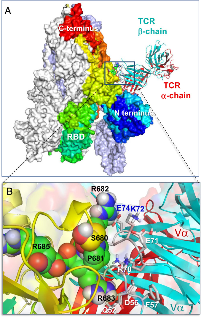
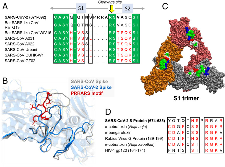
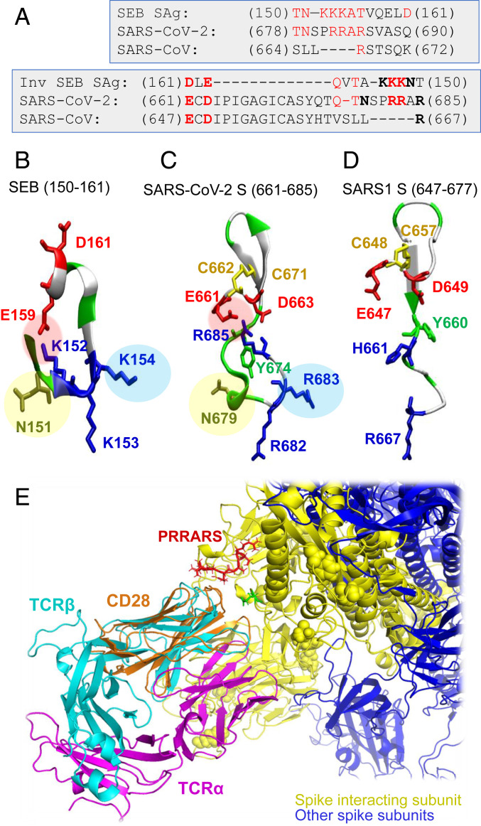
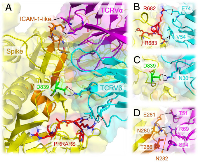
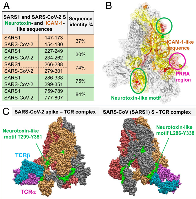
Multisystem Inflammatory Syndrome in Children (MIS-C) associated with COVID-19 is a newly recognized condition in children with recent severe acute respiratory syndrome coronavirus 2 (SARS-CoV-2) infection. These children and adult patients with severe hyperinflammation present with a constellation of symptoms that strongly resemble toxic shock syndrome, an escalation of the cytotoxic adaptive immune response triggered upon the binding of pathogenic superantigens to T cell receptors (TCRs) and/or major histocompatibility complex class II (MHCII) molecules. Here, using structure-based computational models, we demonstrate that the SARS-CoV-2 spike (S) glycoprotein exhibits a high-affinity motif for binding TCRs, and may form a ternary complex with MHCII. The binding epitope on S harbors a sequence motif unique to SARS-CoV-2 (not present in other SARS-related coronaviruses), which is highly similar in both sequence and structure to the bacterial superantigen staphylococcal enterotoxin B. This interaction between the virus and human T cells could be strengthened by a rare mutation (D839Y/N/E) from a European strain of SARS-CoV-2. Furthermore, the interfacial region includes selected residues from an intercellular adhesion molecule (ICAM)-like motif shared between the SARS viruses from the 2003 and 2019 pandemics. A neurotoxin-like sequence motif on the receptor-binding domain also exhibits a high tendency to bind TCRs. Analysis of the TCR repertoire in adult COVID-19 patients demonstrates that those with severe hyperinflammatory disease exhibit TCR skewing consistent with superantigen activation. These data suggest that SARS-CoV-2 S may act as a superantigen to trigger the development of MIS-C as well as cytokine storm in adult COVID-19 patients, with important implications for the development of therapeutic approaches.

Keywords: COVID-19; SARS-CoV-2 spike; TCR binding; superantigen; toxic shock syndrome.


Conflict of interest statement

Competing interest statement: Patent filing process has been started for short peptide sequences to neutralize the superantigenic fragment.

Figures

Fig. 1.
Fig. 1.
Binding of TCR to SARS-CoV-2 spike trimer near the “PRRA” insert. (A) Overall and (B) close-up views of the complex and interfacial interactions. In A, the spike monomers are colored white, ice blue/gray, and spectrally from blue (N-terminal domain) to red, all displayed in surface representation. The N and C termini and RBD of the spectrally colored monomer, which also binds the TCR, are labeled; for better visualization, the S trimer is oriented such that its RBDs are at the bottom. TCR α- and β-chains are in red and cyan ribbons. In B, the segment S680PPRAR685 including the PRRA insert and the highly conserved cleavage site R685 is shown in van der Waals representation (black labels); nearby CDR residues of the TCR Vβ domain are labeled in blue/white. See additional information in SI Appendix, Fig. S1.
Fig. 2.
Fig. 2.
Sequence and structural properties of the insert PRRA. (A and B) SARS-CoV-2 encodes both a cleavage site and SAg-like motifs (20) near the insertion PRRA that distinguishes it from all SARS-related β-CoVs. (A) Sequence alignment of SARS-CoV-2 and multiple SARS-related strains (1) near the insertion PRRA. (B) Structural alignment of SARS-CoV-2 and SARS1 at the same region. The PRRARS motif is shown in red sticks. (C) SARS-CoV-2 S trimer composed of S1 subunits only. The protomers are colored orange, red, and gray, and displayed in van der Waals format. The hydrophobic, hydrophilic, acidic, and basic residues in the protruding motifs E661 to R685 are colored white, green, red, and blue, respectively. (D) Sequence similarity between the close neighborhood of the PRRA insert, neurotoxin motifs reported earlier (20), and HIV-1 gp120 superantigenic motif (63) in the last row.
Fig. 3.
Fig. 3.
The “PRRA” insert in SARS-CoV-2 spike exhibits sequence and structure properties similar to those of the bacterial superantigen SEB. (A) Alignment of SEB superantigenic sequence (21) against the homologous sequence of SARS-CoV-2 spike near the PRRA insert and corresponding SARS1 S segment. Alignments are displayed for both forward (Top) and reverse (Bottom) ordering of the SEB sequence. Note the similarities between the former two, while the third (SARS1 S) shows similarities to SARS-CoV-2, but not to SEB, sequence. Charged amino acids and a critical asparagine that are structurally conserved between SARS-CoV-2 S and SEB, as illustrated in B and C, are written in boldface in the Lower alignment. (B) Structure of the superantigenic peptide (T150-D161) observed in the crystal structure of SEB (64) (PDB ID code 3SEB). (C) Structural model for SARS-CoV-2 S palindromic motif E661-R685. (D) Homologous region in SARS1 S exhibits totally distinctive structural features. Three features (highlighted by pink, blue, and yellow circles) are absent in SARS1 S. The motifs in B and C are polybasic (three lysines and three arginines in the respective cases), whereas SARS1 S counterpart has one basic residue (R667) only; the former two possess a scaffolding ASN, absent in SARS1. (E) Structural alignment of CD28, the receptor binding SEB, onto TCRVβ domain, in support of the adaptability of the putative SAg site to accommodate spike−TCRVβ or SEB−CD28 interactions.
Fig. 4.
Fig. 4.
The interfacial interactions between SARS-CoV-2 spike and αβTCR are further stabilized by the association of an ICAM-1−like motif with TCRVα domain. (A) Interface between SARS-CoV-2 spike and TCR variable domains. Spike is shown in yellow; TCR Vα and Vβ are in magenta and cyan, respectively. The PRRARS insert is highlighted in red; the mutation site D839 identified in recent study (26) is in green; SARS-CoV-2 counterpart of ICAM-1 (CD54)-like motif identified for SARS1 spike (23) is in orange. Residues involved in close interfacial contacts are shown in sticks, with nitrogen and oxygen atoms colored blue and red, respectively. Interactions between atom pairs separated by less than 2.5 Å are indicated by black dashed lines. (B) A close-up view of the interactions between the PRRARS insert/motif and TCR Vβ. (C) Same for the D839 mutation site. (D) Interactions between selected residues on ICAM-1−like motif (labeled, orange) and TCRVα CDRs.
Fig. 5.
Fig. 5.
Neurotoxin-like sequences in SARS-CoV-2 S RBD and their ability to bind TCRs. (A) Comparison of bioactive, neurotoxin-like (green) and ICAM-1 like (orange) segments identified for SARS1 and their SARS-CoV-2 spikes. (B) Loci of two neurotoxin-like regions (enclosed in green circles) and one ICAM-1 region (orange circle; see Fig. 4) conserved between the two CoVs, shown on one monomer (highlighted in yellow) of SARS-CoV-2 S. (C) Binding poses of TCR on SARS-CoV-2 (Left) and SARS1 (Right) S proteins, making contacts with the indicated conserved neurotoxin motif.
Fig. 6.
Fig. 6.
Skewing of TRBV usage in severe/hyperinflammatory COVID-19 patients; 24 repertoires of severe/hyperinflammatory COVID-19 cases versus 42 repertoires of mild/moderate COVID-19 cases were analyzed with and without 23 repertoires of age-matched healthy donors (age-matched to severe/hyperinflammatory COVID-19 group). (A) PCA of TRBV usage. Principal components 1 and 2 are shown; percentage of axis contributions are given in parentheses. Statistical analysis was performed using multivariate analysis of variance (MANOVA) Pillai–Bartlett test. (B) TRBV usage. The fraction of individual TRBV genes per repertoire is shown as mean ± SEM. TRBV genes are sorted to enriched fractions in severe/hyperinflammatory versus mild/moderate COVID-19 disease in ascending order from bottom to top. The top TRBVs enriched in severe/hyperinflammatory COVID-19 patients (TRBV5-6, TRBV14, TRBV13, and TRBV24-1) are enlarged in Inset. (C) PCA of TRBJ usage as described in A. See also SI Appendix, Fig. S7.

Update of

Comment in

References

    1. Walls A. C. et al. ., Structure, function, and antigenicity of the SARS-CoV-2 spike glycoprotein. Cell 181, 281–292.e6 (2020). - PMC - PubMed
    1. Cristiani L. et al. ., Will children reveal their secret? The coronavirus dilemma. Eur. Respir. J. 55, 2000749 (2020). - PMC - PubMed
    1. Tay M. Z., Poh C. M., Rénia L., MacAry P. A., Ng L. F. P., The trinity of COVID-19: Immunity, inflammation and intervention. Nat. Rev. Immunol. 20, 363–374 (2020). - PMC - PubMed
    1. Vabret N. et al. .; Sinai Immunology Review Project , Immunology of COVID-19: Current state of the science. Immunity 52, 910–941 (2020). - PMC - PubMed
    1. Riphagen S., Gomez X., Gonzalez-Martinez C., Wilkinson N., Theocharis P., Hyperinflammatory shock in children during COVID-19 pandemic. Lancet 395, 1607–1608 (2020). - PMC - PubMed

Publication types

MeSH terms