. 2020 Oct 13;117(41):25476-25485.
doi: 10.1073/pnas.2006753117. Epub 2020 Sep 28.

Characterization and engineering of a two-enzyme system for plastics depolymerization

Affiliations

Characterization and engineering of a two-enzyme system for plastics depolymerization

Brandon C Knott et al. Proc Natl Acad Sci U S A. .

Abstract

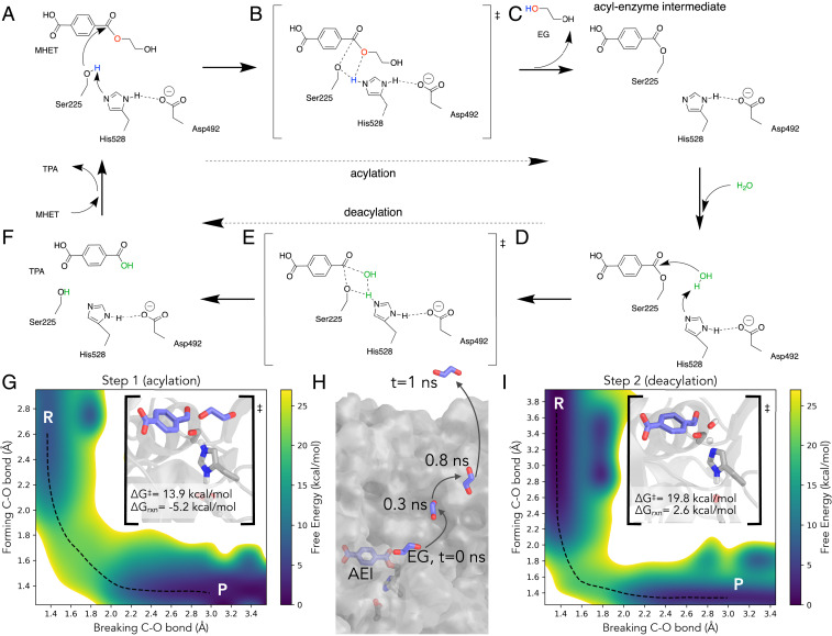
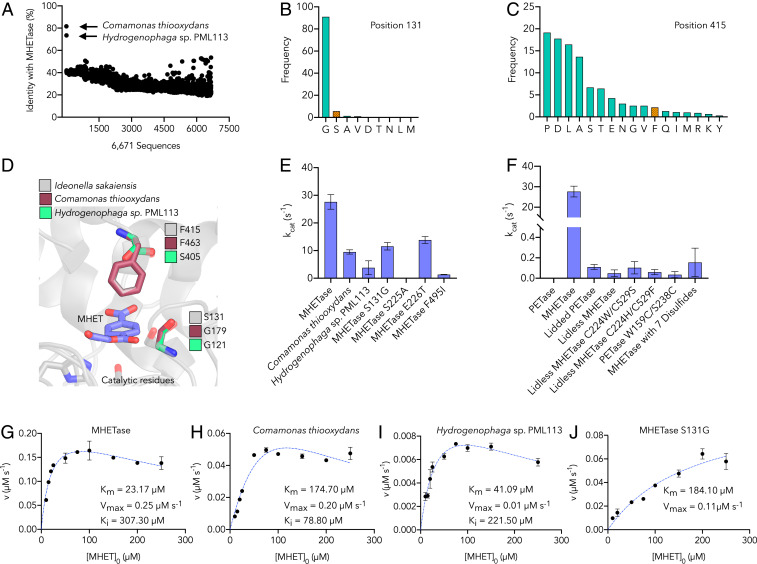
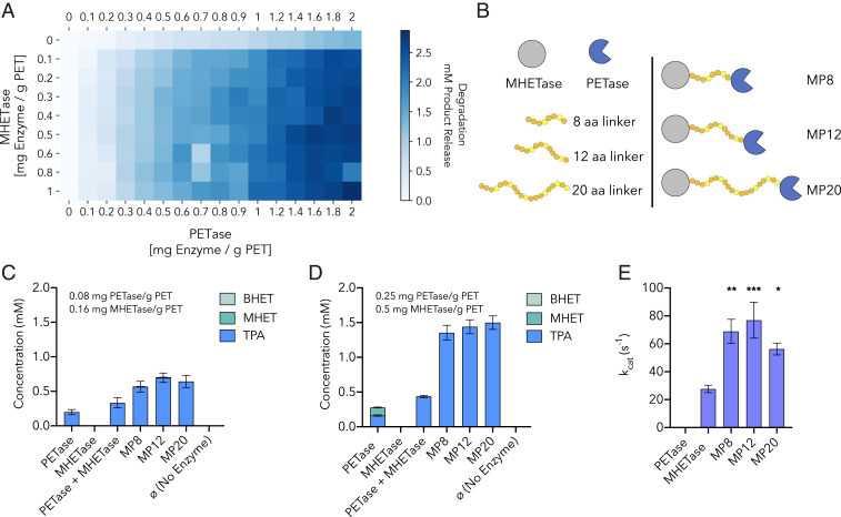
Plastics pollution represents a global environmental crisis. In response, microbes are evolving the capacity to utilize synthetic polymers as carbon and energy sources. Recently, Ideonella sakaiensis was reported to secrete a two-enzyme system to deconstruct polyethylene terephthalate (PET) to its constituent monomers. Specifically, the I. sakaiensis PETase depolymerizes PET, liberating soluble products, including mono(2-hydroxyethyl) terephthalate (MHET), which is cleaved to terephthalic acid and ethylene glycol by MHETase. Here, we report a 1.6 Å resolution MHETase structure, illustrating that the MHETase core domain is similar to PETase, capped by a lid domain. Simulations of the catalytic itinerary predict that MHETase follows the canonical two-step serine hydrolase mechanism. Bioinformatics analysis suggests that MHETase evolved from ferulic acid esterases, and two homologous enzymes are shown to exhibit MHET turnover. Analysis of the two homologous enzymes and the MHETase S131G mutant demonstrates the importance of this residue for accommodation of MHET in the active site. We also demonstrate that the MHETase lid is crucial for hydrolysis of MHET and, furthermore, that MHETase does not turnover mono(2-hydroxyethyl)-furanoate or mono(2-hydroxyethyl)-isophthalate. A highly synergistic relationship between PETase and MHETase was observed for the conversion of amorphous PET film to monomers across all nonzero MHETase concentrations tested. Finally, we compare the performance of MHETase:PETase chimeric proteins of varying linker lengths, which all exhibit improved PET and MHET turnover relative to the free enzymes. Together, these results offer insights into the two-enzyme PET depolymerization system and will inform future efforts in the biological deconstruction and upcycling of mixed plastics.

Keywords: biodegradation; polyethylene terephthalate; recycling; serine hydrolase; upcycling.


Conflict of interest statement

Competing interest statement: A patent application was filed on this work.

Figures

Fig. 1.
Fig. 1.
MHETase structural analysis. (A) MHETase structure (1.6 Å resolution, PDB ID code 6QZ3) highlighting the catalytic triad, five disulfides (in yellow and gray stick representation), benzoate (purple sticks), and calcium ion (green sphere). The lid domain is dark gray, whereas the hydrolase domain is light gray. Main-chain atoms of the linkage residues Tyr252 and Ala469 are colored lime green (also in B). (B) Close-up of the MHETase active site with benzoate bound; catalytic triad, active site disulfide, Ser416, and Arg411 shown as sticks. (C) The concerted movement of residues Gln410 and Phe415 on ligand binding is illustrated with purple arrows in a superposition of the apo enzyme (yellow) with the ligand bound state (gray). The relative position of benzoic acid is depicted in purple. (D) Structural comparison between MHETase (gray) and PETase (PDB ID code 6EQE, in blue), highlighting regions of alignment in the hydrolase domain. A PET tetramer from a prior docking study (29) is shown in yellow sticks (also in E). (E) Electrostatic potential distribution mapped to the solvent-accessible surface of PETase (29) and MHETase as a colored gradient from red (acidic) at −7 kT/e to blue (basic) at 7 kT/e (where k is the Boltzmann’s constant, T is temperature, and e is the charge of an electron). PETase is shown with a bound PET tetramer, and MHETase with benzoate bound from the 6QZ3 structure (yellow). The models are drawn to scale and aligned via their catalytic triad demonstrating their relative size difference.
Fig. 2.
Fig. 2.
The MHETase catalytic mechanism: (A) reactant, (B) transition state, and (C) product of acylation in which His528 transfers a proton from Ser225 to the EG leaving group. In deacylation (DF), His528 plays a similar role and restores the catalytic serine, transferring a proton from a water molecule to Ser225 and generating a free TPA molecule. (G) The free-energy surface for acylation computed along a reaction coordinate described by the breaking and forming C-O bonds. The minimum free energy path is shown in black dashes. (H) Following acylation, EG leaves the active site within 1 ns of a classic MD simulation. (I) The free-energy surface for deacylation, exhibiting a predicted higher barrier than acylation. The MFEP is shown in black dashes.
Fig. 3.
Fig. 3.
Characterization of MHETase, homologs, and mutants. (A) Sequence identity of 6,671 tannase family sequences retrieved by PSI-BLAST compared to MHETase. Sequences (x axis) are in the same order returned by PSI-BLAST. (B and C) Conservation analysis of residue positions 131 (B) and 415 (C) (using MHETase numbering). Frequency of each amino acid is based on a multiple sequence alignment of the 6,671 tannase family sequences. The residue found in MHETase at each position is indicated in orange. (D) Homology model of the MHET-bound active site within 6 Å of the bound substrate comparing MHETase to homology models of the C. thiooxydans and Hydrogenophaga sp. PML113 homologs (generated by SWISS-MODEL) (54), showing sequence variation at residue positions corresponding to Ser131 and Phe415 in MHETase. (E and F) The rate of enzymatic turnover of MHET determined for MHETase, both homologous enzymes, and the indicated MHETase mutants, all of which are active on MHET save the catalytic mutant (S225A) (E), and enzymatic turnover rates for PETase, MHETase, and selected mutants on MHET (F), using 5 nM purified enzyme and 250 µM substrate at 30 °C. (GJ) The initial enzyme reaction velocity as a function of substrate concentration for MHETase, C. thiooxydans, Hydrogenophaga sp. PML113, and the MHETase S131G mutant, respectively. Dashed blue lines represent the Michaelis–Menten kinetic model fit with substrate inhibition (GI) or fit with the simple Michaelis–Menten model (J). Key kinetic parameters are provided in the Inset. Additional parameters and confidence intervals on the listed parameters are provided in SI Appendix, Table S3.
Fig. 4.
Fig. 4.
PETase-MHETase synergy and chimeric enzymes. (A) Heatmap of synergistic degradation by PETase and MHETase on amorphous PET film over 96 h at 30 °C. Total product release in millimolars (sum of BHET, MHET, and TPA); x axis: PETase loading (mg/g PET), y axis: MHETase loading (mg/g PET). (B) Illustrations of three chimeric enzymes. Linkers composed of glycine (orange) and serine (yellow) residues connecting the C terminus of MHETase to the N terminus of PETase. (C and D) Comparison of depolymerization performance of PETase alone, MHETase alone, PETase and MHETase at equimolar loading, and the three chimeric enzymes on amorphous PET film after 96 h at 30 °C. Product release in millimolars resulting from hydrolysis by (C) 0.08 mg PETase/g PET or 0.16 mg MHETase/g PET and (D) 0.25 mg PETase/g PET or 0.5 mg MHETase/g PET. All comparisons are statistically significant with P ≤ 0.0001 based on two-way ANOVA analysis and Tukey’s multiple comparisons test. (E) MHET turnover rate by each chimeric enzyme compared to MHETase alone using 250 µM MHET and 5 nM enzyme. Asterisks indicate statistically significant comparisons between MHETase and each chimera enzyme with *P ≤ 0.01, **P ≤ 0.001, and ***P ≤ 0.0005.

References

    1. Law K. L. et al. ., Plastic accumulation in the North Atlantic subtropical gyre. Science 329, 1185–1188 (2010). - PubMed
    1. Cózar A. et al. ., Plastic debris in the open ocean. Proc. Natl. Acad. Sci. U.S.A. 111, 10239–10244 (2014). - PMC - PubMed
    1. Jambeck J. R. et al. ., Marine pollution. Plastic waste inputs from land into the ocean. Science 347, 768–771 (2015). - PubMed
    1. Lamb J. B. et al. ., Plastic waste associated with disease on coral reefs. Science 359, 460–462 (2018). - PubMed
    1. de Souza Machado A. A., Kloas W., Zarfl C., Hempel S., Rillig M. C., Microplastics as an emerging threat to terrestrial ecosystems. Glob. Change Biol. 24, 1405–1416 (2018). - PMC - PubMed

Publication types

Supplementary concepts

LinkOut - more resources