

Nociceptive behavioural assessments in mouse models of temporomandibular joint disorders
- PMID: 32989215
- PMCID: PMC7522224
- DOI: 10.1038/s41368-020-00095-0
Nociceptive behavioural assessments in mouse models of temporomandibular joint disorders
Abstract
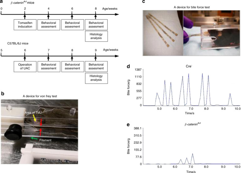
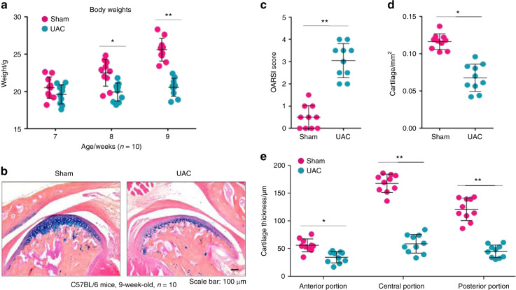
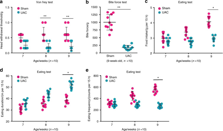
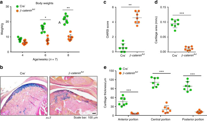
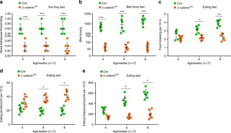
Orofacial pain or tenderness is a primary symptom associated with temporomandibular joint (TMJ) disorders (TMDs). To understand the pathological mechanisms underlying TMDs, several mouse models have been developed, including mechanical stimulus-induced TMD and genetic mouse models. However, a lack of feasible approaches for assessing TMD-related nociceptive behaviours in the orofacial region of mice has hindered the in-depth study of TMD-associated mechanisms. This study aimed to explore modifications of three existing methods to analyse nociceptive behaviours using two TMD mouse models: (1) mechanical allodynia was tested using von Frey filaments in the mouse TMJ region by placing mice in specially designed chambers; (2) bite force was measured using the Economical Load and Force (ELF) system; and (3) spontaneous feeding behaviour tests, including eating duration and frequency, were analysed using the Laboratory Animal Behaviour Observation Registration and Analysis System (LABORAS). We successfully assessed changes in nociceptive behaviours in two TMD mouse models, a unilateral anterior crossbite (UAC)-induced TMD mouse model and a β-catenin conditional activation mouse model. We found that the UAC model and β-catenin conditional activation mouse model were significantly associated with signs of increased mechanical allodynia, lower bite force, and decreased spontaneous feeding behaviour, indicating manifestations of TMD. These behavioural changes were consistent with the cartilage degradation phenotype observed in these mouse models. Our studies have shown reliable methods to analyse nociceptive behaviours in mice and may indicate that these methods are valid to assess signs of TMD in mice.
Conflict of interest statement
The authors declare no competing interests.
Figures
Similar articles
-
Analysis of pain in the rabbit temporomandibular joint after unilateral splint placement.J Oral Facial Pain Headache. 2015 Spring;29(2):193-202. doi: 10.11607/ofph.1371. J Oral Facial Pain Headache. 2015. PMID: 25905538
-
Prevalence of temporomandibular disorder pain, jaw noises and oral behaviours in an adult Italian population sample.J Oral Rehabil. 2019 Aug;46(8):691-698. doi: 10.1111/joor.12803. Epub 2019 May 7. J Oral Rehabil. 2019. PMID: 30993737
-
Executive summary of the Diagnostic Criteria for Temporomandibular Disorders for clinical and research applications.J Am Dent Assoc. 2016 Jun;147(6):438-45. doi: 10.1016/j.adaj.2016.01.007. Epub 2016 Feb 26. J Am Dent Assoc. 2016. PMID: 26922248 Free PMC article.
-
Mouse genetic models for temporomandibular joint development and disorders.Oral Dis. 2016 Jan;22(1):33-8. doi: 10.1111/odi.12353. Epub 2015 Jul 2. Oral Dis. 2016. PMID: 26096083 Free PMC article. Review.
-
Comorbidity between fibromyalgia and temporomandibular disorders: a systematic review.Oral Surg Oral Med Oral Pathol Oral Radiol. 2019 Jul;128(1):33-42. doi: 10.1016/j.oooo.2019.02.023. Epub 2019 Feb 28. Oral Surg Oral Med Oral Pathol Oral Radiol. 2019. PMID: 30981530
Cited by
-
Chemokine Regulation in Temporomandibular Joint Disease: A Comprehensive Review.Genes (Basel). 2023 Feb 4;14(2):408. doi: 10.3390/genes14020408. Genes (Basel). 2023. PMID: 36833336 Free PMC article. Review.
-
The involvement of orexin-1 receptors in modulation of feeding and anxiety-like behavior in rats with complete Freund's adjuvant-induced temporomandibular joint disorder.Odontology. 2025 Apr;113(2):764-775. doi: 10.1007/s10266-024-01021-0. Epub 2025 Jan 23. Odontology. 2025. PMID: 39843662 Free PMC article.
-
Multi-class segmentation of temporomandibular joint using ensemble deep learning.Sci Rep. 2024 Aug 16;14(1):18990. doi: 10.1038/s41598-024-69814-5. Sci Rep. 2024. PMID: 39160234 Free PMC article.
-
Preclinical models of deep craniofacial nociception and temporomandibular disorder pain.Jpn Dent Sci Rev. 2021 Nov;57:231-241. doi: 10.1016/j.jdsr.2021.10.002. Epub 2021 Nov 12. Jpn Dent Sci Rev. 2021. PMID: 34815817 Free PMC article. Review.
-
Antisense Oligonucleotide-Based Therapy on miR-181a-5p Alleviates Cartilage Degradation of Temporomandibular Joint Osteoarthritis via Promoting SIRT1.Front Pharmacol. 2022 Jun 15;13:898334. doi: 10.3389/fphar.2022.898334. eCollection 2022. Front Pharmacol. 2022. PMID: 35784690 Free PMC article.
References
Publication types
MeSH terms
Grants and funding
LinkOut - more resources
Full Text Sources
Medical
