. 2020 Oct;22(10):1197-1210.
doi: 10.1038/s41556-020-0569-x. Epub 2020 Sep 28.

STAT3-BDNF-TrkB signalling promotes alveolar epithelial regeneration after lung injury

Affiliations

STAT3-BDNF-TrkB signalling promotes alveolar epithelial regeneration after lung injury

Andrew J Paris et al. Nat Cell Biol. 2020 Oct.

Abstract

Alveolar epithelial regeneration is essential for recovery from devastating lung diseases. This process occurs when type II alveolar pneumocytes (AT2 cells) proliferate and transdifferentiate into type I alveolar pneumocytes (AT1 cells). We used genome-wide analysis of chromatin accessibility and gene expression following acute lung injury to elucidate repair mechanisms. AT2 chromatin accessibility changed substantially following injury to reveal STAT3 binding motifs adjacent to genes that regulate essential regenerative pathways. Single-cell transcriptome analysis identified brain-derived neurotrophic factor (Bdnf) as a STAT3 target gene with newly accessible chromatin in a unique population of regenerating AT2 cells. Furthermore, the BDNF receptor tropomyosin receptor kinase B (TrkB) was enriched on mesenchymal alveolar niche cells (MANCs). Loss or blockade of AT2-specific Stat3, Bdnf or mesenchyme-specific TrkB compromised repair and reduced Fgf7 expression by niche cells. A TrkB agonist improved outcomes in vivo following lung injury. These data highlight the biological and therapeutic importance of the STAT3-BDNF-TrkB axis in orchestrating alveolar epithelial regeneration.


Figures

Extended Data Fig. 1
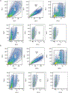
Extended Data Fig. 1. Sorting Strategies for Isolating AT2 cells.
Lungs from C57BL/6 mice were digested into a single-cell suspension. We collected EpCAM positive cells (APC) that were PDPN, CD45, CD34, CD31 and SCA1 negative (all PE).
Extended Data Fig. 2
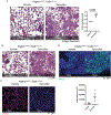
Extended Data Fig. 2. STAT3 is activated following sterile and infectious lung injuries.
Cells staining positive for proSFTPC+ and pSTAT3+ are highlighted with a white box while proSFTPC + pSTAT3− cells are highlighted with a yellow box. Areas that were felt to be indeterminate for cell type or pSTAT3 status are highlighted in blue. Scale bars, 50μm.
Extended Data Fig. 3
Extended Data Fig. 3. Loss of Stat3 diminished alveolar organoid formation.
(A) PCR shows the expected 802bp product when corn oil is administered to SftpcCreERT2:Stat3LoxP/LoxP mice and the recombined 514bp product when tamoxifen is administered to SftpcCreERT2:Stat3LoxP/LoxP mice. The gels in this figure represent n=2 mice per condition (corn oil and tamoxifen). This experiment was repeated 4 times with similar results. (B) AT2 cells from tamoxifen treated SftpcCreERT2-Rosa26tdTomato and SftpcCreERT2-Rosa26tdTomato:Stat3LoxP/LoxP mice were isolated and co-cultured with PDGFRα mesenchymal cells in transwells containing Matrigel for four weeks (n=4 wells per condition). Organoid forming efficiency was significantly decreased in the absence of STAT3 signaling in AT2 cells (p=2.2x10⁻⁸). Data is shown as the mean +/− SEM. Statistical significance was determined using a two-tailed Student’s t-test. Scale bars, 2.5mm.
Extended Data Fig. 4
Extended Data Fig. 4. Loss of Stat3 in AT2 cells does not alter distal lung morphology.
SftpcCreERT2:Stat3LoxP/LoxP mice were given tamoxifen or vehicle and euthanized three months later. Representative H&E stained histological specimens are shown. The Mean linear intercept was unchanged by the loss of Stat3 in these unchallenged mice (n=4 mice per group). Data is shown as the mean +/− SEM. Statistical significance was determined with a two-tailed Student’s t-test. Scale bars, 500μm.
Extended Data Fig. 5
Extended Data Fig. 5. Single-cell expression of TrkB, Ccnd1, Tm4sf1 and Sftpc in isolated AT2 cells.
Plots show relative expression of TrkB, Ccnd1, Tm4sf1 and Sftpc in AT2 cells isolated isolated from uninjured mice and mice 24h after acid-induced lung injury.
Extended Data Fig. 6
Extended Data Fig. 6. Pathways analysis of isolated AT2 cells reveals multiple subsets of cells with multiple functions.
Pathways analysis of differentially expressed genes in the clusters shown in Fig. 4B.
Extended Data Fig 7
Extended Data Fig 7. AT2 cells express Bdnf after acid-induced lung injury.
BdnfCre-Rosa26tdTomato were subjected to acid-induced lung injury (n=4 mice per group). One day after injury the mice were euthanized, and the lungs were probed for Tomato (red) and proSFTPC (green). Autofluorescence delineates tissue structure. Scale bar, 25μm.
Extended Data Fig. 8
Extended Data Fig. 8. Loss of TrkB in mesenchymal cells worsens outcomes following sterile and infectious lung injuries.
Lungs from PdgfraEGFP mice were digested into a single-cell suspension either before or 24h after acid-induced lung injury (n=4/group). We first gated on PDGFR α + and then quantified the percent of GFP+ cells within that population.
Extended Data Fig. 9
Extended Data Fig. 9. Loss of TrkB in mesenchymal cells worsens outcomes following sterile and infectious lung injuries.
(A) H&E staining and ATS lung injury scores of tamoxifen and corn oil exposed PdgfrαCreERT2:TrkBLoxP/LoxP mice 24 hours after acid-induced lung injury (n=4 mice per group). (B-D) H&E (B), PDPN (C) and Krt5 (D) staining of tamoxifen and corn oil exposed PdgfrαCreERT2:TrkBLoxP/LoxP mice 21 days after infection with PR8 influenza (n=3 mice per group). Statistical analysis was performed with a two-tailed Student’s t-test or ANOVA, where appropriate. For panels (A) and (D), data is shown as the mean +/− SEM. Statistical significance was determined with a two-tailed Student’s t-test (A) and (D). Scale bars, 100μm.
Extended Data Fig. 10
Extended Data Fig. 10. The TrkB agonist 7,8-DHF is unable to rescue mice with an AT2-specific Stat3 deletion.
Survival curves for Tamoxifen-exposed SftpcCreERT2:Stat3LoxP/LoxP mice that had been infected with intranasal PR8 influenza (5x10⁻⁵ HAU/mouse) and were given intraperitoneal injections of 7,8-DHF or vehicle every other day (n=5 mice per group). The data analyzed using the Log-rank (Mantel-Cox) test and is not statistically significant.
Figure 1 –
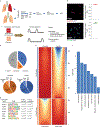
Figure 1 –. The response to injury is marked by significant changes in AT2 chromatin architecture.
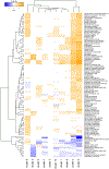
(A) AT2 cells were isolated from single cell suspensions of murine lung tissue (3 uninjured mice, 2 injured mice) using fluorescence activated cell sorting (FACS). Nuclei were isolated and incubated with Tn5 transpose followed by amplification of fragments and next-generation sequencing (NGS). (B) Staining of isolated cells for the AT2 markers proSFTPC and DC-LAMP confirmed >97% isolated cells are AT2 cells (100 cells per stain analyzed in n=3 independent experiments). (C) ATAC-seq was performed on AT2 cells from uninjured mice (quiescent) and from mice 24 hours post acid-induced lung injury (regenerative). NGS reads were aligned and uninjured and injured sequences were compared. (D & E) Quantitation of shared and unique regions of accessible chromatin. (F) Ingenuity pathway analyses of genes associated with differentially accessible chromatin following acute lung injury demonstrated newly accessible areas of the genome encode proteins important for alveolar epithelial regeneration. (G) HOMER analysis of accessible chromatin demonstrated common enrichment for alveolar epithelium-associated transcription factors including NKX2.1, FOXF1 and FOXP1. After injury STAT3, TBX5, and SPIB chromatin accessibility becomes increasingly enriched. For panel (B), data is shown as the mean +/− SEM. Statistical significance was determined using a two-tailed Student’s T-test (B) and with the findMotifGenome.pl script in the HOMER software package with adjustment for multiple comparisons (G). Scale bars, 50μm.
Figure 2 –
Figure 2 –. Stat3 regulates key pathways in alveolar epithelial regeneration.
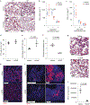
(A) IL-6 is undetectable in uninjured lungs. Levels of IL-6 in BAL fluid peak at 24h after acid-induced lung injury (n=3 mice per time point). (B & C) C57BL/6 mice that underwent acid-induced lung injury demonstrate activated pSTAT3 in AT2 cells (p=2.9x10⁻⁵ for 9 days post infection vs uninjured). There was minimal phosphorylated STAT3 in AT2 cells of uninjured mice (n= 4 mice per group). (D & E) Human samples with diffuse alveolar damage had increased AT2 specific pSTAT3 (p=1.8x10⁻⁶) that was not present in uninjured samples (n=4 patients per group). (F & G) Unbiased pathway analyses demonstrated newly accessible STAT3 binding sites adjacent to genes that control key biological mechanisms including proliferation and migration. For panels (A) (C) and (E), data is shown as the mean +/− SEM. Statistical significance was determined with a two-tailed Student’s t-test (C & E). Scale bars 50μm (B, D) and 25μm (B, D).
Figure 3 –
Figure 3 –. Deletion of Stat3 in AT2 cells worsens outcomes following sterile and infectious lung injuries.
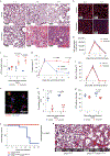
(A) SftpcCreERT2:Stat3LoxP/LoxP mice were given tamoxifen in corn oil or corn oil alone 18 days prior to acid-induced lung injury (3 mice per group). H&E staining shows that control mice had histological injury that resolved by day 5 post-injury. Mice that received tamoxifen which resulted in AT2 specific Stat3 deletion had persistent and pronounced cellular, proteinaceous and hemorrhagic infiltrates that obscured the alveolar structures. (B) PDPN staining revealed that mice lacking Stat3 had decreased AT1 populations at 3 and 5 days following acid-induced lung injury compared to controls (n=3 mice per group). (C) ATS lung injury scores were increased at later time points following acid-induced lung injury in mice lacking Stat3 (3 mice per group per time point ). (D) BAL protein from mice described in (A) showed non-resolving increase in alveolar protein following acid-induced lung injury in mice that lacked AT2 specific Stat3 (n=3 mice per group per time point). (E, F, G) The percent of Ki6+ AT2 cells was decreased in tamoxifen treated SftpcCreERT2:Stat3LoxP/LoxP mice following acid-induced lung injury (n=3 mice per group per time point). (H & I) BAL neutrophils and IL-1β levels following acid-induced lung injury were not changed by the presence or absence of functional AT2 specific Stat3 (n=3 mice per group per time point). (J) SfptcCreERT2 and SftpcCreERT2:Stat3LoxP/LoxP mice were given tamoxifen 18 days prior to intranasal infection with PR8 influenza (5x10⁻⁵ HAU/mouse). Mortality was significantly higher in the mice that lack AT2 specific STAT3 compared to controls (n=10 mice per group). (K) At 21 days post infection the mouse lacking AT2 specific Stat3 (1 mouse) had distorted distal alveolar lung architecture with cellular infiltrate and cystic structures that were not observed in controls (10 mice). For panels (C), (D), (F), (G), (H), and (I), data is shown as the mean +/− SEM. Statistical significance was determined with a two-tailed Student’s t-test (C), (F), and (G), with a two-way ANOVA (D), and with the Log-rank (Mantel-Cox) test (J). Scale bars, 100μm (A, B, K) and 25μm (E).
Figure 4 –
Figure 4 –. BDNF identified as important for alveolar epithelial regeneration.
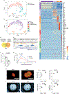
(A) AT2 cells were isolated from an uninjured C57BL/6 mouse and from a C57BL/6 mouse that underwent acid-induced lung injury 24 hours prior. Single-cell transcriptomic analysis was performed and cells were grouped into populations using t-SNE. There was little overlap between AT2 cells from uninjured and injured mice. (B) K-means analysis was used to sub-divide AT2 cells into groups based on their individual gene expression. (C) Genes whose expression was unique to each cluster are shown. Cluster 3 has gene expression from both AT2 and AT1 cells (Sftpc and Pdpn) and AEP marker Tm4sf1. (D) Bdnf is the only bona fide STAT3 target gene with newly accessible chromatin following acid-induced lung injury that is differentially expressed in cluster 3. (E) Data from ATAC-seq (shown in Fig. 1) demonstrate increased accessibility of the Bdnf promotor in cells isolated from mice 24 hours after acid-induced lung injury. (F) AT2 cells from tamoxifen-exposed SftpcCreERT2:Stat3LoxP/LoxP mice express less Bdnf than vehicle exposed mice (n=4 mice per group) (G) BAL fluid from SftpcCreERT2:Stat3LoxP/LoxP mice treated with either tamoxifen or corn oil (vehicle) prior to undergoing acid-induced lung injury shows that mice lacking AT2-specific Stat3 have significantly less BDNF in their BAL fluid (n=3 mice per group per time point). (H) AT2 cells from SftpcCreERT2-Rosa26TdTomato:Hopx3FlagGFP mice were co-cultured with PDGFRα+ mesenchymal cells from C57BL/6 mice in the presence of BDNF which increased alveolar organoid forming efficiency of murine AT2 cells. Right-hand panels show quantification from n=3 distinct cultures per condition. (I) Primary human AT2 cells were isolated and co-cultured with MRC5 fibroblasts for three weeks. We observed increased organoid size and forming efficiency when BDNF was added to the culture media. Right-hand panels show the average organoid forming efficiency and size from n=3 different donors. Individual points are shown with the mean and standard error of the mean depicted. For panels (F), (G), (H), and (I), data is shown as the mean +/− SEM. Statistical significance was determined with a two-tailed Student’s t-test (F), (H), and (I), and with a two-way ANOVA (G). Scale bars, 1mm.
Figure 5 –
Figure 5 –. Loss of AT2 specific BDNF worsens outcomes following sterile and infectious lung injuries.
(A) We quantified tomato positive AT2 cells in BdnfCre-R26TdTomao mice before and 24h after acid-induced lung injury and found a significant increase in co-positive cells following acute lung injury (n=4 mice per group). (B-F) H&E staining and ATS lung injury scores (B), BAL BDNF concentrations (C) BAL protein concentration (D) AT2 cell numbers (E) and number of proliferating AT2 cells (F) of tamoxifen-exposed and vehicle-exposed SftpcCreERT2:BdnfLoxP/LoxP mice 5 days after acid-included lung injury (n= 3 mice per time point for C & D, n=4 mice per group per time point for B, E, & F). (G) H&E and PDPN staining of lung tissue from tamoxifen-exposed and vehicle-exposed SftpcCreERT2:BdnfLoxP/LoxP mice that had been infected with intranasal PR8 influenza (5x10⁻⁵ HAU/mouse) (4 mice per group). (H) KRT5 staining of the mice described in (G) with quantification of KRT5+ pods in both groups (n=4 mice per group). For panels (A), (B), (C), (D), (E), (F) and (H), data is shown as the mean +/− SEM. Statistical significance was determined with a two-tailed Student’s t-test (A), (B), (E), (F), and (H), and with a two-way ANOVA (C) and (D). Scale bars, 100μm (B, G, H) and 10μm (A).
Figure 6 –
Figure 6 –. BDNF-TrkB signaling promotes alveolar epithelial regeneration.
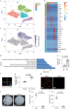
(A-C) Analysis of scRNA-seq of mesenchymal cells from uninjured mice showed that TrkB expression is enriched in mesenchymal alveolar niche cells. (D) Ingenuity pathways analysis show that mesenchymal cells expressing TrkB are enriched with expression of genes that control respiratory system development, organ development and tissue morphology. (E) Expression of TrkB in GFP+ and GFP− cells from TrkBEGFP mice (n=4 mice per group). (F) Expression of TrkB on mesenchymal cells was verified by staining lung tissue form TrkBEGFP mice for PDFRα. (G) Quantification of GFP and PDFRα co-positive cells in TrkBEGFP mice 24 hours after acid-induced lung injury (n=4 mice per group) The gating strategy is shown in Extended Figure 8. (H & I) AT2 cells isolated from a tamoxifen treated SftpcCreERT2-Rosa26mTmG mouse were co-cultured with PDGFRα+ mesenchymal cells for four weeks. ANA-12 abrogated organoid forming efficiency of AT2 cells (n=3 wells per condition). (J & K) The organoid forming capacity of primary human AT2 cells was abrogated by the addition of ANA-12 to the media. Cultures were grown for four weeks. Right-hand panels show average organoid forming efficiency and size from n=3 different donors. For panels (E), (G), (K) and (I), data is shown as the mean +/− SEM. Statistical significance was determined with a two-tailed Student’s t-test (E), (G), (K) and (I). Scale bar, 2.6mm (H, J), 20μm (F).
Figure 7 –
Figure 7 –. The STAT3-BDNF-TrkB axis modulates FGF7 expression by mesenchymal niche cells.
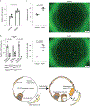
(A) Fgf7 mRNA expression in mesenchymal cells isolated from the organoid culture conditions described in Fig. 4H (n= 5 wells per condition). (B) Fgf7 expression in PDFRα cells isolated from tamoxifen or corn oil exposed SftpcCreERT2:Stat3LoxP/LoxP, SftpcCreERT2:BdnfLoxP/LoxP, and PdgfraCreERT2:TrkBLoxP/LoxP mice 24 hours after acid-induced lung injury (n=4 mice per group). (C) The addition of 0.1μg/mL of recombinant murine BDNF (rmBDNF) caused a significant increase in alveolar organoid forming efficiency and size (n=3 wells per condition). (D) In the setting of acute lung injury STAT3-activating cytokines accumulate in the alveolus and STAT3 becomes activated in the AT2 cell. Accompanying changes in chromatin accessibility within AT2 cells allows for expression of BDNF in transdifferentiating cells which then binds to TrkB on mesenchymal niche cells to stimulate expression of Fgf7, which promotes alveolar epithelial regeneration. For panels (A) (B) and (C), data is shown as the mean +/− SEM. Statistical significance was determined with a two-tailed Student’s t-test (A) (B) and (C). Scale bars, 500μm.
Figure 8 –
Figure 8 –. The TrkB agonist 7,8-dihydroxyflavone improves outcomes following sterile and infectious lung injury.
(A-E) C57BL/6 mice underwent acid-induced unilateral lung injury and were given an intraperitoneal injection of 7,8-DHF or vehicle control at the time of injury (n=3 mice per group). Mice that were euthanized at 48 hours received an additional intraperitoneal dose of 7,8-DHF or vehicle 24 hours after undergoing acid-induced lung injury. Mice that received 7,8-DHF had improved lung histology (A), significantly less BAL protein (B&C) and increased Ki67 and proSFTPC indicating increased AT2 cell proliferation (D&E) at 48 hours after lung injury. No differences in IL-1β or TNFα were observed. (F) Alveolar wall thickness was measured using 10 20x images per lung (means of n=3 mice per group). (G & H) H&E and PDPN staining of lung tissue from C57BL/6 mice infected with intranasal PR8 influenza (5x10⁻⁵ HAU/mouse). Intraperitoneal injections of 7,8-DHF or vehicle every other day (3 mice per group). (I&J) Quantification of KRT5+ pods from mice described in G & H (means of n=3 mice per group). For panels (B), (C), (D), (E), (F) and (J), data is shown as the mean +/− SEM. Statistical significance was determined with a two-tailed Student’s t-test at each time point shown at (B), (C), (D), (E), (F) and (J). Scale bars, 100μm.

Similar articles

Cited by

References

    1. Matthay MA, Robriquet L & Fang X Alveolar epithelium: role in lung fluid balance and acute lung injury. Proc Am Thorac Soc 2, 206–213 (2005). - PubMed
    1. Guillot L et al. Alveolar epithelial cells: master regulators of lung homeostasis. Int J Biochem Cell Biol 45, 2568–2573 (2013). - PubMed
    1. Tyrrell C, McKechnie SR, Beers MF, Mitchell TJ & McElroy MC Differential alveolar epithelial injury and protein expression in pneumococcal pneumonia. Exp Lung Res 38, 266–276 (2012). - PubMed
    1. Herold S, Becker C, Ridge KM & Budinger GR Influenza virus-induced lung injury: pathogenesis and implications for treatment. Eur Respir J 45, 1463–1478 (2015). - PubMed
    1. Herold S et al. Lung epithelial apoptosis in influenza virus pneumonia: the role of macrophage-expressed TNF-related apoptosis-inducing ligand. The Journal of experimental medicine 205, 3065–3077 (2008). - PMC - PubMed

Publication types

MeSH terms