. 2020 Dec;27(12):1115-1124.
doi: 10.1038/s41594-020-0507-4. Epub 2020 Sep 28.

The molecular structure of mammalian primary cilia revealed by cryo-electron tomography

Affiliations

The molecular structure of mammalian primary cilia revealed by cryo-electron tomography

Petra Kiesel et al. Nat Struct Mol Biol. 2020 Dec.

Abstract

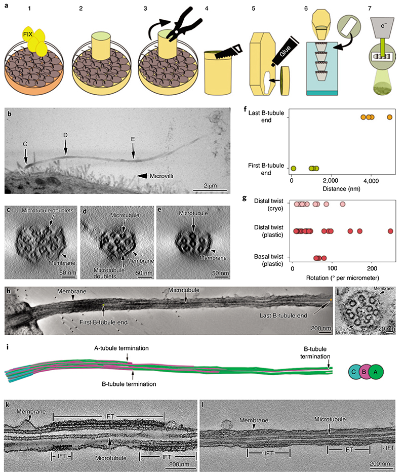
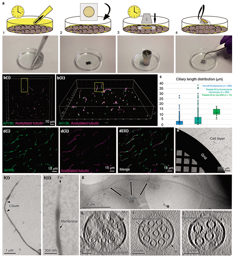
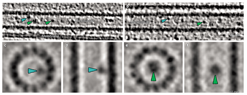
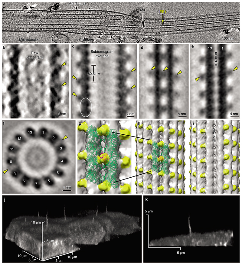
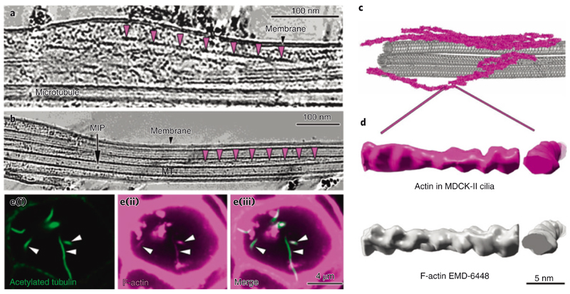
Primary cilia are microtubule-based organelles that are important for signaling and sensing in eukaryotic cells. Unlike the thoroughly studied motile cilia, the three-dimensional architecture and molecular composition of primary cilia are largely unexplored. Yet, studying these aspects is necessary to understand how primary cilia function in health and disease. We developed an enabling method for investigating the structure of primary cilia isolated from MDCK-II cells at molecular resolution by cryo-electron tomography. We show that the textbook '9 + 0' arrangement of microtubule doublets is only present at the primary cilium base. A few microns out, the architecture changes into an unstructured bundle of EB1-decorated microtubules and actin filaments, putting an end to a long debate on the presence or absence of actin filaments in primary cilia. Our work provides a plethora of insights into the molecular structure of primary cilia and offers a methodological framework to study these important organelles.


Figures

Fig. 1
Fig. 1. I Room temperature ET of MDCK-II primary cilia.
a, Description of the steps for resin-embedding and serial sectioning of MDCK-II monolayers. b, Montage of projections from five serial sections covering the entire primary cilium. C, D and E are the areas of the cilium that were digitally sectioned to obtain the images shown in panels c,d and e, respectively. c-e, Representative proximodistal tomographic slices from the same cilium shown in b, showing the axonemal organization of microtubule doublets and singlets. c, Cross-section of the cilium proximal segment showing that 9-fold symmetrical arrangement of the microtubule doublets is already lost. d,e, The number of microtubule doublets (d) and microtubules singlets (e) decreases in the distal part of the cilium. f, Distance measured from the distal side of the basal body to the first and last B-tubule terminations. g, Axonemal twist measured at the basal and distal regions of the cilium. H,i, Tomographic section along a cilium (H), and its microtubule segmentation (i), showing representative positions of first and last B-tubule terminations. J, Ciliary cross-section showing the variety of axonemal structures other than microtubules. k,l, Longitudinal sections through cilia containing IFT train-like particles sandwiched between the ciliary membrane and microtubule singlets. Asterisks indicate locations of unidentified densities.
Fig. 2
Fig. 2. I Cryo-peel off: a method to prepare primary cilia for cryo-ET.
a, Description of the steps followed for the peel off of primary cilia from MDCK-II monolayers. Step 1: removal of the deciliation buffer from the cell culture; step 2: application of poly-L-lysine-coated EM grids, supported by a glass slide, on the apical surface of the cell layer; step 3: pressure was applied over time on the glass slide to favor the adhesion of cilia to the EM grid; step 4: retrieval of the glass slide/EM grid from the cells’ apical surface and consequent primary cilia peel off. b, Laser scanning confocal fluorescence microscopy of the MDCK-II monolayer showing the variability of ciliary lengths after 2d of cell starvation; (b(i)), top view; (b(ii)), tilted view. c, Distribution of lengths of cilia associated with cells (blue), cilia peeled-off by fluorescence microscopy (cyan) and cilia peeled-off by cryo-EM (green). d, Immunofluorescence staining of peeled-off cilia on a glass slide, showing the colocalization of ciliary membrane (d(i)) and microtubules (d(ii) and the combined images (d(iii)).e, A Quantifoil grid on a cell monolayer during the peel off procedure as depicted in a (steps 2 and 3). f(i), Negative staining TEM of a peeled-off primary cilium; f(ii), the zoomed-in view of the ciliary tip from the same cilium shows the preservation of the ciliary membrane. g, Low-magnification cryo-EM image of a peeled-off and plunge-frozen cilium. h-J, Representative proximodistal cryo-tomographic slices of plunge-frozen cilia (h) close to the base central shaft (i) and distal segment (J).
Fig. 3
Fig. 3. I IFT-B-like polymers are visible in cryo-ET of primary cilia.
a, Slice through a denoised tomogram of a primary cilium showing an IFT train (green frame). The train subunits show a similar repeat to the one described for the IFT-B complexes in Chlamydomonas anterograde IFT trains (d). The yellow arrowheads in a indicate a second row of particles placed between the IFT-B-like polymer and the membrane. This location is typical of IFT-A complexes in the anterograde trains of Chlamydomonas. Ring-shaped objects (blue arrowheads) were also present in the area expected for dynein motor cargoes. IFT-A-like (yellow arrowheads) and IFT dynein-like particles (blue arrowheads) appeared partially dissociated from the IFT-B-like polymer, indicating that the structure of the polymer might be altered during the peel off procedure. b, Slice through the same IFT train shown in a after rotating the tomogram by 90° around the microtubule long axis. c, 3D visualization of the relative position of microtubule singlets and two anterograde-like IFT trains from a cryo-tomogram of a primary cilium. d, Subtomogram-averaged model showing the repeat of IFT-B, IFT-A and IFT dynein in the anterograde IFT trains in Chlamydomonas (modified from Jordan et al.). e, StA of IFT-B-like particle repeat from MDCK-II primary cilia. Masked cross-correlation coefficient between the structures shown in e and IFT-B-like in d was approximately 0.59.
Fig. 4
Fig. 4. Proteins are present in the microtubule lumen in MDCK-II primary cilia (MIPs).
a,b, Cryo-electron tomograms of primary cilia show MIPs associated with the internal walls of ciliary microtubules (blue arrowheads) and ‘floating’ in the lumen of the microtubule (green arrowheads).c,d, Subtomogram-averaged structure of microtubule wall-associated MIPs. c, Digital slice perpendicular to the microtubule axis. d, Digital slice parallel to the microtubule axis. e,f, Subtomogram-averaged structure of MIPs located in the center of the microtubule. e, Digital slice perpendicular to the microtubule axis. f, Digital slice parallel to the microtubule axis.
Fig. 5
Fig. 5. I Cryo-ET of primary cilia shows decorations of microtubule singlets by EB1.
a, Representative longitudinal tomography section of a plunge-frozen primary cilium showing EB1-decorated microtubule singlets. Also visible are some MIPs and filaments. b, Close-up view of the EB1 microtubule singlet decoration as seen in a raw tomogram. c-i, Longitudinal slice along a subtomogram-averaged model (c) of the electron density map of microtubule singlets from primary cilia showing the localization (dashed line) and repeat of the tubulin dimer and the associated EB1 pattern. The digital slice (d) and isosurface visualization (h) show the localization of EB1 between protofilaments, recapitulating a microtubule B-type lattice. The digital slice (e) and isosurface visualization (i) show the absence of EB1 along the microtubule seam. f, Orthogonal slice showing 13 protofilaments and some EB1 particles. g, Zoom-in on two protofilaments of the microtubule shown in h. J,k, Live confocal microscopy of MDCK-II cells stably expressing mNeonGreen-tagged EB1 (j). The EB1 signal is visible in the cilium and cytoplasm. Lateral view of a single cilium extending from the apical surface of a MDCK-II cell (k). The continuous EB1 signal is stronger at the base and progressively decreases towards the tip, probably because of the reduction in the number of microtubules.
Fig. 6
Fig. 6. I Primary cilia contain actin filaments.
a,b, Slice through a denoised tomogram of a primary cilium showing numerous actin filaments in the space between the axoneme and the membrane. The repeats of actin filament half-twists are indicated by the magenta arrowheads in a and b. b, Actin filaments are also found in between microtubule singlets. c, 3D visualization of microtubule singlets and some actin filaments from the tomogram in a. d, Comparison of a subtomogram-averaged model of F-actin from the primary cilium (magenta) with a deposited structure (EMDB-6448) (left, longitudinal view; right, cross view). e, Immunofluorescence microscopy of MDCK-II cysts showing the colocalization of acetylated tubulin (green) (i) and F-actin (magenta) (ii) in primary cilia. The merged images are shown in (iii).

Comment in

References

    1. Badano JL, Mitsuma N, Beales PL, Katsanis N. The ciliopathies: an emerging class of human genetic disorders. Annu Rev Genomics Hum Genet. 2006;7:125–148. - PubMed
    1. Fliegauf M, Benzing T, Omran H. When cilia go bad: cilia defects and ciliopathies. Nat Rev Mol Cell Biol. 2007;8:880–893. - PubMed
    1. Waters AM, Beales PL. Ciliopathies: an expanding disease spectrum. Pediatr Nephrol. 2011;26:1039–1056. - PMC - PubMed
    1. Afzelius BA. A human syndrome caused by immotile cilia. Science. 1976;193:317–319. - PubMed
    1. Mitchison HM, Valente EM. Motile and non-motile cilia in human pathology: from function to phenotypes. J Pathol. 2017;241:294–309. - PubMed

Publication types

Substances