. 2020 Oct;52(10):1046-1056.
doi: 10.1038/s41588-020-0695-1. Epub 2020 Sep 28.

Mutations disrupting neuritogenesis genes confer risk for cerebral palsy

Sheng Chih Jin #  1   2   3 Sara A Lewis #  4   5 Somayeh Bakhtiari #  4   5 Xue Zeng #  1   2 Michael C Sierant  1   2 Sheetal Shetty  4   5 Sandra M Nordlie  4   5 Aureliane Elie  4   5 Mark A Corbett  6 Bethany Y Norton  4   5 Clare L van Eyk  6 Shozeb Haider  7 Brandon S Guida  4   5 Helen Magee  4   5 James Liu  4   5 Stephen Pastore  8 John B Vincent  8 Janice Brunstrom-Hernandez  9 Antigone Papavasileiou  10 Michael C Fahey  11 Jesia G Berry  6 Kelly Harper  6 Chongchen Zhou  12 Junhui Zhang  1 Boyang Li  13 Hongyu Zhao  13 Jennifer Heim  4 Dani L Webber  6 Mahalia S B Frank  6 Lei Xia  14 Yiran Xu  14 Dengna Zhu  14 Bohao Zhang  14 Amar H Sheth  1 James R Knight  15 Christopher Castaldi  15 Irina R Tikhonova  15 Francesc López-Giráldez  15 Boris Keren  16 Sandra Whalen  17 Julien Buratti  16 Diane Doummar  18 Megan Cho  19 Kyle Retterer  19 Francisca Millan  19 Yangong Wang  20 Jeff L Waugh  21 Lance Rodan  22 Julie S Cohen  23 Ali Fatemi  23 Angela E Lin  24 John P Phillips  25 Timothy Feyma  26 Suzanna C MacLennan  27 Spencer Vaughan  28 Kylie E Crompton  29 Susan M Reid  29 Dinah S Reddihough  29 Qing Shang  12 Chao Gao  30 Iona Novak  31 Nadia Badawi  31 Yana A Wilson  31 Sarah J McIntyre  31 Shrikant M Mane  15 Xiaoyang Wang  14   32 David J Amor  29 Daniela C Zarnescu  28 Qiongshi Lu  33 Qinghe Xing  20 Changlian Zhu  14   32 Kaya Bilguvar  1   15 Sergio Padilla-Lopez  4   5 Richard P Lifton  1   2 Jozef Gecz  6 Alastair H MacLennan  6 Michael C Kruer  34   35
Affiliations

Mutations disrupting neuritogenesis genes confer risk for cerebral palsy

Sheng Chih Jin et al. Nat Genet. 2020 Oct.

Erratum in

  • Author Correction: Mutations disrupting neuritogenesis genes confer risk for cerebral palsy.
    Jin SC, Lewis SA, Bakhtiari S, Zeng X, Sierant MC, Shetty S, Nordlie SM, Elie A, Corbett MA, Norton BY, van Eyk CL, Haider S, Guida BS, Magee H, Liu J, Pastore S, Vincent JB, Brunstrom-Hernandez J, Papavasileiou A, Fahey MC, Berry JG, Harper K, Zhou C, Zhang J, Li B, Zhao H, Heim J, Webber DL, Frank MSB, Xia L, Xu Y, Zhu D, Zhang B, Sheth AH, Knight JR, Castaldi C, Tikhonova IR, López-Giráldez F, Keren B, Whalen S, Buratti J, Doummar D, Cho M, Retterer K, Millan F, Wang Y, Waugh JL, Rodan L, Cohen JS, Fatemi A, Lin AE, Phillips JP, Feyma T, MacLennan SC, Vaughan S, Crompton KE, Reid SM, Reddihough DS, Shang Q, Gao C, Novak I, Badawi N, Wilson YA, McIntyre SJ, Mane SM, Wang X, Amor DJ, Zarnescu DC, Lu Q, Xing Q, Zhu C, Bilguvar K, Padilla-Lopez S, Lifton RP, Gecz J, MacLennan AH, Kruer MC. Jin SC, et al. Nat Genet. 2021 Mar;53(3):412. doi: 10.1038/s41588-021-00780-8. Nat Genet. 2021. PMID: 33432185 No abstract available.

Abstract

In addition to commonly associated environmental factors, genomic factors may cause cerebral palsy. We performed whole-exome sequencing of 250 parent-offspring trios, and observed enrichment of damaging de novo mutations in cerebral palsy cases. Eight genes had multiple damaging de novo mutations; of these, two (TUBA1A and CTNNB1) met genome-wide significance. We identified two novel monogenic etiologies, FBXO31 and RHOB, and showed that the RHOB mutation enhances active-state Rho effector binding while the FBXO31 mutation diminishes cyclin D levels. Candidate cerebral palsy risk genes overlapped with neurodevelopmental disorder genes. Network analyses identified enrichment of Rho GTPase, extracellular matrix, focal adhesion and cytoskeleton pathways. Cerebral palsy risk genes in enriched pathways were shown to regulate neuromotor function in a Drosophila reverse genetics screen. We estimate that 14% of cases could be attributed to an excess of damaging de novo or recessive variants. These findings provide evidence for genetically mediated dysregulation of early neuronal connectivity in cerebral palsy.


Figures

Extended Data Fig. 1 |
Extended Data Fig. 1 |. Brain MRI features of idiopathic cerebral palsy.
F050: bilateral periventricular leukomalacia; F055: right sided porencephaly; F057: normal (equivocal putaminal rim hyperintensity); F063: mildly globally diminished cerebral volume; F066: normal; F068, bilateral mild periventricular leukomalacia, white matter thinning and colpocephaly; F069: diminished cortical more than cerebellar volumes; F074: normal; F076: ex vacuo ventriculomegaly; bilateral periventricular leukomalacia, and bilateral perisylvian pachygyria; F077: mild periventricular leukomalacia; F082: scattered subcortical T2 hyperintensities; F084: normal; F085: colpocephaly, thinning of periventricular white matter, hypoplastic corpus callosum, diminished left cerebellar hemispheric volume; F093: normal; F124: normal; F162: normal; F217: equivocal ex vacuo ventriculomegaly; F218: normal; F300: bilateral periventricular leukomalacia with thin corpus callosum; F306: scattered bilateral subcortical punctate t2/FLAIr hyperintensities; F309: simplified gyral pattern; F311: normal; F312: normal; F313: normal; F342: diminished cortical volume, thinning and t2/FLAIr signal hyperintensity of periventricular white matter, thin corpus callosum; F356: bilateral perisylvian polymicrogyria; F357: thin corpus callosum; F377: equivocally simplified gyri with ‘open opercula’; F383: bilateral occipital horn heterotopias; F385: hydrocephalus and periventricular leukomalacia; F393: periventricular leukomalacia; F433: normal; F439: increased frontotemporal extra-axial fluid spaces and thin corpus callosum; F444: normal (equivocally thickened corpus callosum); F468: slight ex vacuo ventriculomegaly; F470: equivocally diminished cortical volume; F606: bilateral perislyvian pachygyria; F609: bi hemispheric periventricular leukomalacia; F617: ex vacuo ventriculomegaly; F623: dysplastic corpus callosum, bitemporal diminished cortical volumes; F629: thin corpus callosum, colpocephaly, with periventricular leukomalacia; F648: periventricular leukomalacia; F658: right sided encephalomalacia affecting putamen and thalamus.
Extended Data Fig. 2 |
Extended Data Fig. 2 |. De novo mutation rate closely approximates Poisson distribution in cases and controls.
Observed number of de novo mutations per subject (bars) compared to the numbers expected (line) from the Poisson distribution in the case (red) and control (blue) cohorts. Here, ‘P’ denotes chi-squared P-value.
Extended Data Fig. 3 |
Extended Data Fig. 3 |. De novo mutation in TUBA1A encoding α-tubulin.
a, TUBA1A functional domains schematic with locations of previously-described pathogenic variants (red) compared to those from this work (black). b, Phylogenetic conservation of reference amino acid at each mutated position described in this work. c, Sanger-verified mutated base (red arrow) with the corresponding reference bases. d, MRI of the brain (F356) demonstrates evidence of bilateral perisylvian pachygyria (blue arrows). conserved Domain Annotations: TNBDL (AA 1–244) as IPro36525; SD (AA 418–451) annotated as per.
Extended Data Fig. 4 |
Extended Data Fig. 4 |. De novo mutations in CTNNB1 encoding β-catenin.
a, CTNNB1 functional domain with location of previously reported pathogenic variants (red) and those identified in this work (black). (Given the loss-of-function nature of the identified variants, phylogenetic alignments were not performed; however, 100% identify is seen at these loci (p.E54, p.F99, and p.R449) in primates). b, Sanger-verified mutated base (red arrow) with corresponding reference bases. c, Brain MRI (F066) was unremarkable. conserved Domain Annotations: ARM, Armadillo/beta-catenin-like repeats from UniProtKB/Swiss-Prot (P35222.1); SCRIB, interaction with SCRIB (AA 772–781, by similarity, experimental evidence); BCL9, interaction with BCL9 (AA 156–178, by similarity, experimental evidence); VCL, interaction with VCL (AA 2–23, by similarity, experimental evidence).
Extended Data Fig. 5 |
Extended Data Fig. 5 |. De novo mutations in ATL1 encoding atlastin-1.
a, ATL1 functional domain with location of previously reported variants (red) as well as those identified in this work (black). b, Phylogenetic conservation of reference amino acid at each affected position. c, Sanger-verified mutated base (red arrow) with the corresponding reference bases. d, Brain MRI images from F050 and F609 demonstrate mild periventricular T2 hyperintensity (blue arrows). conserved Domain Annotations: GBP (AA 43–314) as pfam02263; Membrane localization domain (AA 448–558) from UniProtKB (Q8WXF7.1).
Extended Data Fig. 6 |
Extended Data Fig. 6 |. De novo mutations in SPAST encoding spastin.
a, SPAST functional domains with location of CP-associated damaging variants identified in this study (black); 277 pathological mutations have previously been identified in SPAST with the majority (82%) located within the conserved domains (red). b, Phylogenetic conservation of wild-type amino acid at each mutated position. c, Sanger-verified mutated base indicated by red arrow with corresponding reference bases. d, Brain MRI (F082) showed mild subcortical T2 hyperintensities (blue arrows). conserved Domain Annotations: MIT (AA 116–196) as CDD:239142; Microtubule binding domain (AA 270–328) from UniProtKB/Swiss-Prot (Q9UbP0.1); ATPase AAA core and Lid domains (378–567) from IPR003959 and IPR041569, respectively.
Extended Data Fig. 7 |
Extended Data Fig. 7 |. De novo mutations in DHX32 encoding the DEAH box polypeptide 32.
a, DHX32 functional domains with location of CP-associated damaging variants from this work (black). Germline DHX32 variants have not been previously associated with human disease although somatic variants (>40) have been associated with variants cancers (COSMIC). b, Phylogenetic conservation of wild-type amino acid at each mutated position. c, Sanger-verified mutated base indicated by red arrow with corresponding reference bases. d, Brain MRI (F063) showed diffusely diminished cortical volume. conserved Domain Annotations: Helicase and DEAD domains overlap (72–378 and 146–403) from IPR014001 and cd17912, respectively; HA2 domain (AA 458–547) as IPR007502; Helicase associated domain of unknown function (AA 616–696) from IPR011709.
Extended Data Fig. 8 |
Extended Data Fig. 8 |. De novo mutations in ALK encoding the anaplastic lymphoma kinase.
a, ALK functional domain with location of previously reported pathogenic variants associated with susceptibility to neuroblastoma (OMIM# 613014) (red) as well as CP-associated damaging variants identified in this work (black). b, Phylogenetic conservation of wild-type amino acid at each mutated position. c, Sanger-verified mutated base indicated by red arrow with corresponding reference bases. d, Brain MRI (F306) demonstrates punctate subcortical T2 hyperintensities of both hemispheres. conserved Domain Annotations: Signal Peptide (AA 1–18) by SignalP 4.0; MAM (AA 266–427, 480–636) as pfam #00629; LDLa (AA 441–467) as smart#00192; Fxa (AA 987–1021) as pfam#14670; PtKc ALK LTK (AA 1109–1385) as CDD#05036.
Extended Data Fig. 9 |
Extended Data Fig. 9 |. Additional locomotor phenotypes of loss of function mutations in Drosophila orthologs of candidate cerebral palsy risk genes.
Drosophila mutant and control genotypes are shown in Supplementary Table 9. a, Turning time, a measure of coordinated movements, is increased in larva with mutations in AKT3 and PNPLA7 orthologs, but not in MAP2K4. b-o, Distance threshold assay examining negative geotaxis climbing defects in for 14 day-old adult flies with mutations in orthologs of AGAP1 (b), AKT3 (c), ANKS1A (d), ARHGEF17 (e), DIAPH2 (f), HSPG2 (g), KIDINS220 (h), MAP2K4 (i), MPP1 (j), PNPLA7 (k), PRICKLE1 (l), SYNGAP1 (m), TBC1D17 (n), and TENM1 (o). Impairments in the climbing assay was detected for males with mutations in AKT3 and PRICKLE1 (c,l) and for both sexes with mutations in MAP2K4 and MPP1 (i,j) orthologs. climbing phenotype mapped to gene using deficiency chromosome for AGAP1 (b), but did not map for TENM1 (o). there was no locomotor impairment in the two negative control genotypes, ARHGEF15 and ANKS1A, where the patient variant did not pass our deleteriousness filters (d). For larval turning, box indicates 75th and 25th percentile with median line; whiskers indicate 10th and 90th percentile (n = 50 larvae). Locomotor curve represents average of all trials and bars indicate standard error (n = 10–21 trials). Statistics between larval turning times determined using unpaired 2-tailed t-test. Locomotor curves considered to be significantly different from each other if P < 0.05 for Kolomogrov-Smirnov test in addition to a significant difference at one or more time bins by Mann-Whitney rank sum 2-tailed test. *P < 0.05, ****P < 1 ×10⁻⁶. exact genotypes, n, and P values are provided in Supplementary Table 9.
Extended Data Fig. 10 |
Extended Data Fig. 10 |. Cerebral palsy gene discovery projections.
a, estimation of the number of cerebral palsy risk genes via de novo mechanism. Monte carlo simulation performed was performed based on observed damaging de novo mutations in 3,049 loss-of-function intolerant genes (pLI ≥ 0.9 in gnomAD (v2.1.1)) using 20,000 iterations. We estimate that the number of risk genes via de novo events to be ∼75 (95% confidence interval = (26.5, 123.5)). b, estimation of the number of recurrent genes. the number of trios and the number of genes with more than one damaging de novo mutation are specified on the x and y-axis, respectively. We modeled the expected rate of damaging de novo mutations given an increasing sample size. A total of 10,000 iterations were performed to estimate the number of genes with more than one damaging de novo mutations taking into account of the damaging de novo mutation probability. WeS of 2,500 and 7,500 trios are expected to yield a 65.3% and 91.8% saturation rate, respectively, for all cerebral palsy risk genes.
Fig. 1 |
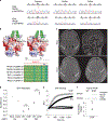
Fig. 1 |. Functional validation of the CP-associated RHOB variant S73F.
a, Sanger traces of the mother, father and proband from families F064 and F244 verify de novo inheritance and the position of the variant (red arrow). b, Top: Poisson–Boltzmann electrostatic maps of wild-type RHOB (left) and the F73 variant (right) showing changes to the kinase-binding site (arrow) and the surface charge of the protein. bottom: alignment of human Rho family proteins shows high conservation of the RHOB 73 residue in the Switch II domain. the site of S73/F73 has been labeled (X). c, Top: brain MRI from F064 demonstrates bilateral periventricular T2/FLAIR hyperintensity (arrows) on axial imaging (left), while the sagittal view (right) reveals equivocal thinning of the isthmus of the corpus callosum (asterisk). bottom: MRI from F244 demonstrates T2 hyperintensity of the posterior limb of the internal capsule and optic radiations (arrows; left image) and hyperintensity of the periventricular white matter (arrows; right image). d, GTP hydrolysis is enhanced ∼1.5-fold in the S73F RHOB variant in a GAP assay. the plot shows absorbance measurements of hydrolyzed GTP in the presence of either a low (5 µg; P = 0.003) or high (13 µg; P = 5.6 × 10⁻⁵) level of RHOA GAP. there was no change in the endogenous GTPase activity with the S73F variant without GAP added (not shown; n = 3). e, GTP binding is enhanced in the S73F RHOB variant in a GEF assay. the N-methylantraniloyl–GTP fluorophore increases its fluorescence emission when bound to Rho family GTPases, indicating nucleotide uptake by the GTPase. both the wild type and S73F have low endogenous GTP binding (bottom curves). In the presence of the GEF protein Dbs, GTP binding is enhanced, and the Michaelis constant (Km) of S73F is significantly reduced compared to that of wild-type RHOB (n = 5; mean 243 versus 547 s, P = 0.0017; top curves). f, S73F GTP binding is increased fourfold in a pulldown assay with rhotekin, an interactor with active GTP-bound Rho proteins. Top: a sample western blot cropped to show RHOB from the bead-bound fraction and the total input detected using an antibody against the V5 tag. bottom: quantification of the ratio of rhotekin-bound/total RHOB (n = 5), P = 0.001. RFU, relative fluorescence units (106) at 360 nm excitation. the statistics were determined by a two-tailed unpaired t-test. **P < 0.003. Full-length blots are provided as source data.
Fig. 2 |
Fig. 2 |. Functional validation of the CP-associated FBXO31 variant p.Asp334Asn shows alterations in cyclin D regulation.
a, Sanger traces of the mother, father and proband from families F218 and F699 verify de novo inheritance and the position of the variant (red arrow). b, Poisson–Boltzmann electrostatic maps of wild-type FbXO31 (left) and the p.Asp334Asn variant (right). D334 is positioned around the cyclin D1 (green)-binding pocket on FbXO31. the mutation alters the surface electrostatic charge around the cyclin D1-binding site with a predicted effect on cyclin D1 binding to FbXO31. the site of D334/N334 has been labeled (arrow). the bottom panels are magnified views showing the alterations to the surface charge in the cyclin D1-binding site. c, A representative western blot cropped to show the decreased cyclin D expression in patient-derived fibroblasts with the FBXO31 p.Asp334Asn variant. Quantification of cyclin D is normalized to in-lane β-tubulin and the within-experiment control GMO8398. both patients had reduced cyclin D compared to pooled controls. the data are averaged for three independent cell culture experiments (n = 7 controls, n = 6 patient measurements). the box indicates the 75th and 25th percentiles with a center line indicating the median; the whiskers indicate the 10th and 90th percentiles. **P = 0.004 calculated using a two-tailed unpaired t-test. Full-length blots are provided as source data.
Fig. 3 |
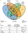
Fig. 3 |. Genetic overlap among common NDDs.
a, A Venn diagram showing the number of overlapping genes between candidate CP genes and genes linked to other NDDs, ID, epilepsy and ASD. CP risk genes were identified as having one or more damaging variants across modes of inheritance with overlap determined using DisGeNET. b, Overlap between CP and other NDDs was significant by hypergeometric two-tailed test, while overlap between CP and Alzheimer’s disease was not. total number of genes in DisGeNET = 17,549; total number of genes in our gene set = 439.
Fig. 4 |
Fig. 4 |. Locomotor phenotypes of LoF mutations in Drosophila orthologs of candidate CP risk genes.
a, Turning time, a measure of coordinated movements, is increased in larvae with mutations in AGAP1, SEMA4A and TENM1 orthologs. Drosophila mutant and control genotypes are provided in Supplementary Table 9. bi, 14-day-old adult flies have locomotor impairments. be, Negative geotaxis climbing defects in distance threshold assays for flies with mutations in orthologs of DOCK11 (b), RABEP1 (c), PTK2B (d) and ATL1 (e). Some genotypes have a male-specific locomotor defect (c). f,g, Increased number of falls for flies with mutations in SYNGAP1 (f) and TBC1D17 (g) orthologs, although the percentage reaching the threshold distance was normal (extended Data Fig. 10). h,i, Impairments in the average distance traveled of flies with mutations in MKL1 (h) and ZDHHC15 (i) orthologs. related GO terms for genes are shown in bold. For the box and whisker plots, the box indicates the 75th and 25th percentiles with a median line, and the whiskers indicate the 10th and 90th percentiles. the locomotor curve represents the average of all trials and the error bars indicate standard error. n = 50 larvae, n = 10–21 trials for falls and distance traveled assays, and n = 10–21 trials for locomotor curves. the differences in larval turning times, distances traveled and numbers of falls were determined by unpaired two-tailed t-tests. the locomotor curves were considered to be significantly different from each other if P < 0.05 for a Kolmogorov–Smirnov test in addition to a significant difference at one or more time bins by a Mann–Whitney rank sum two-tailed test. *P < 0.05, **P < 0.005, ***P < 0.001, ****P < 1 × 10⁻⁶. exact genotypes, n and P values are provided in Supplementary Table 9. j, Enrichment of locomotor phenotypes detected in studies of putative CP genes (observed) compared to genome-wide rates annotated in https://flybase.org (expected, 3.1%). The P value was calculated by Fisher’s exact two-tailed test.

Similar articles

Cited by

References

    1. Christensen D et al. Prevalence of cerebral palsy, co-occurring autism spectrum disorders, and motor functioning - Autism and Developmental Disabilities Monitoring Network, USA, 2008. Dev. Med. Child Neurol. 56, 59–65 (2014). - PMC - PubMed
    1. Oskoui M, Coutinho F, Dykeman J, Jette N & Pringsheim T An update on the prevalence of cerebral palsy: a systematic review and meta-analysis. Dev. Med. Child Neurol. 55, 509–519 (2013). - PubMed
    1. Cans C Surveillance of cerebral palsy in Europe: a collaboration of cerebral palsy surveys and registers. Dev. Med. Child Neurol. 42, 816–824 (2000). - PubMed
    1. Longo LD & Ashwal S William Osler, Sigmund Freud and the evolution of ideas concerning cerebral palsy. J. Hist. Neurosci. 2, 255–282 (1993). - PubMed
    1. Panteliadis C, Panteliadis P & Vassilyadi F Hallmarks in the history of cerebral palsy: from antiquity to mid-20th century. Brain Dev. 35, 285–292 (2013). - PubMed

Publication types

MeSH terms