Melatonin prevents the binding of vascular endothelial growth factor to its receptor and promotes the expression of extracellular matrix-associated genes in nucleus pulposus cells
- PMID: 32989385
- PMCID: PMC7517348
- DOI: 10.3892/etm.2020.9227
Melatonin prevents the binding of vascular endothelial growth factor to its receptor and promotes the expression of extracellular matrix-associated genes in nucleus pulposus cells
Abstract
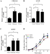
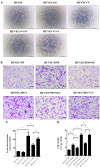
The mechanisms of intervertebral disc degeneration (IDD) involve numerous factors, including loss of the extracellular matrix (ECM) and vascular ingrowth. Melatonin has been reported to protect intervertebral discs (IVDs) from degeneration and to exert a potential anti-angiogenic effect. The aim of the present study was to investigate the anti-angiogenic and anabolic effects of melatonin in IVDs. Human nucleus pulposus (NP) and degenerative nucleus pulposus (DNP) cells were isolated and treated with melatonin. The results indicated that melatonin promoted ECM synthesis and NP cell proliferation. In addition, an NP/DNP and human umbilical vein endothelial cell (HUVEC) co-culture model was used to investigate the anti-angiogenesis effect of melatonin. Melatonin was indicated to suppress tube formation and migration of HUVECs in culture with NP cell-conditioned medium, as well as in an NP cell co-culture model. Fluorescence-labeled vascular endothelial growth factor (VEGF) was used to study the binding between VEGF and its receptor. The results of the present study indicated that melatonin exerts an angiogenic effect via inhibition of the binding of VEGF to its receptor in HUVECs. Taken together, these results suggest that melatonin is a potential agent to prevent IDD.
Keywords: extracellular matrix; intervertebral disc degeneration; melatonin; nucleus pulposus.
Copyright: © Shen et al.
Figures


Similar articles
-
Influence of rabbit notochordal cells on symptomatic intervertebral disc degeneration: anti-angiogenic capacity on human endothelial cell proliferation under hypoxia.Osteoarthritis Cartilage. 2017 Oct;25(10):1738-1746. doi: 10.1016/j.joca.2017.06.003. Epub 2017 Jun 21. Osteoarthritis Cartilage. 2017. PMID: 28647468
-
Melatonin inhibits nucleus pulposus (NP) cell proliferation and extracellular matrix (ECM) remodeling via the melatonin membrane receptors mediated PI3K-Akt pathway.J Pineal Res. 2017 Oct;63(3). doi: 10.1111/jpi.12435. Epub 2017 Aug 16. J Pineal Res. 2017. PMID: 28719035
-
Co-culturing nucleus pulposus mesenchymal stem cells with notochordal cell-rich nucleus pulposus explants attenuates tumor necrosis factor-α-induced senescence.Stem Cell Res Ther. 2018 Jun 26;9(1):171. doi: 10.1186/s13287-018-0919-9. Stem Cell Res Ther. 2018. PMID: 29941029 Free PMC article.
-
Growth and differentiation factor-5 contributes to the structural and functional maintenance of the intervertebral disc.Cell Physiol Biochem. 2015;35(1):1-16. doi: 10.1159/000369670. Epub 2015 Jan 2. Cell Physiol Biochem. 2015. PMID: 25547527 Review.
-
Differentiation of Pluripotent Stem Cells into Nucleus Pulposus Progenitor Cells for Intervertebral Disc Regeneration.Curr Stem Cell Res Ther. 2019;14(1):57-64. doi: 10.2174/1574888X13666180918095121. Curr Stem Cell Res Ther. 2019. PMID: 30227822 Review.
Cited by
-
Progress of melatonin in the treatment of intervertebral disc degeneration.Front Physiol. 2025 May 14;16:1529315. doi: 10.3389/fphys.2025.1529315. eCollection 2025. Front Physiol. 2025. PMID: 40438255 Free PMC article. Review.
-
Therapeutic potential of melatonin in the intervertebral disc degeneration through inhibiting the ferroptosis of nucleus pulpous cells.J Cell Mol Med. 2023 Aug;27(16):2340-2353. doi: 10.1111/jcmm.17818. Epub 2023 Jun 16. J Cell Mol Med. 2023. PMID: 37329158 Free PMC article.
-
The Role of Microvascular Variations in the Process of Intervertebral Disk Degeneration and Its Regulatory Mechanisms: A Literature Review.Orthop Surg. 2024 Nov;16(11):2587-2597. doi: 10.1111/os.14209. Epub 2024 Aug 28. Orthop Surg. 2024. PMID: 39205477 Free PMC article. Review.
References
LinkOut - more resources
Full Text Sources
Miscellaneous
