

miR-4295 promotes cell proliferation, migration and invasion of osteosarcoma through targeting interferon regulatory factor 1
- PMID: 32989394
- PMCID: PMC7517570
- DOI: 10.3892/ol.2020.12123
miR-4295 promotes cell proliferation, migration and invasion of osteosarcoma through targeting interferon regulatory factor 1
Abstract
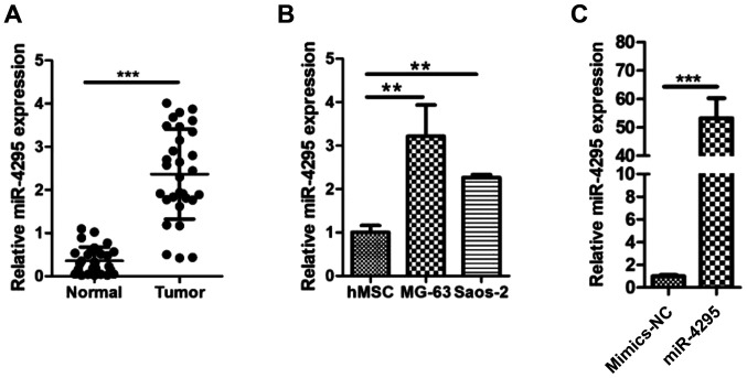
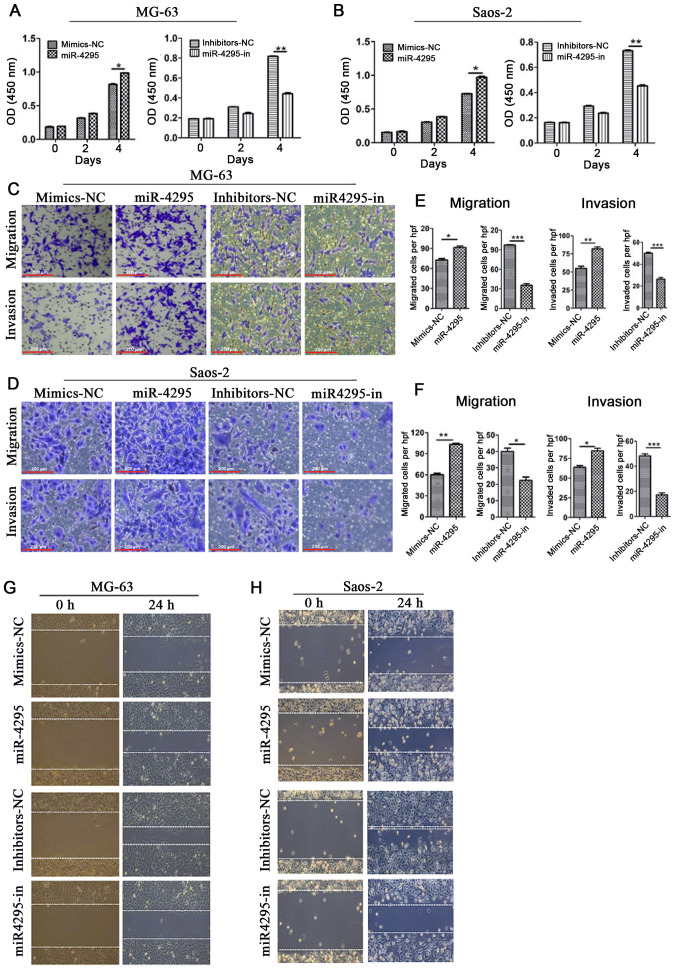
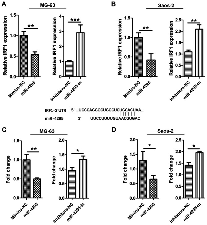
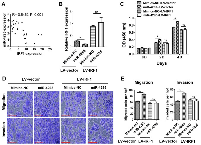
Osteosarcoma (OS) is the most common form of primary malignant bone tumor. Despite encouraging progress in the treatment of OS, the survival rate for patients with OS has remained unchanged over the past 40 years. It has been established that miRNA plays a crucial regulatory role in the progression and development of OS. To explore the potential association of miRNAs with OS, bioinformatics techniques were used to screen for differentially expressed miRNA genes in OS in the Gene Expression Omnibus database. In the GSE70367 database, it was revealed that miR-4295 expression was abnormally elevated in the expression of OS cells. To characterize the potential function of miR-4295 in OS, the expression levels of miR-4295 in 30 samples of OS and adjacent normal tissues was examined. The results revealed that the expression of miR-4295 was significantly increased in OS tissues compared with the paired normal tissues. Moreover, the expression levels of miR-4295 in OS cell lines (MG-63 and Saos-2) were significantly higher compared with those in the normal human mesenchymal stem cells. In addition, miR-4295 was associated with OS cell proliferation, migration and invasion. Furthermore, it was demonstrated that the expression of interferon regulatory factor (IRF)1, a tumor suppressor, was regulated by miR-4295 directly in OS cells. Taken together, the present results revealed that miR-4295 may act as a tumor activator by targeting IRF1 during the progression of OS. Investigating miR-4295 may provide novel insight into the mechanisms of OS metastasis, and inhibition and targeting miR-4295 may be a novel therapeutic strategy for the treatment of OS.
Keywords: interferon regulatory factor 1; invasion; miR-4295; migration; osteosarcoma; proliferation.
Copyright © 2020, Spandidos Publications.
Figures
Similar articles
-
MiR-134, Mediated by IRF1, Suppresses Tumorigenesis and Progression by Targeting VEGFA and MYCN in Osteosarcoma.Anticancer Agents Med Chem. 2020;20(10):1197-1208. doi: 10.2174/1871520620666200402074752. Anticancer Agents Med Chem. 2020. PMID: 32238141
-
MicroRNA-567 inhibits cell proliferation, migration and invasion by targeting FGF5 in osteosarcoma.EXCLI J. 2018 Jan 15;17:102-112. doi: 10.17179/excli2017-932. eCollection 2018. EXCLI J. 2018. PMID: 29743851 Free PMC article.
-
Circ_0001174 facilitates osteosarcoma cell proliferation, migration, and invasion by targeting the miR-186-5p/MACC1 axis.J Orthop Surg Res. 2022 Mar 12;17(1):159. doi: 10.1186/s13018-022-03059-8. J Orthop Surg Res. 2022. PMID: 35279159 Free PMC article.
-
MiR-205 functions as a tumor suppressor via targeting TGF-α in osteosarcoma.Exp Mol Pathol. 2016 Feb;100(1):160-6. doi: 10.1016/j.yexmp.2015.12.010. Epub 2015 Dec 17. Exp Mol Pathol. 2016. PMID: 26708425
-
miR-18a-5p promotes cell invasion and migration of osteosarcoma by directly targeting IRF2.Oncol Lett. 2018 Sep;16(3):3150-3156. doi: 10.3892/ol.2018.9032. Epub 2018 Jun 27. Oncol Lett. 2018. PMID: 30127908 Free PMC article.
Cited by
-
Long Non-Coding RNA TPTEP1 Exerts Tumor Suppressive Functions via Sequestering miR-4295 to Regulate Growth Arrest and DNA Damage-Inducible 45α Expression in Acute Myeloid Leukemia.Cancer Manag Res. 2025 Jun 4;17:1059-1072. doi: 10.2147/CMAR.S486875. eCollection 2025. Cancer Manag Res. 2025. PMID: 40491712 Free PMC article.
-
The multiple roles of interferon regulatory factor family in health and disease.Signal Transduct Target Ther. 2024 Oct 9;9(1):282. doi: 10.1038/s41392-024-01980-4. Signal Transduct Target Ther. 2024. PMID: 39384770 Free PMC article. Review.
-
The Double Life of microRNAs in Bone Sarcomas: Oncogenic Drivers and Tumor Suppressors.Int J Mol Sci. 2025 May 17;26(10):4814. doi: 10.3390/ijms26104814. Int J Mol Sci. 2025. PMID: 40429954 Free PMC article. Review.
-
MicroRNA signatures in osteosarcoma: diagnostic insights and therapeutic prospects.Mol Cell Biochem. 2025 Apr;480(4):2065-2075. doi: 10.1007/s11010-024-05135-5. Epub 2024 Oct 17. Mol Cell Biochem. 2025. PMID: 39419925 Free PMC article. Review.
-
Identification of LTF as a Prognostic Biomarker for Osteosarcoma.J Oncol. 2022 Jan 21;2022:4656661. doi: 10.1155/2022/4656661. eCollection 2022. J Oncol. 2022. PMID: 35096061 Free PMC article.
References
-
- Bacci G, Briccoli A, Rocca M, Ferrari S, Donati D, Longhi A, Bertoni F, Bacchini P, Giacomini S, Forni C, et al. Neoadjuvant chemotherapy for osteosarcoma of the extremities with metastases at presentation: Recent experience at the Rizzoli Institute in 57 patients treated with cisplatin, doxorubicin, and a high dose of methotrexate and ifosfamide. Ann Oncol. 2003;14:1126–1134. doi: 10.1093/annonc/mdg286. - DOI - PubMed
-
- Kimura K, Nakano T, Park YB, Tani M, Tsuda H, Beppu Y, Moriya H, Yokota J. Establishment of human osteosarcoma cell lines with high metastatic potential to lungs and their utilities for therapeutic studies on metastatic osteosarcoma. Clin Exp Metastasis. 2002;19:477–485. doi: 10.1023/A:1020395816633. - DOI - PubMed
-
- Morishita T, Mii Y, Miyauchi Y, Miura S, Honoki K, Aoki M, Kido A, Tamai S, Tsutsumi M, Konishi Y. Efficacy of CDDP and AGM-1470 chemotherapy against lung metastasis in rat osteosarcoma depends on the timing of combined administration. Jpn J Clin Oncol. 1997;27:236–239. doi: 10.1093/jjco/27.4.236. - DOI - PubMed
LinkOut - more resources
Full Text Sources
Miscellaneous