. 2020 Nov;20(5):270.
doi: 10.3892/ol.2020.12133. Epub 2020 Sep 21.

Agaricus blazei extract (FA-2-b-β) induces apoptosis in chronic myeloid leukemia cells

Affiliations

Agaricus blazei extract (FA-2-b-β) induces apoptosis in chronic myeloid leukemia cells

Yanqing Sun et al. Oncol Lett. 2020 Nov.

Abstract

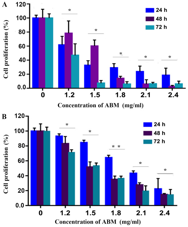
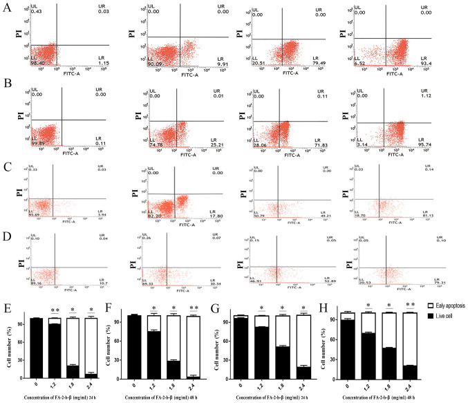
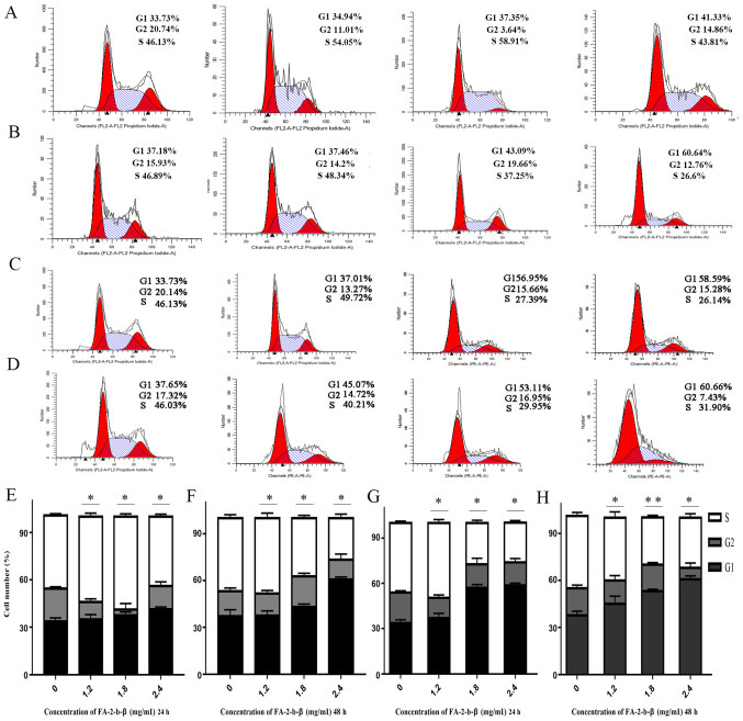
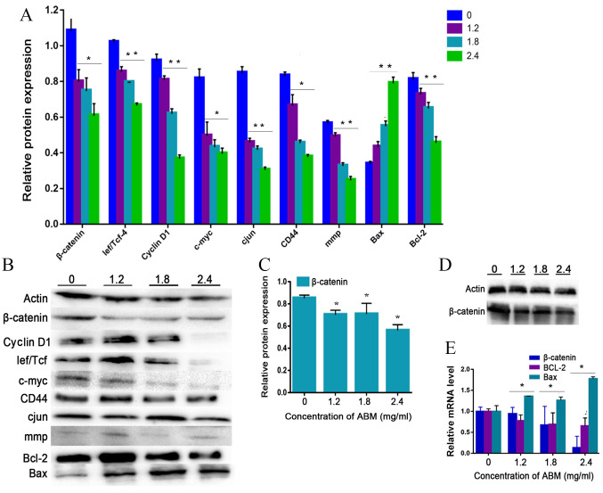
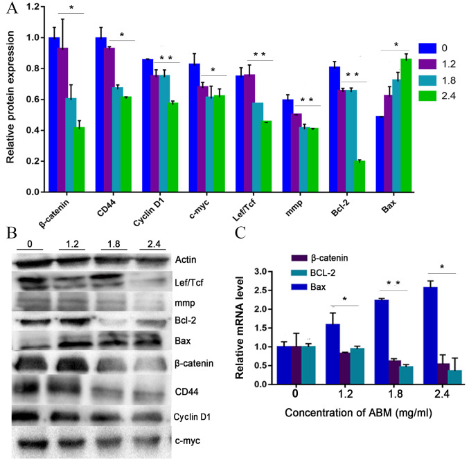
Agaricus blazei Murill (AbM) is a mushroom belonging to the Basidiomycetes family, which is believed to have antitumor and antioxidative activities. Proteoglycans and ergosterol are considered the key compounds of AbM for antitumor properties and so are used in complementary and alternative medicine as an anticancer drug. AbM is used to avoid serious side effects that would inevitably affect patients. Currently, the efficacy of AbM against chronic myeloid leukemia (CML) has not been established. The present study aimed to investigate the antitumor activities of the acidic RNA protein complex, FA-2-b-β, extracted from wild edible AbM. The CML K562 cells or primary CML bone marrow (BM) cells were treated with FA-2-b-β at different concentrations and time points. CML cell line proliferation and apoptosis were determined using the CCK-8 assay or Annexin V/propidium iodide (PI) labeling, RT-qPCR and western blotting was performed to determine the involvement of the Wnt/β-catenin-associated apoptotic pathway. The results of the present study demonstrated that FA-2-b-β has a high anti-proliferative potency and strong pro-apoptotic effects. Thus, daily intake of mushrooms containing FA-2-b-β may be an adequate source as an alternative medicine in the management of CML, and may provide useful information for the development of a novel therapeutic target in this area.

Keywords: AbM; CML; Wnt/β-catenin signaling pathway; apoptosis cycle.


Figures

Figure 1.
Figure 1.
AbM inhibits the cell proliferation of K562 or primary CML BM cells. Cell proliferation was inhibited in (A) K562 or (B) primary CML BM cells at the indicated time points according to the CCK-8 assay. The plots represent the mean ± SD of five replicates (after 24, 48 and 72 h).
Figure 2.
Figure 2.
Flow cytometry analysis reveals AbM increased the apoptotic K562 cells and primary CML BM cells percentages in a concentration-dependent manner. K562 cells and primary CML BM cells were treated with different concentrations of AbM (0, 1.2, 1,8 and 2.4 mg/ml). K562 cells treated with different concentrations of AbM for (A) 24 and (B) 48 h. Primary CML BM cells treated with different concentrations of AbM for (C) 24 and (D) 48 h. Quantification of K562 cells treated with different concentrations of AbM for (E) 24 and (F) 48 h. Quantification of primary CML BM cells treated with different concentrations of AbM for (G) 24 and (H) 48 h. Data shown are representative of three independent experiments with K562 cells and primary CML BM cells. Data are presented as the mean ± SD. *P<0.05; **P<0.01 vs. 0 mg/ml.
Figure 3.
Figure 3.
AbM promotes K562 cells and primary CML BM cells cycle. AbM induced cell cycle G1 phase arrest. K562 cells and primary CML BM cells were treated with different concentrations of AbM (0, 1.2, 1,8 and 2.4 mg/ml). Representative flow-cytograms are shown. Bar graphs indicate the mean percentages. (A) K562 cells and (B) primary CML BM cells treated with different concentrations of AbM for 24 h. (C) K562 cells and (D) primary CML BM cells treated with different concentrations of AbM for 48 h. Quantification of K562 cells treated with different concentrations of AbM for (E) 24 and (F) 48 h. Quantification of primary CML BM cells treated with different concentrations of AbM for (G) 24 and (H) 48 h. Data shown are representative of three independent experiments. K562 and primary CML BM cells data analysis revealed the proportion of cells in each phase of the cell cycle. Data are presented as the mean ± SD. *P<0.05, **P<0.01 vs. 0 mg/ml.
Figure 4.
Figure 4.
AbM induces apoptosis of K562 cells. K562 cells were treated with different concentrations of AbM (0, 1.2, 1.8 and 2.4 mg/ml) for 48 h and the expression of apoptosis-associated genes and proteins was determined using western blot analysis for (A and B) 24 h (*P<0.05 and **P<0.001) or (C and D) 48 h and (E) reverse transcription-quantitative PCR. The relative expression levels of β catenin, Bax and Bcl-2 genes in K562 cells were quantified according to the gene expression levels of actin. The relative expression levels of cyclin D1, Lef/Tcf, c-myc, β catenin, CD44, c-Jun, mmp, Bax and Bcl-2 proteins in K562 cells were quantified according to the protein expression levels of actin. Data are representative of three independent experiments and are expressed as the mean ± SD. *P<0.05 vs. 0 mg/ml.
Figure 5.
Figure 5.
AbM induces apoptosis of CML cells. Primary CML BM cells were treated with different concentrations of AbM (0, 1.2, 1.8 and 2.4 mg/ml) for 24 h and the expression of apoptosis-related genes and proteins was determined using (A and B) western-blot analysis (*P<0.05 and **P<0.001) and (C) reverse transcription-quantitative PCR. The relative expression levels of β catenin, Bax and Bcl-2 genes in primary CML BM cells were quantified according to the expression levels of actin. The relative expression levels of cyclin D1, Lef/Tcf, c-myc, β catenin, CD44, c-Jun, mmp, Bax and Bcl-2 proteins in primary CML BM cells were quantified according to the expression levels of Actin. Data are representative of three independent experiments and are expressed as the mean ± SD. *P<0.05, **P<0.001.

Similar articles

Cited by

References

    1. Bamodu OA, Kuo KT, Yuanc LP, et al. HDAC inhibitor suppresses proliferation and tumorigenicity of drug-resistant chronic myeloid leukemia stem cells through regulation of hsa-miR-196a targeting BCR/ABL1. Exp Cell Res. 2018;370:519–530. doi: 10.1016/j.yexcr.2018.07.017. - DOI - PubMed
    1. Zhou H, Mak PY, Mu H, Mak DH, Zeng Z, Cortes J, Liu Q, Andreeff M, Carter BZ. Combined inhibition of β-catenin and Bcr-Abl synergistically targets tyrosine kinase inhibitor-resistant blast crisis chronic myeloid leukemia blasts and progenitors in vitro and in vivo. Leukemia. 2017;31:2065–2074. doi: 10.1038/leu.2017.87. - DOI - PMC - PubMed
    1. Xiao X, Liu P, Li D, Xia Z, Wang P, Zhang X, Liu M, Liao L, Jiao B, Ren R. Combination therapy of BCR-ABL-positive B cell acute lymphoblastic leukemia by tyrosine kinase inhibitor dasatinib and c-JUN N-terminal kinase inhibition. J Hematol Oncol. 2020;13:80. doi: 10.1186/s13045-020-00912-3. - DOI - PMC - PubMed
    1. Deshpande A, Buske C. Knocking the Wnt out of the sails of leukemia stem cell development. Cell Stem Cell. 2007;1:597–598. doi: 10.1016/j.stem.2007.11.006. - DOI - PubMed
    1. Zovko A, Yektaei-Karin E, Daniel S, Nilsson A, Wallvik J, Stenke L. Montelukast, a cysteinyl leukotriene receptor antagonist, inhibits the growth of chronic myeloid leukemia cells through apoptosis. Oncol Rep. 2018;40:902–908. - PubMed