. 2021 Mar;478(3):471-485.
doi: 10.1007/s00428-020-02934-1. Epub 2020 Sep 28.

Fibrotic progression and radiologic correlation in matched lung samples from COVID-19 post-mortems

Affiliations

Fibrotic progression and radiologic correlation in matched lung samples from COVID-19 post-mortems

Emanuela Barisione et al. Virchows Arch. 2021 Mar.

Abstract

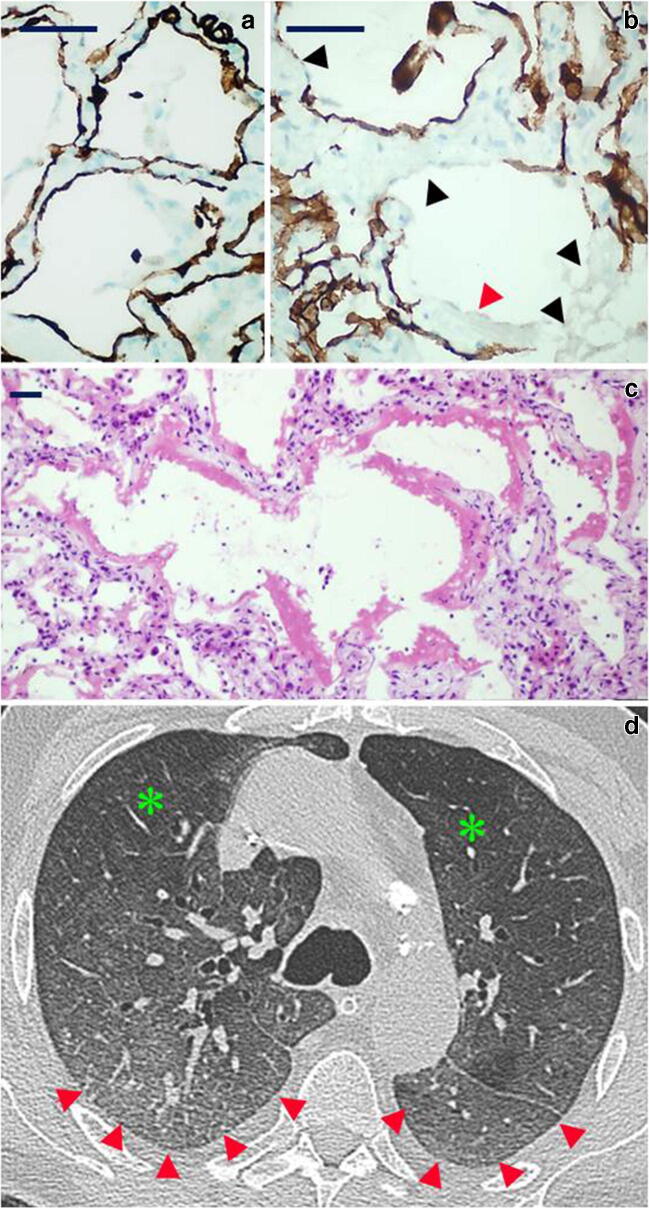
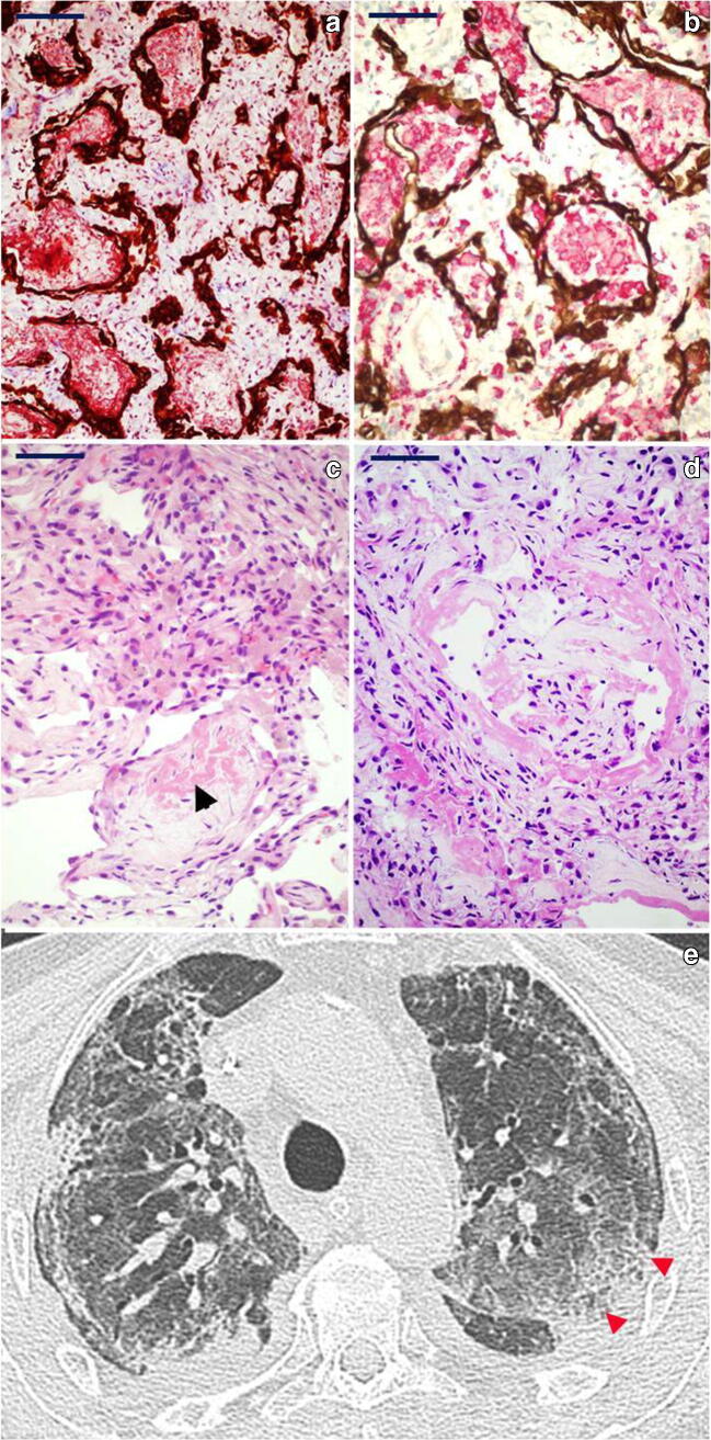
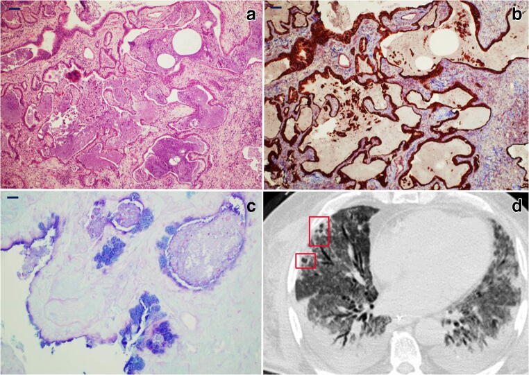
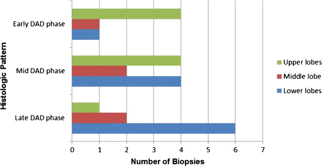
Data on the pathology of COVID-19 are scarce; available studies show diffuse alveolar damage; however, there is scarce information on the chronologic evolution of COVID-19 lung lesions. The primary aim of the study is to describe the chronology of lung pathologic changes in COVID-19 by using a post-mortem transbronchial lung cryobiopsy approach. Our secondary aim is to correlate the histologic findings with computed tomography patterns. SARS-CoV-2-positive patients, who died while intubated and mechanically ventilated, were enrolled. The procedure was performed 30 min after death, and all lung lobes sampled. Histopathologic analysis was performed on thirty-nine adequate samples from eight patients: two patients (illness duration < 14 days) showed early/exudative phase diffuse alveolar damage, while the remaining 6 patients (median illness duration-32 days) showed progressive histologic patterns (3 with mid/proliferative phase; 3 with late/fibrotic phase diffuse alveolar damage, one of which with honeycombing). Immunohistochemistry for SARS-CoV-2 nucleocapsid protein was positive predominantly in early-phase lesions. Histologic patterns and tomography categories were correlated: early/exudative phase was associated with ground-glass opacity, mid/proliferative lesions with crazy paving, while late/fibrous phase correlated with the consolidation pattern, more frequently seen in the lower/middle lobes. This study uses an innovative cryobiopsy approach for the post-mortem sampling of lung tissues from COVID-19 patients demonstrating the progression of fibrosis in time and correlation with computed tomography features. These findings may prove to be useful in the correct staging of disease, and this could have implications for treatment and patient follow-up.

Keywords: COVID-19; Diffuse alveolar damage; Lung fibrosis; Radiologic patterns; Transbronchial lung cryobiopsy.


Conflict of interest statement

All the authors declare no conflicts of interest.

Figures

Fig. 1
Fig. 1
Elementary lesions commonly seen in the organizing phase of DAD: a Hyperplastic type II pneumocytes line an alveolus inside which an aggregate of macrophages can be seen (H&E, × 60; scale bar 50 μm); b atypical pneumocytes with occasional multi-nucleation line a residual alveolar lumen surrounded by early fibrosis (H&E, × 60; scale bar 50 μm). c Intracytoplasmic eosinophilic Mallory-like inclusions in type 2 pneumocytes covering alveolar residues surrounded by fibroblastic fibrosis (H&E, × 60; scale bar 50 μm). d Fibroblastic foci (arrowheads) are characteristic of the late/organizing phase of DAD and associated with complete derangement/obliteration of alveolar structure (H&E, × 40; scale bar 50 μm). e CD61 immuno-stained section showing a platelet microthrombus in a small-sized vessel (CD61, × 20; scale bar 50 μm). f Interstitial proliferation of smooth muscle fibres (red) in a micro-honeycombing area; CK7 highlights the bronchiolization of the alveolar epithelium (CK7 immunoperoxidase with Mallory trichrome counterstain, × 20; scale bar 50 μm)
Fig. 2
Fig. 2
Histology and radiology of early diffuse alveolar damage (DAD) pattern: a CK7 highlights the continuity of the alveolar epithelial lining in an unaffected area (CK7 immunoperoxidase, × 60; scale bar 30 μm). b Loss of the alveolar epithelium (black arrowheads) is accompanied by the appearance of hyaline membranes (red arrowhead) (CK7 immunoperoxidase, × 60; scale bar 30 μm). c Intra-alveolar hyaline membranes in the absence of interstitial fibrosis and alveolar distortion (H&E, × 20; scale bar 50 μm). d CT findings of diffuse bilateral ground-glass opacities (red arrowheads) defined as hazy, increased opacity of lung, with preservation of bronchial and vascular margins. Normal lung for comparison is identified by green asterisk
Fig. 3
Fig. 3
Histology and radiology of mid-phase DAD pattern: a CK7 and Mallory trichrome, respectively, highlight marked intra-alveolar fibrinous exudates (red) and hyperplasia of the epithelial lining (brown) (CK7 immunoperoxidase and Mallory trichrome counterstain, × 40; scale bar 50 μm). b Numerous intra-alveolar macrophages are stained by CD163 (red), while CK7 highlights the epithelial lining (brown); also note the interstitial thickening (CK7 immunoperoxidase and CD163 alkaline phosphatase red; × 40; scale bar 50 μm). c Mural incorporation of the intra-alveolar fibrinous exudate (arrowhead) (H&E, × 40; scale bar 50 μm). d Intra-alveolar organization of hyaline membrane residues with organizing pneumonia (OP)–like pattern (H&E, × 40; scale bar 50 μm). e CT showing a “crazy paving” pattern (red arrowheads), characterized by thickened interlobular septa and intralobular lines superimposed on a background of GGO resembling irregularly shaped paving stones. Peripheral fibrotic bands and traction bronchiolectasia are also present
Fig. 4
Fig. 4
Histology and radiology of late/organizing phase of DAD pattern: aspects of progressive derangement/obliteration of alveolar structure by interstitial fibroblast proliferation. a–b Alveoli are still recognizable although distorted (a, H&E, × 20; b, CK7 immunoperoxidase with Mallory trichrome counterstain, × 20; scale bar 50 μm). c–d Alveoli become slit-like and compressed by fibrosis and are only readily seen by CK7 immunostaining (c, H&E, × 20; d, CK7 immunoperoxidase with Mallory trichrome counterstain, × 20; scale bar 50 μm). e CT shows advanced parenchymal alterations with predominant dorsal consolidations (homogeneous increase in pulmonary parenchymal attenuation that obscures the margins of vessels and airway walls as opposed to GGO—red asterisks); peripheral crazy paving pattern is seen anteriorly
Fig. 5
Fig. 5
Histology and radiology of micro-honeycombing pattern: a Complete re-modelling of parenchymal structure by fibrosis and mucinous filling of the alveolar spaces (H&E, × 10; scale bar 100 μm). b Mallory trichrome shows collagen deposition (light blue), and CK7 highlights diffuse bronchiolization of epithelial lining (brown) (CK7 immunoperoxidase with Mallory trichrome counterstain, × 10; scale bar 100 μm). c Alcian blue/PAS stain highlights diffuse mucinous metaplasia of the epithelial lining (alcian blue/PAS, × 40; scale bar 50 μm). d CT shows parenchymal diffuse GGO alterations with initial honeycombing pattern (subpleural clustered cystic air spaces, characterized by well-defined walls) in the middle lobe (red boxes); bilateral pleural effusion and mild consolidations in the posterior lower lobes, with bronchial enlargement, are also seen
Fig. 6
Fig. 6
Histogram showing relation between histologic phase and lung lobe involvement. The exudative phase is more represented in the upper lobes while the late phase prevails in the lower lobes
Fig. 7
Fig. 7
SARS-CoV-2 nucleocapsid protein detection by immunohistochemistry. a Immunopositivity in the cytoplasm of bronchial cells and as a rim on their luminal border (× 60; scale bar 50 μm). b Immunopositivity in the cytoplasm of pneumocytes lining the alveolar space in early exudative phase DAD (× 100; scale bar 25 μm). c Immunoreactivity of hyaline membranes in early exudative phase DAD (× 20; scale bar 100 μm). d) Immunopositive alveolar macrophages in exudative phase DAD (× 40; scale bar 50 μm)
Fig. 8
Fig. 8
Correlation between histologic and radiologic CT patterns (see also Table 3). Early damage was found either in apparently healthy lung regions at imaging or associated with ground-glass opacities. Crazy paving was the most characteristic feature of the mid DAD phase, while consolidation and honeycombing were associated with the late-phase histologic changes

References

    1. Zhu N, Zhang D, Wang W, Li X, Yang B, Song J, Zhao X, Huang B, Shi W, Lu R, Niu P, Zhan F, Ma X, Wang D, Xu W, Wu G, Gao GF, Tan W, China Novel Coronavirus Investigating and Research Team A novel coronavirus from patients with pneumonia in China, 2019. N Engl J Med. 2020;382:727–733. doi: 10.1056/NEJMoa2001017. - DOI - PMC - PubMed
    1. Huang C, Wang Y, Li X, Ren L, Zhao J, Hu Y, Zhang L, Fan G, Xu J, Gu X, Cheng Z, Yu T, Xia J, Wei Y, Wu W, Xie X, Yin W, Li H, Liu M, Xiao Y, Gao H, Guo L, Xie J, Wang G, Jiang R, Gao Z, Jin Q, Wang J, Cao B. Clinical features of patients infected with 2019 novel coronavirus in Wuhan, China. Lancet. 2020;395:497–506. doi: 10.1016/S0140-6736(20)30183-5. - DOI - PMC - PubMed
    1. Wang D, Hu B, Hu C, Zhu F, Liu X, Zhang J, Wang B, Xiang H, Cheng Z, Xiong Y, Zhao Y, Li Y, Wang X, Peng Z. Clinical characteristics of 138 hospitalized patients with 2019 novel coronavirus-infected pneumonia in Wuhan, China. JAMA. 2020;323:1061–1069. doi: 10.1001/jama.2020.1585. - DOI - PMC - PubMed
    1. Grasselli G, Pesenti A, Cecconi M. Critical care utilization for the COVID19 outbreak in Lombardy Italy: early experience and forecast during emergency response. Crit Care. 2020;24:175–1546. doi: 10.1001/jama.2020.4031. - DOI - PubMed
    1. Bao C, Liu X, Zhang H, Li Y, Liu J. Coronavirus disease 2019 (COVID-19) CT findings: a systematic review and meta-analysis. J Am Coll Radiol. 2020;119:982–989. doi: 10.1016/j.jacr.2020.03.006. - DOI - PMC - PubMed