

Eliminating computed tomography imaging artifacts through 3D printed radiotherapy head supports
- PMID: 32989881
- PMCID: PMC7592983
- DOI: 10.1002/acm2.13027
Eliminating computed tomography imaging artifacts through 3D printed radiotherapy head supports
Abstract
Purpose: The geometry of an immobilization device such as a headrest can cause undesired computed tomography (CT) artifacts that may affect both volume definition and dosimetry in radiotherapy of the brain. The purpose of this work was to reduce CT artifacts caused by a standard hard plastic hollow radiotherapy headrest. This was to be achieved through design and prototyping of a custom-made head support.
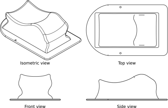
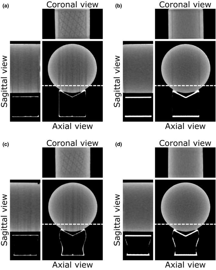
Methods: A series of CT scans were acquired of both a water phantom and an anthropomorphic head phantom which were resting on custom-made three-dimensional (3D) printed supports. All custom-made supports were made of polylactic acid (PLA) plastic filament and printed by fused deposition modeling (FDM) 3D printing technology. Initial designs were studied with a water phantom using a simplified support with straight and curved shapes both at the edges and as infill patterns. Imaging of a 3D printed clinical prototype was then compared to our standard headrest using an anthropomorphic head phantom.
Results: The presence of dark streaks inside both phantoms was seen on the CT images for headrests involving supports with straight shapes at the edges or as infill patterns. Such artifacts were ascribed to the exponential edge-gradient effect (EEGE). No such artifact was observed when the support was designed with a combination of curved edges and infill patterns.
Conclusion: When developing immobilization accessories for use in CT scanners, more attention could be paid to artifact attenuating design elements. This work illustrates the usefulness of 3D printing in prototyping radiotherapy accessories and solving concrete clinical problems.
Keywords: 3D printing; CT artifact; Head CT scan; Immobilization.
© 2020 The Authors. Journal of Applied Clinical Medical Physics published by Wiley Periodicals LLC on behalf of American Association of Physicists in Medicine.
Conflict of interest statement
No conflicts of interest.
Figures


References
-
- Joseph PM, Spital RD. The exponential edge‐gradient effect in X‐ray computed tomography. Phys Med Biol. 1981;26:473–487. - PubMed
-
- Buzug TM. Computed Tomography ‐ From Photon Statistics to Modern Cone‐Beam CT, 2nd edn Berlin, Germany: Springer; 2008.
