. 2020 Dec;9(1):2269-2277.
doi: 10.1080/22221751.2020.1830715.

Establishment of replication-competent vesicular stomatitis virus-based recombinant viruses suitable for SARS-CoV-2 entry and neutralization assays

Affiliations

Establishment of replication-competent vesicular stomatitis virus-based recombinant viruses suitable for SARS-CoV-2 entry and neutralization assays

Hongyue Li et al. Emerg Microbes Infect. 2020 Dec.

Abstract

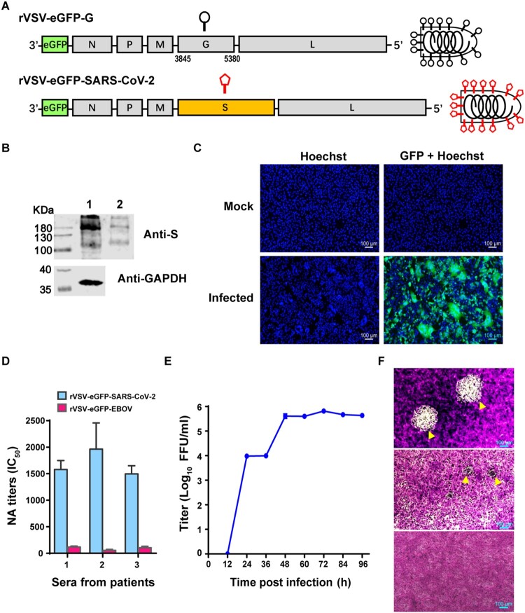
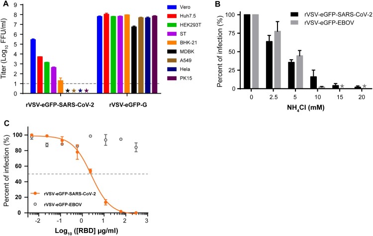
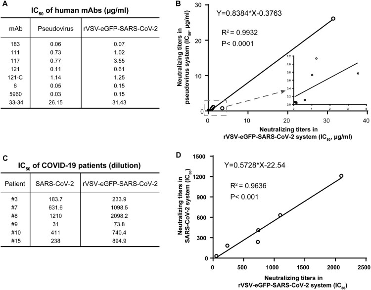
Replication-competent vesicular stomatitis virus (VSV)-based recombinant viruses are useful tools for studying emerging and highly pathogenic enveloped viruses in level 2 biosafety facilities. Here, we used a replication-competent recombinant VSVs (rVSVs) encoding the spike (S) protein of SARS-CoV-2 in place of the original G glycoprotein (rVSV-eGFP-SARS-CoV-2) to develop a high-throughput entry assay for SARS-CoV-2. The S protein was incorporated into the recovered rVSV-eGFP-SARS-CoV-2 particles, which could be neutralized by sera from convalescent COVID-19 patients. The recombinant SARS-CoV-2 also displayed entry characteristics similar to the wild type virus, such as cell tropism and pH-dependence. The neutralizing titers of antibodies and sera measured by rVSV-eGFP-SARS-CoV-2 were highly correlated with those measured by wild-type viruses or pseudoviruses. Therefore, this is a safe and convenient screening tool for SARS-CoV-2, and it may promote the development of COVID-19 vaccines and therapeutics.

Keywords: SARS-CoV-2; VSV; entry; neutralization assay; replication-competent.


Conflict of interest statement

No potential conflict of interest was reported by the author(s).

Figures

Figure 1.
Figure 1.
Characterization of rVSV-eGFP-SARS-CoV-2. (A) Schematic diagrams showing genome organization in rVSV vectors. The original VSV G gene was replaced by SARS-CoV-2 S gene to generate rVSV-eGFP-SARS-CoV-2. eGFP was inserted into the first position of the genome. (B) Lysate of rVSV-eGFP-SARS-CoV-2 producing cells (Lane 1) and purified rVSV-eGFP-SARS-CoV-2 (Lane 2) were analysed by western blot using an antibody recognizing the RBD domain of the S protein. (C) Images of mock- (upper panels) or rVSV-eGFP-SARS-CoV-2-infected (lower panels) Vero cells stained with Hoechst to label the nuclei (blue). The GFP signal is shown in green. The overlay of blue and green is shown to visualize syncytia. (D) The titers of neutralizing antibodies (NAbs) in three serum samples from COVID-19 convalescent patients were determined against rVSV-eGFP-SARS-CoV-2 by a focus reduction neutralization test. rVSV-eGFP-EBOV (Ebola virus) were used as a control virus. (E) Growth kinetics of rVSV-eGFP-SARS-CoV-2 in Vero cells (MOI=0.0l). Viral titers were measured by a focus-forming assay and expressed as focus forming units per ml (FFU/ml). Error bars in (D-E) indicate standard deviation of the mean (n=3). (F) Plaque morphology of rVSVs in Vero cells. The plaques of rVSV-eGFP-EBOV (upper panel) or rVSV-eGFP-SARS-CoV-2 (middle panel) at 72 h post-infection indicated by yellow arrowhead were visualized under microscope. Lower panel: Mock-infected cells (control). The data represent three independent experiments.
Figure 2.
Figure 2.
Characteristics of rVSV-eGFP-SARS-CoV-2 entry. (A) Infectivity of rVSV-eGFP-SARS-CoV-2 and rVSV-eGFP-G on various cell types. Infectivity was quantified by counting GFP positive cells in the above nine cell lines. The infectious titers were expressed as log10 FFU/ml. The limit of detection was 10 FFU/ml. Titers below 10 FFU/ml (★) are indicated. (B) Inhibition of rVSV-eGFP-SARS-CoV-2 infection by NH4Cl. Vero cells were infected with rVSV-eGFP-SARS-CoV-2 or rVSV-eGFP-EBOV at MOI of 0.01 after treatment with different concentrations of NH4Cl. The results are expressed as the percentage of the control (without NH4Cl treatment). (C) Effect of SARS-CoV-2 RBD fragment on the infectivity of rVSV-eGFP-SARS-CoV-2. All infection experiments were performed in triplicate. Error bars in (A–C) indicate standard deviation of the mean (n=3).
Figure 3.
Figure 3.
Comparison of rVSV-eGFP-SARS-CoV-2-based neutralization assay with pseudotype and live viruses. (A) IC50 values for 8 human mAbs against rVSV-eGFP-SARS-CoV-2 and VSV pseudotype. NAb titers of the mAbs against rVSV-eGFP-SARS-CoV-2 was evaluated by a focus reduction neutralization test. NAb titers against pseudovirus was evaluated by pseudovirus based neutralization assay (PBNA). IC50 was calculated by the Reed-Muench method. (B) Correlation of IC50 values in (A) between pseudovirus- and rVSV-based assays. (C) IC50 values for six serum samples from COVID-19 convalescent patients against rVSV-eGFP-SARS-CoV-2 and SARS-CoV-2. NAb titers of the serum samples against SARS-CoV-2 was determined by plaque reduction neutralization test, and IC50 was calculated by the Reed-Muench method. (D) Correlation of IC50 values in COVID-19 patients measured by rVSV-eGFP-SARS-CoV-2 and SARS-CoV-2.
Figure 4.
Figure 4.
Validation of rVSV-eGFP-SARS-CoV-2-based neutralization assay. (A) Representative inhibition curves of two positive and two negative human serum samples. (B) The limit of detection (LOD) of the assay. A subset of negative serum samples from 20 mice and 4 humans was tested against rVSV-eGFP-SARS-CoV-2 using the focus reduction neutralization test. (C) Reproducibility of the neutralization assay. Pooled positive serum samples (n=2) were run six times per experiment in total of three experiments to measure reproducibility. Individual IC50 values were plotted to evaluate the intra- and inter-assay variability. Error bars in (A–C) indicate standard deviation of the mean.

Similar articles

Cited by

References

    1. Zhu N, Zhang D, Wang W, et al. . A novel coronavirus from patients with Pneumonia in China, 2019. N Engl J Med. 2020 Feb 20;382(8):727–733. - PMC - PubMed
    1. Zhou P, Yang XL, Wang XG, et al. . A pneumonia outbreak associated with a new coronavirus of probable bat origin. Nature. 2020 Mar;579(7798):270–273. - PMC - PubMed
    1. Lu R, Zhao X, Li J, et al. . Genomic characterisation and epidemiology of 2019 novel coronavirus: implications for virus origins and receptor binding. Lancet. 2020 Feb 22;395(10224):565–574. - PMC - PubMed
    1. Ou X, Liu Y, Lei X, et al. . Characterization of spike glycoprotein of SARS-CoV-2 on virus entry and its immune cross-reactivity with SARS-CoV. Nat Commun. 2020 Mar 27;11(1):1620. - PMC - PubMed
    1. Hoffmann M, Kleine-Weber H, Schroeder S, et al. . SARS-CoV-2 cell entry depends on ACE2 and TMPRSS2 and Is blocked by a clinically proven protease inhibitor. Cell. 2020 Apr 16;181(2):271–280. e8. - PMC - PubMed

Publication types

Substances