. 2020 Nov;26(6):531-540.
doi: 10.5152/dir.2020.19569.

Hepatocellular carcinoma with macrovascular invasion: multimodality imaging features for the diagnosis

Affiliations

Hepatocellular carcinoma with macrovascular invasion: multimodality imaging features for the diagnosis

Roberto Cannella et al. Diagn Interv Radiol. 2020 Nov.

Abstract

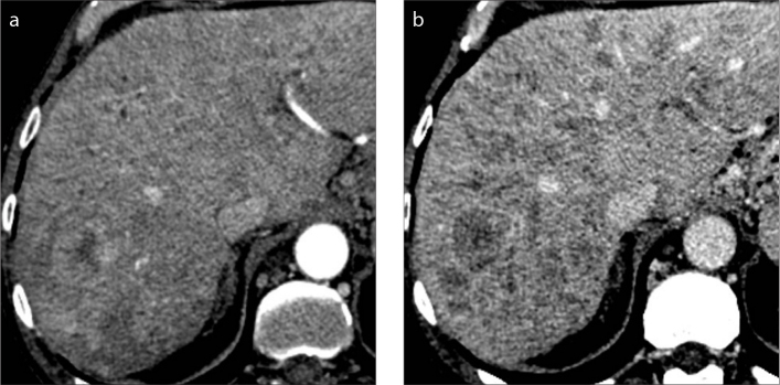
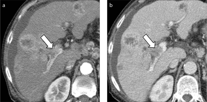
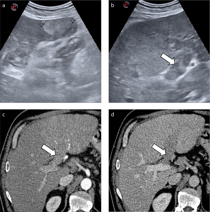
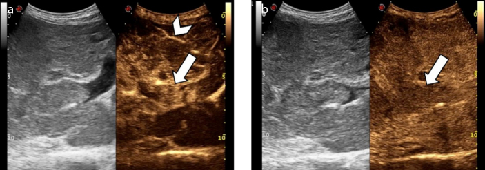
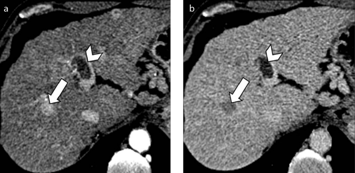
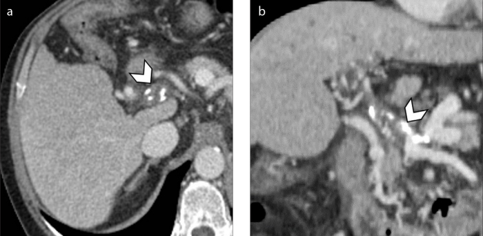
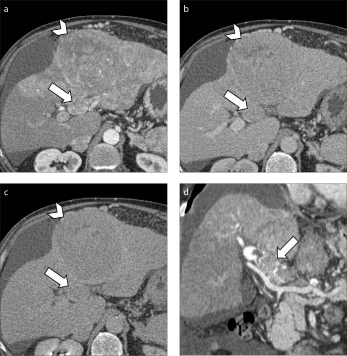
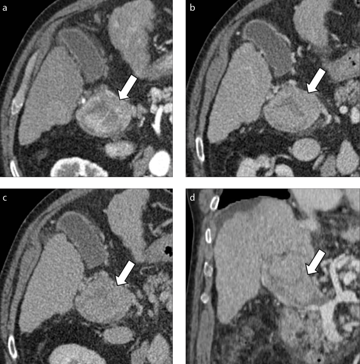
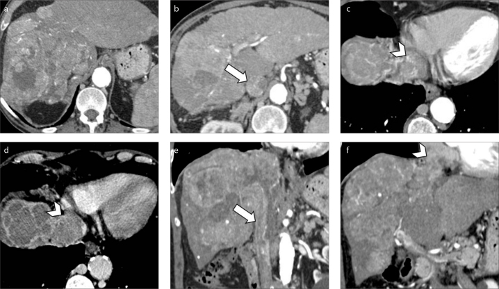
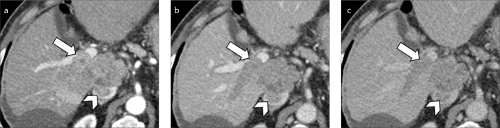
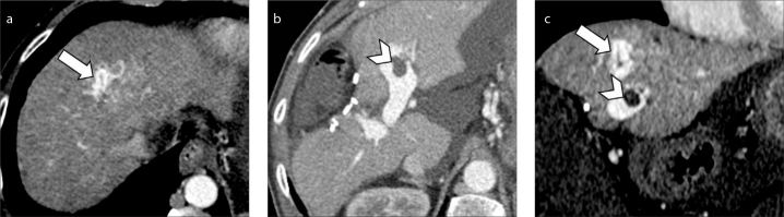
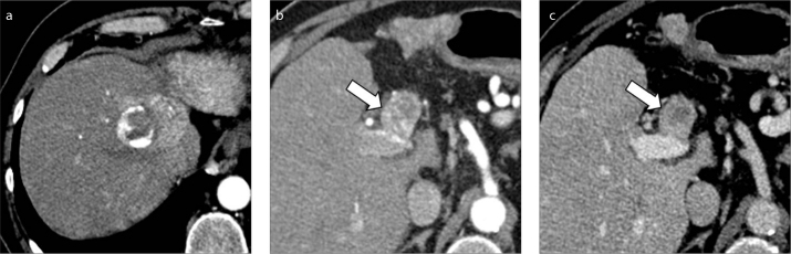
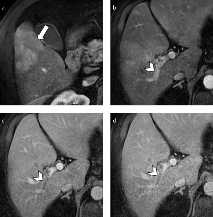
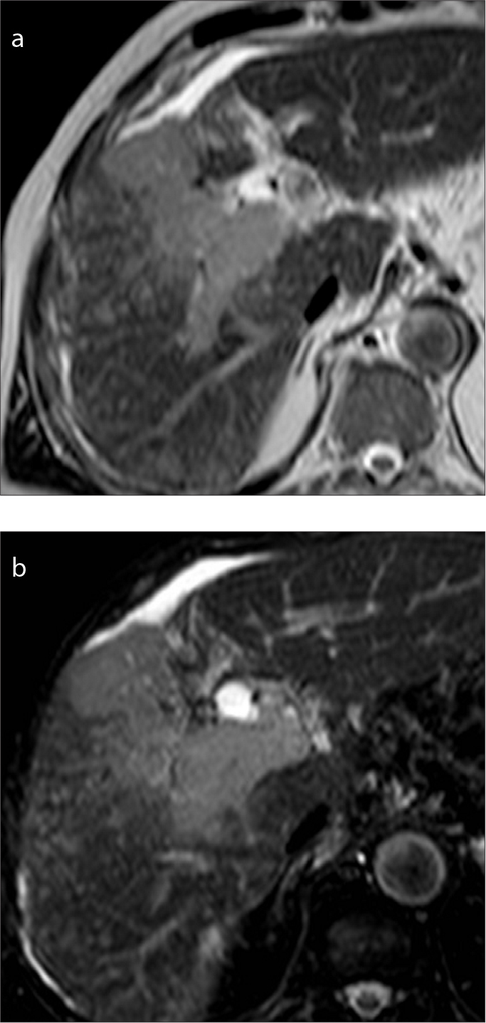
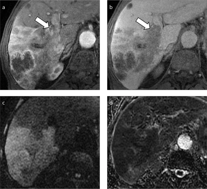
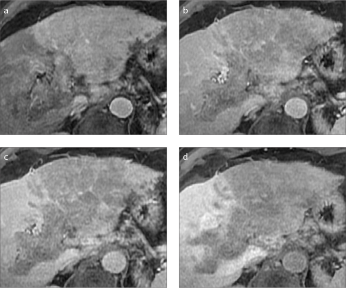
Hepatocellular carcinoma (HCC) is frequently associated with macrovascular invasion of the portal vein or hepatic veins in advanced stages. The accurate diagnosis of macrovascular invasion and the differentiation from bland non-tumoral thrombus has significant clinical and management implications, since it narrows the therapeutic options and it represents a mandatory contraindication for liver resection or transplantation. The imaging diagnosis remains particularly challenging since the imaging features of HCC with macrovascular invasion may be subtle, especially in lesions showing infiltrative appearance. However, each radiologic imaging modality may provide findings suggesting the presence of tumor thrombus rather than bland thrombus. The purpose of this paper is to review the current guidelines and imaging appearance of HCC with macrovascular invasion. Knowledge of the most common imaging features of HCC with macrovascular invasion may improve the diagnostic confidence of tumor thrombus in clinical practice and help to guide patients' management.


Conflict of interest statement

Conflict of interest disclosure

The authors declared no conflicts of interest.

Figures

Figure 1. a, b
Figure 1. a, b
A 61-year-old woman with hepatitis C virus (HCV)-related cirrhosis and infiltrative HCC. Contrast-enhanced CT on hepatic arterial (a) and portal venous (b) phases show innumerable tumor nodules with permeative growth spreading through the liver parenchyma with mild heterogeneous enhancement and washout on portal venous phase.
Figure 2. a, b
Figure 2. a, b
A 70-year-old man with cirrhosis and multifocal intrahepatic cholangiocarcinoma. Contrast-enhanced CT on hepatic arterial (a) and portal venous (b) phases demonstrate a hepatic mass with rim arterial phase hyperenhancement and macrovascular invasion in the main portal vein (arrows).
Figure 3. a–d
Figure 3. a–d
A 47-year-old man with alcohol-related cirrhosis. Conventional ultrasound examination depicts the presence of a hyperechoic lesion in the left hepatic lobe (a) and a heterogeneously hyperechoic thrombus (b, arrow) expanding the left portal vein. Subsequent contrast-enhanced CT (c, d) confirmed the presence of a tumor thrombus in the left hepatic vein (arrows).
Figure 4. a, b
Figure 4. a, b
An 84-year-old man with hepatitis B virus (HBV)-related cirrhosis and tumor thrombus. Contrast-enhanced ultrasound image (a) at 13 seconds after the intravenous administration of contrast agent demonstrates the presence of an enhancing thrombus within the main portal vein (arrow), simultaneously with the intra-hepatic artery (arrowhead). Image acquired at 180 seconds (b) shows washout of the tumor thrombus (arrow).
Figure 5. a, b
Figure 5. a, b
A 52-year-old man with HCV-related cirrhosis and multifocal HCC. Contrast-enhanced CT depicts a 1.5 cm HCC with arterial phase hyperenhancement (a, arrow) and washout (b, arrow) on delayed phase. In the same images, a coexistent nonenhancing bland thrombus (arrowheads) is seen in the left portal branch, not in contact with the HCC lesion.
Figure 6. a, b
Figure 6. a, b
Axial (a) and coronal (b) contrast-enhanced CT on portal venous phase of a 60-year-old man with HCV-related cirrhosis shows chronic thrombosis of the main portal vein with wall calcification (arrowheads) and associated cavernous transformation of the portal vein at the level of hepatic hilum.
Figure 7. a–d
Figure 7. a–d
A 75-year-old man with HCV-related cirrhosis and HCC with macrovascular invasion. Contrast-enhanced hepatic arterial (a), portal venous (b) and delayed (c) phase CT images show macrovascular invasion (arrows) of the portal vein adjacent to a 12 cm HCC lesion (arrowheads). Coronal image (d) of the same patient demonstrates enhancing soft tissue (arrow) within the main portal vein.
Figure 8. a–d
Figure 8. a–d
A 73-year-old woman with nonalcoholic steatohepatitis (NASH)-related cirrhosis and tumor thrombus. Contrast-enhanced CT shows increased diameter of the main portal vein caliber with unequivocal enhancing soft tissue within the vein (a, arrow) and subsequent washout (arrows) during portal venous (b) and delayed (c) phases consistent with tumor thrombus. Coronal image on portal venous phase (d) shows the extension of the macrovascular invasion involving the main portal vein (arrow) and portal confluence.
Figure 9. a–f
Figure 9. a–f
A 73-year-old man with cirrhosis and HCC with macrovascular invasion. Contrast-enhanced CT image (a) demonstrates a massive HCC involving the whole right hepatic lobe. The HCC is extending into the right hepatic vein along with inferior vena cava (b, arrow) and right atrium (c, arrowhead). Portal venous phase (d) depicts tumor thrombus involving the vast majority of the right atrium (arrowhead). Coronal images (e, f) show the massive macrovascular tumor invasion of the inferior vena cava (arrow) and right atrium (arrowhead).
Figure 10. a–c
Figure 10. a–c
An 80-year-old man with HBV-related cirrhosis and HCC. Contrast-enhanced CT on hepatic arterial (a), portal venous (b) and delayed (c) phases demonstrate a 4.5 cm HCC in the caudate lobe with macrovascular invasion on both right portal vein branch (arrows) and inferior vena cava (arrowheads).
Figure 11. a–c
Figure 11. a–c
A 75-year-old man with HCV-related cirrhosis, history of treated HCC and co-existence of bland and tumor thrombi. Contrast-enhanced CT on hepatic arterial phase shows enhancing tumor thrombus (a, arrow) in the upper branch of the left portal vein and bland non-enhancing thrombus (b, arrowhead) in the left portal vein. Coronal images (c) better demonstrate the co-existence of tumor (arrow) and bland (arrowhead) thrombus in the same patient.
Figure 12. a–c
Figure 12. a–c
A 79-year-old man with HCV-related cirrhosis and history of HCC treated with transarterial chemoembolization (TACE). Contrast-enhanced CT on hepatic arterial phase (a) shows treated HCC with TACE with adjacent residual enhancing tumor. CT images on hepatic arterial (b) and portal venous (c) phases at the level of portal vein bifurcation demonstrate macrovascular invasion of the left portal vein (arrows), not present at prior examinations (not shown).
Figure 13. a–d
Figure 13. a–d
A 70-year-old man with NASH-related cirrhosis and HCC. MRI on hepatic arterial phase (a) shows a 6.8 cm arterial phase hyperenhancing HCC (arrow). The lesion invades the right portal vein, which demonstrates enhancing tumor thrombus (b, arrowhead) with subsequent washout on portal venous (c) and delayed (d) phases (arrowheads).
Figure 14. a, b
Figure 14. a, b
A 71-year-old man with HBV-related cirrhosis and HCC with macrovascular invasion on the right portal vein. The tumor thrombus shows mild-to-moderate hyperintensity on T2-weighted (a) and SPIR images (b).
Figure 15. a–d
Figure 15. a–d
An 82-year-old man with cirrhosis and history of HCC. MRI images on hepatic arterial (a) and portal venous (b) phases show a large HCC with infiltrative imaging appearance and macrovascular invasion (arrows) in the right portal vein. DWI image at b= 800 s/mm2 (c) demonstrates diffusion restriction of the liver mass and hypointensity on ADC map (d).
Figure 16. a–d
Figure 16. a–d
A 70-year-old woman with cryptogenic cirrhosis and HCC with infiltrative appearance. Gadoxetic acid-enhanced MRI shows a large infiltrative HCC with macrovascular invasion of the left portal vein demonstrating mild arterial phase hyperenhancement (a), washout on portal venous phase (b), and hypointensity on 3 minutes transitional (c) and 20 minutes (d) hepatobiliary phases.

References

    1. Ferlay J, Soerjomataram I, Dikshit R, et al. Cancer incidence and mortality worldwide: sources, methods and major patterns in GLOBOCAN 2012. Int J Cancer. 2015;136:E359–386. doi: 10.1002/ijc.29210. - DOI - PubMed
    1. Llovet JM, Brú C, Bruix J. Prognosis of hepatocellular carcinoma: the BCLC staging classification. Semin Liver Dis. 1999;19:329–338. doi: 10.1055/s-2007-1007122. - DOI - PubMed
    1. European Association for the Study of the Liver. EASL Clinical Practice Guidelines: Management of hepatocellular carcinoma. J Hepatol. 2018;69:182–236. - PubMed
    1. Marrero JA, Kulik LM, Sirlin CB, et al. Diagnosis, staging, and management of hepatocellular carcinoma: 2018 practice guidance by the American Association for the Study of Liver Diseases. Hepatology. 2018;68:723–750. doi: 10.1002/hep.29913. - DOI - PubMed
    1. Reynolds AR, Furlan A, Fetzer DT, et al. Infiltrative hepatocellular carcinoma: what radiologists need to know. Radiographics. 2015;35:371–386. doi: 10.1148/rg.352140114. - DOI - PubMed

MeSH terms