

Tubular gastric adenocarcinoma: machine learning-based CT texture analysis for predicting lymphovascular and perineural invasion
- PMID: 32990246
- PMCID: PMC7664741
- DOI: 10.5152/dir.2020.19507
Tubular gastric adenocarcinoma: machine learning-based CT texture analysis for predicting lymphovascular and perineural invasion
Abstract
Purpose: Lymphovascular invasion (LVI) and perineural invasion (PNI) are associated with poor prognosis in gastric cancers. In this work, we aimed to investigate the potential role of computed tomography (CT) texture analysis in predicting LVI and PNI in patients with tubular gastric adenocarcinoma (GAC) using a machine learning (ML) approach.
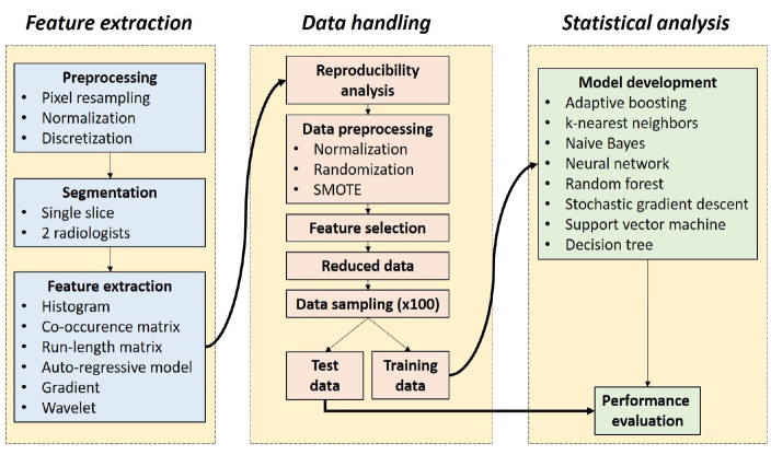
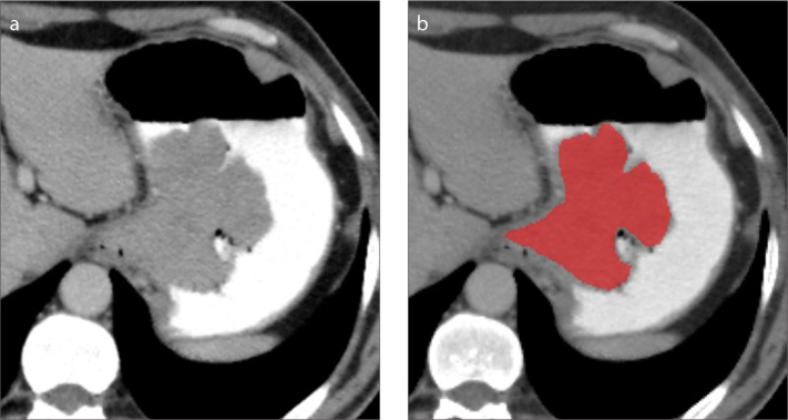
Methods: Sixty-eight patients who underwent total gastrectomy with curative (R0) resection and D2-lymphadenectomy were included in this retrospective study. Texture features were extracted from the portal venous phase CT images. Dimension reduction was first done with a reproducibility analysis by two radiologists. Then, a feature selection algorithm was used to further reduce the high-dimensionality of the radiomic data. Training and test splits were created with 100 random samplings. ML-based classifications were done using adaptive boosting, k-nearest neighbors, Naive Bayes, neural network, random forest, stochastic gradient descent, support vector machine, and decision tree. Predictive performance of the ML algorithms was mainly evaluated using the mean area under the curve (AUC) metric.
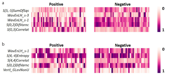
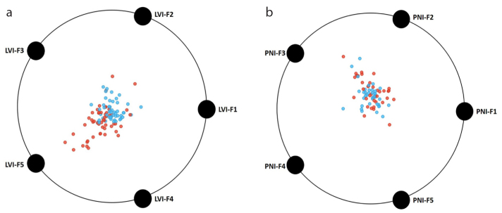
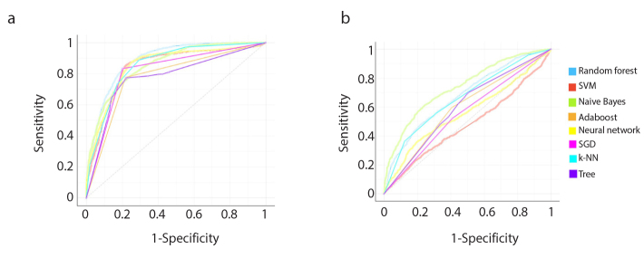
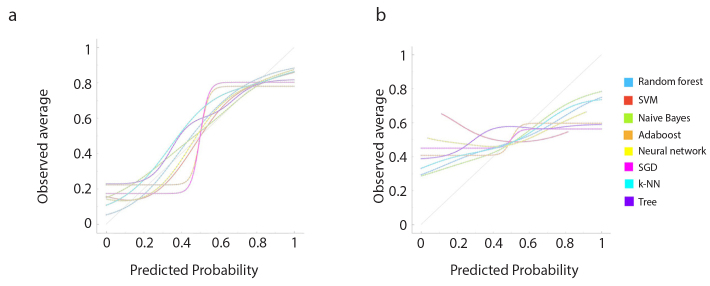
Results: Among 271 texture features, 150 features had excellent reproducibility, which were included in the further feature selection process. Dimension reduction steps yielded five texture features for LVI and five for PNI. Considering all eight ML algorithms, mean AUC and accuracy ranges for predicting LVI were 0.777-0.894 and 76%-81.5%, respectively. For predicting PNI, mean AUC and accuracy ranges were 0.482-0.754 and 54%-68.2%, respectively. The best performances for predicting LVI and PNI were achieved with the random forest and Naive Bayes algorithms, respectively.
Conclusion: ML-based CT texture analysis has a potential for predicting LVI and PNI of the tubular GACs. Overall, the method was more successful in predicting LVI than PNI.
Conflict of interest statement
The authors declared no conflicts of interest.
Figures
Similar articles
-
Preoperative prediction of lymphovascular and perineural invasion in gastric cancer using spectral computed tomography imaging and machine learning.World J Gastroenterol. 2024 Feb 14;30(6):542-555. doi: 10.3748/wjg.v30.i6.542. World J Gastroenterol. 2024. PMID: 38463023 Free PMC article.
-
Radiogenomics of lower-grade gliomas: machine learning-based MRI texture analysis for predicting 1p/19q codeletion status.Eur Radiol. 2020 Feb;30(2):877-886. doi: 10.1007/s00330-019-06492-2. Epub 2019 Nov 5. Eur Radiol. 2020. PMID: 31691122
-
Hypovascular pancreas head adenocarcinoma: CT texture analysis for assessment of resection margin status and high-risk features.Eur Radiol. 2020 May;30(5):2853-2860. doi: 10.1007/s00330-019-06583-0. Epub 2020 Jan 17. Eur Radiol. 2020. PMID: 31953662
-
Clear Cell Renal Cell Carcinoma: Machine Learning-Based Quantitative Computed Tomography Texture Analysis for Prediction of Fuhrman Nuclear Grade.Eur Radiol. 2019 Mar;29(3):1153-1163. doi: 10.1007/s00330-018-5698-2. Epub 2018 Aug 30. Eur Radiol. 2019. PMID: 30167812
-
Prediction of Benign and Malignant Solid Renal Masses: Machine Learning-Based CT Texture Analysis.Acad Radiol. 2020 Oct;27(10):1422-1429. doi: 10.1016/j.acra.2019.12.015. Epub 2020 Feb 1. Acad Radiol. 2020. PMID: 32014404
Cited by
-
The diagnostic value of a nomogram based on enhanced CT radiomics for differentiating between intrahepatic cholangiocarcinoma and early hepatic abscess.Front Mol Biosci. 2024 Aug 23;11:1409060. doi: 10.3389/fmolb.2024.1409060. eCollection 2024. Front Mol Biosci. 2024. PMID: 39247207 Free PMC article.
-
Spectral CT-based nomogram for preoperative prediction of perineural invasion in locally advanced gastric cancer: a prospective study.Eur Radiol. 2023 Jul;33(7):5172-5183. doi: 10.1007/s00330-023-09464-9. Epub 2023 Feb 24. Eur Radiol. 2023. PMID: 36826503
-
Radiomics as a New Frontier of Imaging for Cancer Prognosis: A Narrative Review.Diagnostics (Basel). 2021 Sep 29;11(10):1796. doi: 10.3390/diagnostics11101796. Diagnostics (Basel). 2021. PMID: 34679494 Free PMC article. Review.
-
Machine learning model based on enhanced CT radiomics for the preoperative prediction of lymphovascular invasion in esophageal squamous cell carcinoma.Front Oncol. 2024 Feb 23;14:1308317. doi: 10.3389/fonc.2024.1308317. eCollection 2024. Front Oncol. 2024. PMID: 38549935 Free PMC article.
-
Preoperative prediction of perineural invasion and lymphovascular invasion with CT radiomics in gastric cancer.Eur J Radiol Open. 2024 Jan 25;12:100550. doi: 10.1016/j.ejro.2024.100550. eCollection 2024 Jun. Eur J Radiol Open. 2024. PMID: 38314183 Free PMC article.
References
-
- Hwang J-E, Hong J-Y, Kim JE, et al. Prognostic significance of the concomitant existence of lymphovascular and perineural invasion in locally advanced gastric cancer patients who underwent curative gastrectomy and adjuvant chemotherapy. Jpn J Clin Oncol. 2015;45:541–546. doi: 10.1093/jjco/hyv031. - DOI - PubMed
MeSH terms
LinkOut - more resources
Full Text Sources
Medical
