. 2020 Dec;47(12):1476-1484.
doi: 10.1111/jcpe.13371. Epub 2020 Nov 9.

Formation and regeneration of a Wnt-responsive junctional epithelium

Affiliations

Formation and regeneration of a Wnt-responsive junctional epithelium

Xue Yuan et al. J Clin Periodontol. 2020 Dec.

Abstract

Aim: To identify the molecular mechanisms mediating the persistent defensive functions of the self-renewing junctional epithelium (JE).

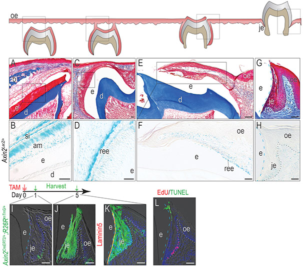
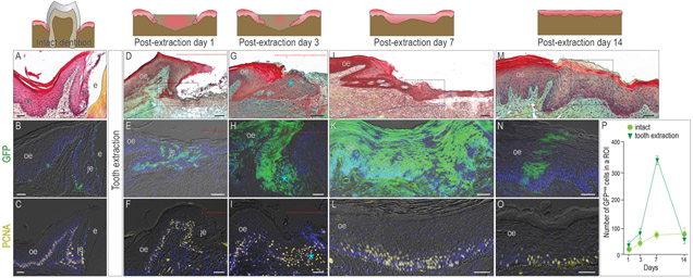
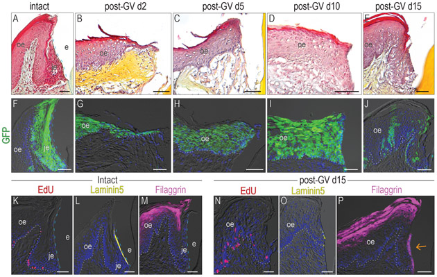
Materials and methods: Two strains of Wnt reporter mice, Axin2CreErt2/+ ;R26RmTmG/+ and Axin2LacZ/+ , were employed, along with three clinically relevant experimental scenarios where the function of the JE is disrupted: after tooth extraction, after a partial gingivectomy, and after a complete circumferential gingivectomy.

Results: Using transgenic Wnt reporter strains of mice, we established the JE is a Wnt-responsive epithelium beginning at the time of its formation and that it maintains this status into adulthood. After tooth extraction, progeny of the initial Wnt-responsive JE population directly contributed to healing and ultimately adopted an oral epithelium (OE) phenotype. In the traditional partial gingivectomy model, the JE completely regenerated and did so via progeny of the original Wnt-responsive population. However, following circumferential gingivectomy, the OE was incapable of re-establishing a functional JE.

Conclusions: A Wnt-responsive niche at the interface between tooth and oral epithelia is required for a functional JE.

Keywords: epithelial attachment; gingiva; gingivectomy; oral epithelium.


Conflict of interest statement

Conflict of Interest Statement

All authors declare that no conflicts of interest exist.

Figures

Figure 1.
Figure 1.. The JE originates from Wnt-responsive cells and maintains the Wnt-responsiveness.
(A) Masson’s trichrome staining showing the OE covering the unerupted maxillary first molar on P9. (B) In Axin2LacZ/+ mice, Xgal staining showing Xgal+ve Wnt-responsive cells in the JE on P9. (C) By P13, the enamel was completely formed, and the inner enamel epithelium reduced to a few layers of flat cuboidal cells called REE. At this stage, the REE started to fuse with OE. (D) Xgal staining showing Xgal+ve Wnt-responsive cells on P13. (E) On P14, the maxillary first molar penetrated to the oral cavity and the JE started to form. (F)Xgal staining showing Xgal+ve Wnt-responsive cells on P14. (G) Masson’s trichrome staining of the first maxillary molar in adult mice. (H) Xgal staining showing Wnt-responsive cells in adults. One dose of tamoxifen was given to adult Axin2CreERT2/+;R26RmTmG/+ mice and GFP+ve cells were analyzed (I) 1 day and (J) 5 days later. (K) Co-staining of 5-day chase GFP (green) with Laminin 5 (red) to show the attachment to the tooth surface. (L) TUNEL staining (green) showing apoptosis and EdU staining (red) showing cell proliferation in the JE. Dashed blue lines indicate the shape of enamel. Dotted black and dotted white lines indicate the demarcation between the epithelium and the connective tissue. Abbreviations: e, enamel space; je, junctional epithelium; oe, oral epithelium; b, bone; d, dentin; ree, reduced enamel epithelium; am, ameloblasts. Blue dashed lines indicate the edge of the enamel and white dotted lines indicate the boundary between the epithelium and the connective tissue. Scale bars: 50μm.
Figure 2.
Figure 2.. After tooth extraction, the Wnt-responsive JE disappears.
In (A) an intact dentition, (B) one-day lineage tracing reveals a few Wnt-responsive cells in the JE and OE. (C) PCNA staining showing the proliferative cells in the OE and JE. One day after tooth extraction, (D) the JE remnant, (E) the progeny of Wnt-responsive cells, and (F) the cell proliferation were analyzed. By post-extraction day 3, (G) the enlarged JE, (H) burst of the progeny of Wnt-responsive cells, and (I) massive cell proliferation were observed. By post-extraction day 7, (J) the re-epithelization of the extraction socket, (K) the distribution of the Wnt-responsive progeny, and (L) the cell proliferation were analyzed. By post-extraction day 14, (M) the re-epithelization of the extraction socket, (N) the distribution of Wnt-responsive progeny, and (O) the cell proliferation were analyzed. (P) Quantification of Wnt-responsive cells (n=4). The data were expressed as mean ± SD. Dotted Red lines indicate the extraction site. Dotted white lines indicate the demarcation between the epithelium and the connective tissue. Dashed blue lines indicate the shape of enamel. Abbreviations: je, junctional epithelium; oe, oral epithelium; e, enamel space. Scale bars: 50 μm.
Figure 3.
Figure 3.. After a partial gingivectomy, a regenerated JE re-establishes its Wnt-responsiveness.
Pentachrome staining was used to analyze the morphology with the samples from (A) intact, post-GV day (B)1, (C) 3, (D) 5, (E) 7, and (F) 14. (G) One dose of tamoxifen was given to Axin2CreERT2/+;R26RmTmG/+ mice and GFP+ve cells were analyzed 5 days later. (H) EdU staining was performed to visualize proliferating cells. On post-GV day 1, (I) the progeny of Wnt-responsive cells, and (J) the cell proliferation were analyzed. On post-GV day 3, (K) the progeny of Wnt-responsive cells, and (L) the cell proliferation were analyzed. On post-GV day 5, (M) the progeny of Wnt-responsive cells, and (N) the cell proliferation were analyzed. On post-GV day7, (Q) the progeny of Wnt-responsive cells, (P) the cell proliferation, and (Q) Laminin 5 expression were analyzed. On post-GV day 14, (R) the progeny of Wnt-responsive cells and cell proliferation, and (S) Laminin 5 expression were analyzed. (T) An illustration of the healing process after a gingivectomy. (U) Pentachrome staining showing the two epithelial populations co-existed on post-GV day 3. The blue arrow points the non-keratinized epithelium tissue around the tooth. (V) lineage tracing shows two GFP+ve populations co-existed on post-GV day 3. The green star indicates the GFP+ve population around the tooth. (W) An illustration showing both JE and OE contribute to the regeneration after a gingivectomy. Dashed blue lines indicate the shape of enamel. Dotted black and dotted white lines indicate the demarcation between the epithelium and the connective tissue. Abbreviations: GV, gingivectomy; M1, molar 1; je, junctional epithelium; oe, oral epithelium; e, enamel space. Scale bars: 50 μm.
Figure 4.
Figure 4.. A functional JE cannot be solely regenerated from the OE.
Pentachrome staining was used to analyze the morphology with the samples from (A) intact group, post-GV day (B)2, (C) 5, (D) 10, and (E) 15. (F) One dose of tamoxifen was given to Axin2CreERT2/+;R26RmTmG/+ mice, GFP+ve cells were analyzed 5 days later. Lineage tracing showing the distribution of Wnt-responsive cells on post-GV day (G)2, (H) 5, (I) 10, (J) 15. In the intact samples, (K) EdU+ve cells, (L) Laminin 5 expression, and (M) Filaggrin expression pattern was examined. In the gingivectomy group, (N) EdU+ve cells, (O) Laminin 5 expression, and (P) Filaggrin expression pattern was examined on post-GV day 15. The orange arrow indicates the abnormal Filaggrin expression on the interface between the epithelium and tooth surface. Dashed blue lines indicate the shape of enamel. Dotted white lines indicate the demarcation between the epithelium and the connective tissue. Abbreviations: GV, gingivectomy; je, junctional epithelium; oe, oral epithelium; e, enamel space. Scale bars: 50 μm.

References

    1. Al Alam D, Green M, Tabatabai Irani R, Parsa S, Danopoulos S, Sala FG, … Bellusci S (2011). Contrasting expression of canonical Wnt signaling reporters TOPGAL, BATGAL and Axin2(LacZ) during murine lung development and repair. PLoS ONE, 6(8), e23139. doi:10.1371/journal.pone.0023139 - DOI - PMC - PubMed
    1. Atsuta I, Yamaza T, Yoshinari M, Mino S, Goto T, Kido MA, … Tanaka T (2005). Changes in the distribution of laminin-5 during peri-implant epithelium formation after immediate titanium implantation in rats. Biomaterials, 26(14), 1751–1760. doi:10.1016/j.biomaterials.2004.05.033 - DOI - PubMed
    1. Bosshardt DD, & Lang NP (2005). The junctional epithelium: from health to disease. J Dent Res, 84(1), 9–20. doi:10.1177/154405910508400102 - DOI - PubMed
    1. Engler WO, Ramfjord SP, & Hiniker JJ (1966). Healing following simple gingivectomy. A tritiated thymidine radioautographic study. I. Epithelialization. J Periodontol, 37(4), 298–308. - PubMed
    1. Fujiseki M, Matsuzaka K, Yoshinari M, Shimono M, & Inoue T (2003). An experimental study on the features of peri-implant epithelium: immunohistochemical and electron-microscopic observations. Bull Tokyo Dent Coll, 44(4), 185–199. doi:10.2209/tdcpublication.44.185 - DOI - PubMed

Publication types