. 2020 Sep 29;7(1):447-473.
doi: 10.1146/annurev-virology-021920-113833.

Vaccines to Emerging Viruses: Nipah and Hendra

Affiliations

Vaccines to Emerging Viruses: Nipah and Hendra

Moushimi Amaya et al. Annu Rev Virol. .

Abstract

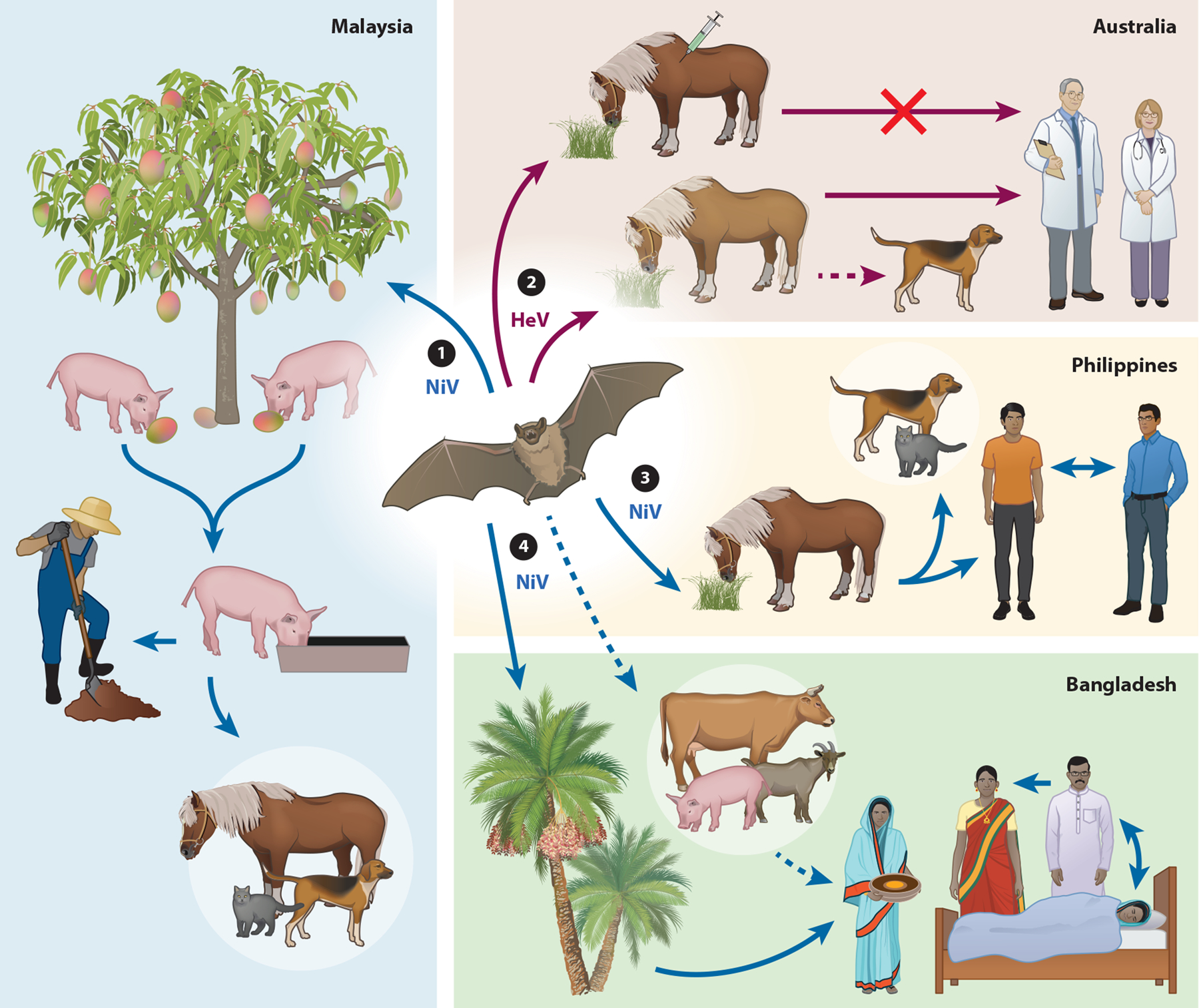
Hendra virus (HeV) and Nipah virus (NiV) are bat-borne zoonotic para-myxoviruses identified in the mid- to late 1990s in outbreaks of severe disease in livestock and people in Australia and Malaysia, respectively. HeV repeatedly re-emerges in Australia while NiV continues to cause outbreaks in South Asia (Bangladesh and India), and these viruses have remained transboundary threats. In people and several mammalian species, HeV and NiV infections present as a severe systemic and often fatal neurologic and/or respiratory disease. NiV stands out as a potential pandemic threat because of its associated high case-fatality rates and capacity for human-to-human transmission. The development of effective vaccines, suitable for people and livestock, against HeV and NiV has been a research focus. Here, we review the progress made in NiV and HeV vaccine development, with an emphasis on those approaches that have been tested in established animal challenge models of NiV and HeV infection and disease.

Keywords: Hendra virus; Nipah virus; henipavirus; henipavirus countermeasures; subunit vaccine; vaccine.


Conflict of interest statement

DISCLOSURE STATEMENT

C.C.B. is a US federal employee and co-inventor on US and foreign patents pertaining to soluble forms of HeV and NiV G and F glycoproteins and monoclonal antibodies against HeV and NiV whose assignee is the United States as represented by the Henry M. Jackson Foundation for the Advancement of Military Medicine (Bethesda, Maryland). The soluble forms of the HeV and NiV G glycoproteins are licensed to Zoetis, Inc., and Aurobindo Pharma USA Inc. M.A. declares no competing interests.

Figures

Figure 1
Figure 1
Nipah virus (NiV) and Hendra virus (HeV) modes of transmission in different countries. The transmission routes of NiV in Malaysia (left), Philippines (middle right), Bangladesh (bottom right), and HeV (top right) are depicted. Solid lines represent transmission that has been observed and documented, and dashed lines represent suspected transmission in natural conditions. Fruit bats are the natural reservoirs of NiV and HeV. ① Pigs are infected by consuming partially eaten or contaminated fruit from infected bats (urine, saliva) and transmit NiV to other pigs, pig farmers, or other animals (dogs, cats, and horses) through close or direct contact. ② Horses can be infected from grazing in contaminated pastures and transmit HeV to humans and on occasion domestic dogs through close contact. A One Health vaccine approach was developed for vaccination of horses in Australia with the dual purpose of saving horses from lethal HeV infection and preventing HeV transmission from horses to humans. ③ NiV is transmitted to humans through close contact with infected horses. NiV transmission to humans, cats, and dogs appears to have occurred following close contact with or consumption of infected horse meat. Human-to-human NiV transmission can occur through close contact. ④ Bat-to-human NiV transmission occurs through consumption of contaminated date palm sap. Human-to-human transmission can occur through close contact with infected patients. Humans may also become infected through contact with infected animals. Figure adapted with permission from Reference .
Figure 2
Figure 2
Henipavirus structure and genome organization and models of the G and F glycoprotein soluble ectodomains, Hendra virus (HeV-sG) and Nipah virus (NiV-sF), respectively, and their complexes with respective NiV and HeV cross-reactive neutralizing monoclonal antibodies m102.3 (anti-G) and 5B3 (anti-F). (a) Schematic representation of a henipavirus particle with the structural proteins depicted in different colors (left) and the henipavirus genome (right). HeV and NiV P genes encode 3 nonstructural proteins: The C protein is expressed from an alternative start site, and the V and W proteins are expressed following the addition of one or two G residues at the messenger RNA editing site, respectively (right). (b, left) HeV-sG shown as a dimer solvent-accessible surface view with one monomer (cyan) overlaid with the monoclonal antibody m102.3 CDR-H3 loop (red) at the receptor binding site, and the other monomer (magenta) in complex with m102.3 Fab, which has an identical heavy chain and a similar light chain, that was used in place of the m102.4 monoclonal antibody (mAb) in the structural solution of the complex (109). The HeV-sG consists of amino acids 76–604, and the structures of the two globular head domains of HeV-sG are derived from the crystal structure (103, 172). The stalk regions of each G monomer (residues 77–136) are modeled (173). The light chain of m102.3 Fab is colored in yellow, and the heavy chain is colored in red. (b, middle) The HeV-sG tetramer surface view is modeled with one dimer (cyan and magenta) in front and the other dimer (blue and green) in back. N-linked glycans are gray spheres. (b, right) Structural model of the NiV-sF trimer in complex with the 5B3 Fab derived from the cryo–electron microscopy structure (110). The NiV-sF consists of amino acid residues 1–494 with a FLAG tag (DYKDDDK) introduced between residues L104-V105 and a C-terminal GCN4 motif. Each monomer of NiV-sF is in a different shade of blue, 5B3 heavy chain is in red, and light chain is in gold. N-linked glycans are illustrated in gray.

References

    1. Eaton BT, Broder CC, Middleton D, Wang LF. 2006. Hendra and Nipah viruses: different and dangerous. Nat. Rev. Microbiol 4:23–35 - PMC - PubMed
    1. Wang L-F, Mackenzie JS, Broder CC. 2013. Henipaviruses. In Fields Virology, ed. Knipe DM, Howley PM, pp. 1070–85. Philadelphia: Lippincott Williams & Wilkins
    1. Harcourt BH, Lowe L, Tamin A, Liu X, Bankamp B, et al. 2005. Genetic characterization of Nipah virus, Bangladesh, 2004. Emerg. Infect. Dis 11:1594–97 - PMC - PubMed
    1. Marsh GA, de Jong C, Barr JA, Tachedjian M, Smith C, et al. 2012. Cedar virus: a novel Henipavirus isolated from Australian bats. PLOS Pathog. 8:e1002836. - PMC - PubMed
    1. Drexler JF, Corman VM, Gloza-Rausch F, Seebens A, Annan A, et al. 2009. Henipavirus RNA in African bats. PLOS ONE 4:e6367. - PMC - PubMed

Publication types

MeSH terms

LinkOut - more resources