Review
. 2020 Sep 29;7(1):37-61.
doi: 10.1146/annurev-virology-122019-070009.

Actinobacteriophages: Genomics, Dynamics, and Applications

Affiliations
Review

Actinobacteriophages: Genomics, Dynamics, and Applications

Graham F Hatfull. Annu Rev Virol. .

Abstract

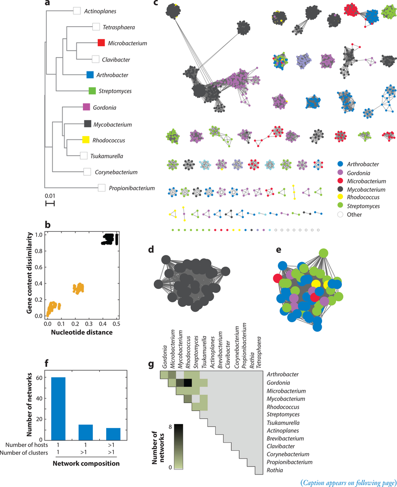
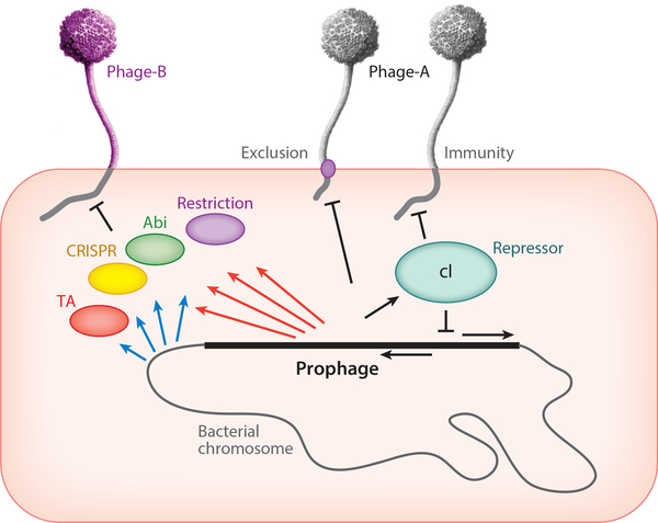
Actinobacteriophages are viruses that infect bacterial hosts in the phylum Actinobacteria. More than 17,000 actinobacteriophages have been described and over 3,000 complete genome sequences reported, resulting from large-scale, high-impact, integrated research-education initiatives such as the Science Education Alliance Phage Hunters Advancing Genomics and Evolutionary Sciences (SEA-PHAGES) program. Their genomic diversity is enormous; actinobacteriophages comprise many architecturally mosaic genomes with distinct DNA sequences. Their genome diversity is driven by the highly dynamic interactions between phages and their hosts, and prophages can confer a variety of systems that defend against attack by genetically distinct phages; phages can neutralize these defense systems by coding for counter-defense proteins. These phages not only provide insights into diverse and dynamic phage populations but also have provided numerous tools for mycobacterial genetics. A case study using a three-phage cocktail to treat a patient with a drug-resistant Mycobacterium abscessus suggests that phages may have considerable potential for the therapeutic treatment of mycobacterial infections.

Keywords: bacteriophage; genomics; mycobacterium; phage therapy.


Figures

Figure 1
Figure 1
Organization and structure of the Science Education Alliance Phage Hunters Advancing Genomics and Evolutionary Sciences (SEA-PHAGES) program. SEA-PHAGES program administrators (yellow box, top) oversee support components critical to program implementation (green box, upper middle). The typical two-term course structure (pink box, lower middle) includes phage isolation through comparative genomics; additional characterization includes electron microscopy and PCR/restriction analysis. Sequence and annotation quality control are shared with SEA-PHAGES faculty teams.
Figure 2
Figure 2
Actinobacteriophage virion morphologies. Electron microscope images are shown for phages (a) Monty, (b) Jordan, (c) MooMoo, (d) Jasmine, and (e) Wheeheim. Monty and MooMoo are examples of siphoviral morphotypes with long flexible tails, but MooMoo has a prolate (elongated) head, whereas Monty has an isometric head. Jordan, Jasmine, and Wheeheim are exmplaes of myoviral, podoviral, and tectiviral morphotypes.
Figure 3
Figure 3
Temperate phage systems for prophage maintenance. The central parts of three Cluster A2 phage genomes are shown that vary in their prophage maintenance mechanisms. Rightward- and leftward-transcribed genes are shown as boxes above and below the genome markers, respectively, with gene numbers shown in the boxes. Numbers above or below the genes indicate their assignment into gene phamilies, with the numbers of phamily members shown in parentheses. Pairwise nucleotide similarity is indicated as spectrum-colored shading between genomes, with violet reflecting closest similarity. The ends of the structural gene operons (tail protein genes) and the early gene operons are indicated. Between these, phage NothingSpecial has an integration cassette including a tyrosine-integrase (Int-Y) and attP site, whereas phages BobSwaget and Lokk have partitioning cassettes encoding parAB genes. Lokk also codes for a RepA-like protein for extrachromosomal prophage replication, whereas BobSwaget likely replicates using an alternative mechanism.
Figure 4
Figure 4
A network phylogeny of actinobacteriophages. A randomly chosen phage from each subcluster and nonsubclustered cluster together with the singletons were compared by their gene contents and the relationships displayed using SplitsTree (132). Each phage node is indicated by a colored circle indicating the genus of the bacterial host used for isolation. Clusters containing 100 or more individual phages are circled and the cluster indicated.
Figure 5
Figure 5
Heterogeneous genomic diversity of actinobacteriophages. (a) Phylogenetic tree of several actinobacterial host genera. (b) Representative genomic similarity plot comparing gene content dissimilarity and nucleotide distance (46) between phages in Cluster L (n = 38) and not in Cluster L (n = 2,384). Each data point represents a pairwise comparison involving a phage within Cluster L and another phage within (gold) or without (black) Cluster L. (c) Genome networks (n = 87) for all sequenced actinobacteriophages (n = 2,422). A node represents a phage genome and is colored according to its host genus. Two nodes connected by an edge reflect phages with an intracluster genomic relationship, having gene content dissimilarity <0.89 and nucleotide distance <0.42 (46). A network consists of a group of phages that contain at least one edge to another phage in the group and no edges to any phages outside of the group. (d) Enlarged representative network from panel c containing phages (n = 38) from a single cluster (Cluster L) and a single host genus (Mycobacterium). (e) Enlarged representative network from panel c containing phages (n = 52) from multiple clusters (Clusters AM, AU, BI, CC, DJ, and EL and Singleton RosaAsantewaa) and multiple host genera. (f) Histogram reflecting phage diversity based on the composition of the networks in panel c. The number of clusters and host genera represented within each network were quantified, and the number of networks containing the indicated number of clusters and host genera were reported. For this analysis, singletons were treated as clusters. (g) Heatmap reflecting phage relationships within networks from panel c by unique host genera (n = 14). Within each network, host genera pairs connected by an edge were identified. For each possible host genera pair in the database, the number of networks containing at least one edge connecting the two host genera was quantified and represented by a color spectrum. Panel a adapted with permission from Reference .
Figure 6
Figure 6
Transcription of the D29 genome. Strand-specific RNA sequencing analysis of D29 (Multiplicity of Infection = 3) infected Mycobacterium smegmatis me2 155. Time points after adsorption are indicated on the upper right: 15 min (teal), 30 min (blue), 60 min (purple), and 150 min (red). At the left are scale maxima and indications of top or bottom strand. The D29 map is shown at the bottom. Figure adapted from Reference .
Figure 7
Figure 7
Phage-host dynamics. A lysogenic cell is depicted carrying a prophage integrated into the bacterial chromosome (not to scale). The prophage genome is derived from Phage-A and encodes a repressor protein (cI) that shuts down lytic genes of both the integrated prophage and superinfecting Phage-A particles. Some prophages may express membrane proteins that prevent superinfection by the same phage (Phage-A) or closely related phages. The bacterial chromosome may express a variety of systems to defend against viral attack (blue arrows), including restriction, various abortive infection systems (abi), CRISPR-Cas, and toxin-antitoxin (TA) systems. Prophages can express analogous systems (red arrows) that defend against infection by heterotypic (i.e., unrelated) phages, such as Phage-B.

References

    1. Barka EA, Vatsa P, Sanchez L, Gaveau-Vaillant N, Jacquard C, et al. 2016. Taxonomy, physiology, and natural products of Actinobacteria. Microbiol. Mol. Biol. Rev 80:1–43 - PMC - PubMed
    1. Durand GA, Raoult D, Dubourg G. 2019. Antibiotic discovery: history, methods and perspectives. Int. J. Antimicrob. Agents 53:371–82 - PubMed
    1. Hatfull GF. 2018. Mycobacteriophages. Microbiol. Spectr 6. 10.1128/microbiolspec.GPP3-0026-2018 - DOI - PMC - PubMed
    1. Froman S, Will DW, Bogen E. 1954. Bacteriophage active against Mycobacterium tuberculosis I. Isolation and activity. Am. J. Pub. Health 44:1326–33 - PMC - PubMed
    1. Murohashi T, Tokunaga T, Mizuguchi Y, Maruyama Y. 1963. Phage typing of slow-growing mycobacteria. Am. Rev. Respir. Dis 88:664–69 - PubMed

Publication types

LinkOut - more resources