Comparative Study
. 2020 Dec;44(12):1677-1684.
doi: 10.1097/PAS.0000000000001575.

Recurrent YAP1 and MAML2 Gene Rearrangements in Retiform and Composite Hemangioendothelioma

Affiliations
Comparative Study

Recurrent YAP1 and MAML2 Gene Rearrangements in Retiform and Composite Hemangioendothelioma

Cristina R Antonescu et al. Am J Surg Pathol. 2020 Dec.

Abstract

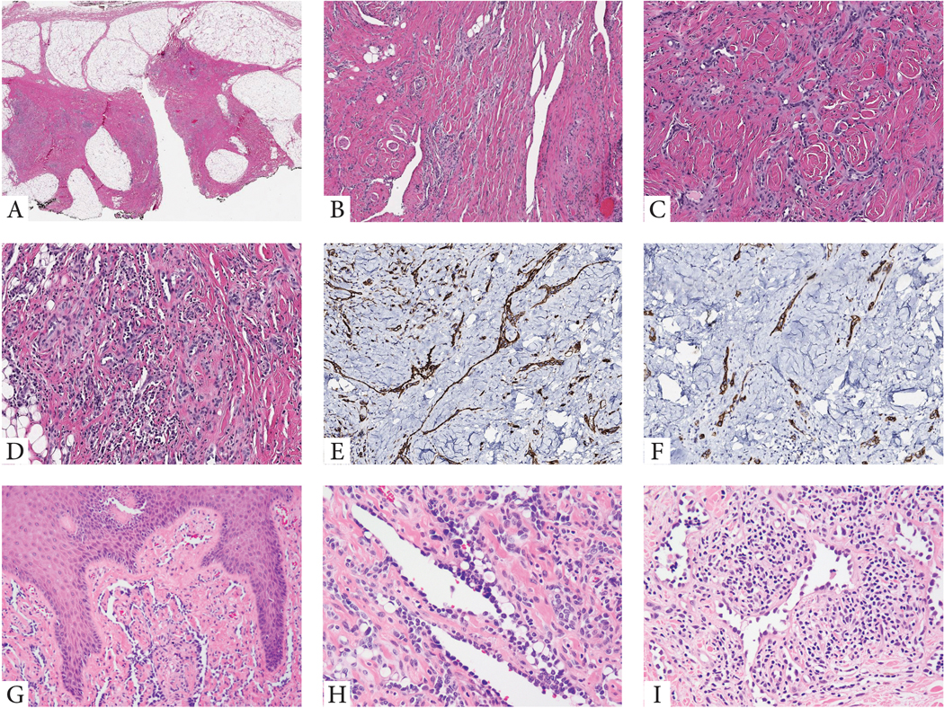
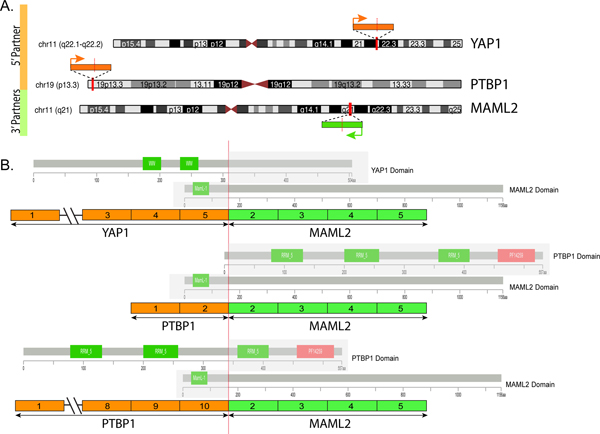
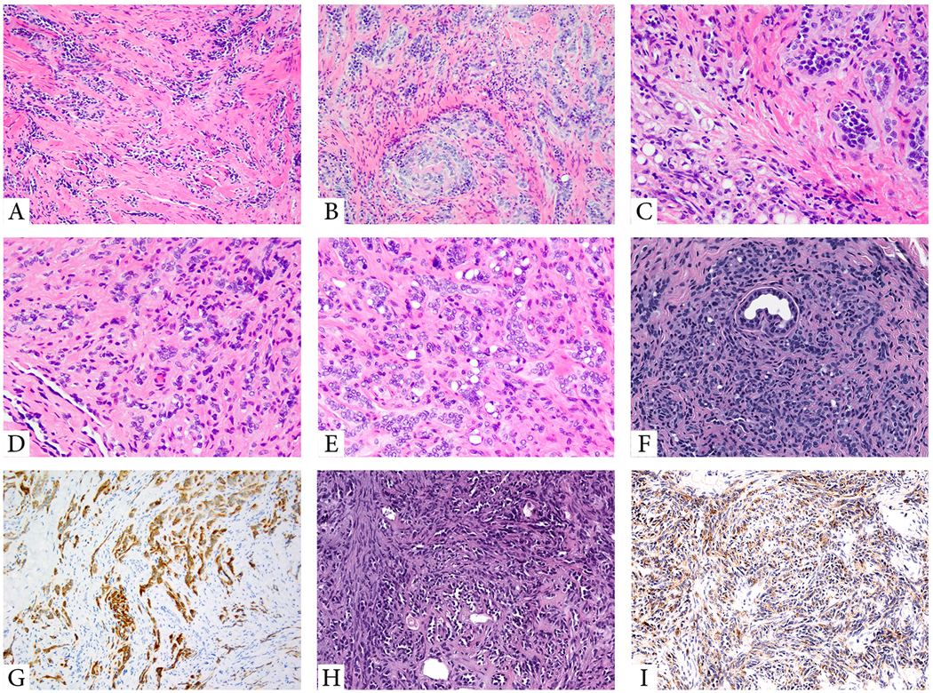
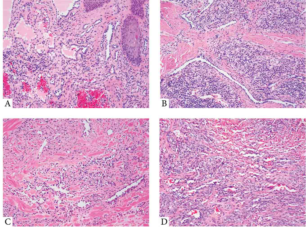
Retiform and composite hemangioendotheliomas (CHEs) are both locally aggressive, rarely metastasizing vascular neoplasms characterized by arborizing vascular channels lined by endothelial cells with a hobnail morphology. CHE displays additional cytologic and architectural components, including often vacuolated epithelioid cells, solid areas, or features reminiscent of well-differentiated angiosarcoma. Triggered by an index case of a soft tissue retiform hemangioendothelioma (RHE) which revealed a YAP1-MAML2 gene fusion by targeted RNA sequencing, we sought to investigate additional cases in this morphologic spectrum for this genetic abnormality. A total of 24 cases, 13 RHE and 11 CHE involving skin and soft tissue were tested by fluorescence in situ hybridization using custom BAC probes for rearrangements involving these genes. An additional visceral CHE with neuroendocrine differentiation was tested by targeted RNA sequencing. Among the soft tissue cohort, 5/13 (38%) RHE and 3/11 (27%) CHE showed YAP1 gene rearrangements, with 5 cases showing a YAP1-MAML2 fusion, including all 3 CHE. The single neuroendocrine CHE showed the presence of a PTBP1-MAML2 fusion. All YAP1-positive CHE lesions occurred in female children at acral sites, compared with fusion-negative cases which occurred in adults, with a wide anatomic distribution. YAP1-positive RHE occurred preferentially in males and lower limb, compared with negative cases. These results suggest that RHE and CHE represent a morphologic continuum, sharing abnormalities in YAP1 and MAML2 genes. In contrast, the neuroendocrine CHE occurring in a 37-year-old male harbored a distinct PTBP1-MAML2 fusion and showed aggressive clinical behavior (pancreatic mass with multiple liver and lung metastases). These preliminary findings raise the possibility that neuroendocrine CHE may be genetically distinct from the conventional RHE/CHE spectrum. Further studies are needed to investigate the pathogenetic relationship of fusion-negative cases with this subset and, less likely, with other members of the HE family of tumors.


Conflict of interest statement

Conflict of interests: none

Figures

Figure 1.
Figure 1.. Morphologic features of RHE with YAP1 gene rearrangements.
(A-F). Index case of a superficial soft tissue lesion from the knee in a 10 year-old male (case 1). Low power view showing a vague nodular and infiltrating growth pattern within subcutis which appears heavily fibrotic (A). Medium power shows densely sclerotic stromal background with either elongated, staghorn vessels (B), or shorter, retiform vascular network, lined by small endothelial cells with scant cytoplasm (C), with patchy prominent lymphocytic infiltrate, obscuring the vascular proliferation (D). Tumor cells typically co-express CD31 (E) and podoplanin (D2-40) (F). G-H. Cutaneous example showing elongated vascular channels infiltrating through the superficial dermis, lined by hobnailed endothelial cells (case 4, 10/M, buttock). I. Another RHE skin lesion in an adult (50/F, knee) showing angulated vessels with dilated lumina and protruding endothelial cells, some appearing detached and reminiscent of micropapillae. The vascular channels are embedded in a stroma with abundant lymphocytic infiltrate (case 5).
Figure 2.
Figure 2.. Diagrammatic representation of the YAP1-MAML2 and PTBP1-MAML2 fusions.
(A) Chromosomal location of YAP1 gene locus in 11q22.1-q22.2, PTBP1 in 19p13.3 and MAML2 in 11q21; red vertical lines depict the genomic breakpoint locus. Arrows show the direction of transcription of each gene. (B) Upper panel depicts the YAP1-MAML2 transcript, composed of the first 5 exons of YAP1 fused to most of MAML2 (exons 2-5). Middle and lower panels show the two transcript isoforms of PTBP1-MAML2, composed of either the first 2 exons or the first 10 exons of PTBP1 gene fused to MAML2 gene exons 2-5. The protein domains of each of the genes involved are also schematically depicted.
Figure 3.
Figure 3.. Morphologic features of YAP1-MAML2 fusion-positive soft tissue CHE and PTBP1-MAML2-positive neuroendocrine CHE.
(A-C; Case 6, 9/F; foot). Low power showing a predominant retiform component (A), with other areas showing solid sheets or cords of epithelioid cells, embedded in a diffusely fibrotic stroma (B). High power showing epithelioid cells and vacuolated cells as well as small vessels lined by plump epithelioid cells, obstructing the lumina (C). (D-G; Case 9, 37/M, liver biopsy). Low power showing a small retiform area with predominantly epithelioid cells arranged in cords and single cells in a sclerotic stroma (D). Numerous vacuolated cells can be seen (E). The lesion lacks a lobular growth pattern, with cords and single tumor cells encasing adnexal structures (F). Tumor is positive for D2–40 (G) and ERG (not shown). (H-I; case 8, 7/F, middle finger). (H). Neuroendocrine CHE with a predominant retiform architecture lined by uniform hobnailed endothelial cells. (I) Tumor showed positivity for synaptophysin.
Figure 4.
Figure 4.. Histologic features of the molecular negative subset of RHE and CHE.
(A-B) YAP1-negative RHE. (A; 14/M, shoulder) Low power view showing dilated blood vessels within dermis lined by hobnail endothelial cells. (B; 55/M, thigh) Elongated, tubular vascular channels lined by cuboidal hyperchromatic endothelial cells, embedded in a stroma with abundant lymphocytic infiltrate. (C,D) CHE lacking YAP1 or MAML2 gene rearrangements. (C; 24/F, scalp) Predominant retiform areas with a focal epithelioid component arranged in single files. (D; 36/F, scalp) A variegated appearance showing solid, single cells as well as hemangioma-like areas.

References

    1. Rubin BP. Composite haemangioendothelioma In: Fletcher CD, Bridge JA, Pancras CW, et al. [eds]. World Health Organization Classification of Tumours. Pathology and Genetics of Tumours of Soft Tissue and Bone. Lyon, France: IARC Press; 2013.
    1. Nayler SJ, Rubin BP, Calonje E, et al. Composite hemangioendothelioma: a complex, low-grade vascular lesion mimicking angiosarcoma. Am J Surg Pathol. 2000;24:352–361. - PubMed
    1. Perry KD, Al-Lbraheemi A, Rubin BP, et al. Composite hemangioendothelioma with neuroendocrine marker expression: an aggressive variant. Mod Pathol. 2017;30:1589–1602. - PubMed
    1. Kirchner M, Neumann O, Volckmar AL, et al. RNA-Based Detection of Gene Fusions in Formalin-Fixed and Paraffin-Embedded Solid Cancer Samples. Cancers (Basel). 2019;11. - PMC - PubMed
    1. Shang Leen SL, Fisher C, Thway K. Composite hemangioendothelioma: clinical and histologic features of an enigmatic entity. Adv Anat Pathol. 2015;22:254–259. - PubMed

Publication types

MeSH terms