. 2020 Oct 29;183(3):739-751.e8.
doi: 10.1016/j.cell.2020.09.032. Epub 2020 Sep 15.

Structural and Functional Analysis of the D614G SARS-CoV-2 Spike Protein Variant

Affiliations

Structural and Functional Analysis of the D614G SARS-CoV-2 Spike Protein Variant

Leonid Yurkovetskiy et al. Cell. .

Abstract

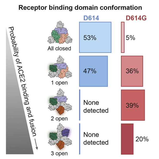
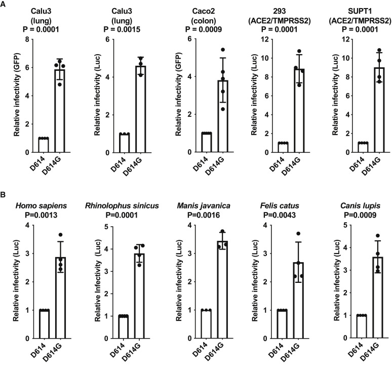
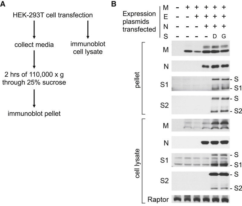
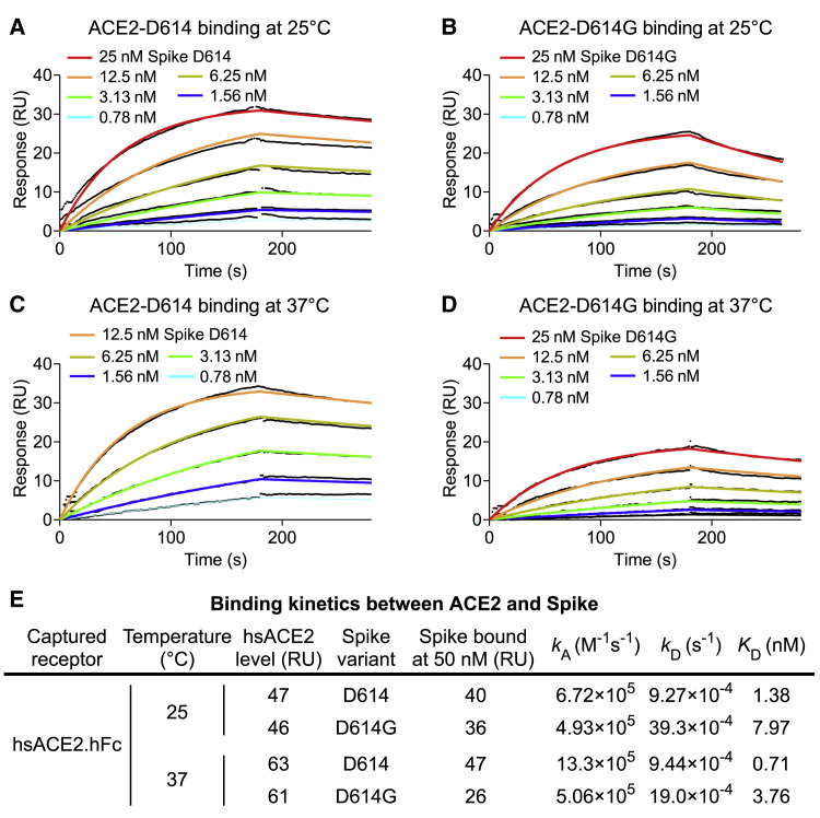
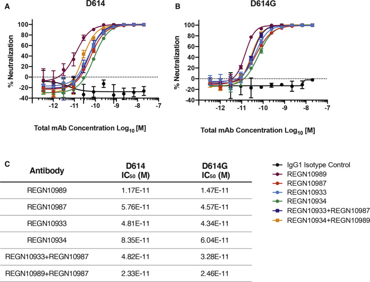
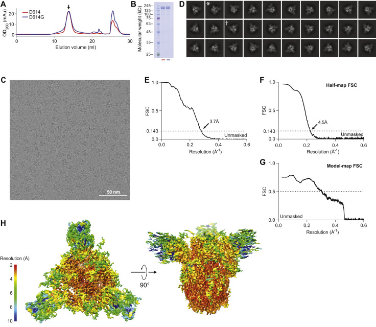
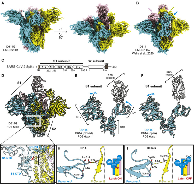
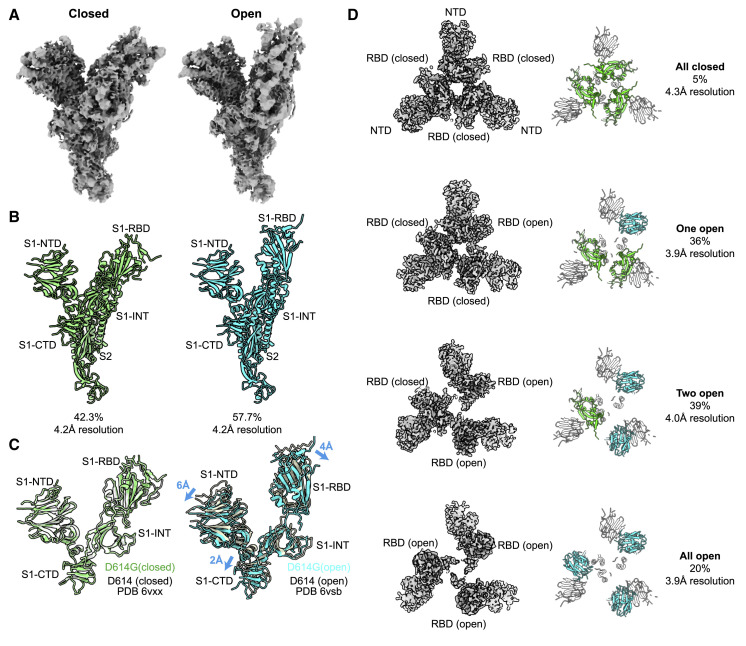
The SARS-CoV-2 spike (S) protein variant D614G supplanted the ancestral virus worldwide, reaching near fixation in a matter of months. Here we show that D614G was more infectious than the ancestral form on human lung cells, colon cells, and on cells rendered permissive by ectopic expression of human ACE2 or of ACE2 orthologs from various mammals, including Chinese rufous horseshoe bat and Malayan pangolin. D614G did not alter S protein synthesis, processing, or incorporation into SARS-CoV-2 particles, but D614G affinity for ACE2 was reduced due to a faster dissociation rate. Assessment of the S protein trimer by cryo-electron microscopy showed that D614G disrupts an interprotomer contact and that the conformation is shifted toward an ACE2 binding-competent state, which is modeled to be on pathway for virion membrane fusion with target cells. Consistent with this more open conformation, neutralization potency of antibodies targeting the S protein receptor-binding domain was not attenuated.

Keywords: ACE2; COVID-19; SARS-CoV-2; Spike protein; coronavirus; cryo-electron microscopy; infectivity; neutralizing antibody; pandemic.


Conflict of interest statement

Declaration of Interests P.C.S. is a co-founder and shareholder of Sherlock Biosciences and a board member and shareholder of Danaher Corporation. J.E.L. consulted for Sherlock Biosciences. C.A.K., K.E.P., and A.B. are employed by Regeneron Pharmaceuticals and own stock options in the company. C.A.K. is an officer at Regeneron. X.W., A.B., and N.D. are employees of Thermo Fisher Scientific.

Figures

None
Graphical abstract
Figure 1
Figure 1
The Frequency of the SARS-CoV-2 S Protein D614G Variant over the Course of the Pandemic Has Increased Nearly to Fixation (A) The global frequency of the S protein D614G variant over time in the GISAID SARS-CoV-2 database as of June 25, 2020. The filled blue plot represents a seven-day rolling average of the fraction of sequences bearing the D614G variant for each collection date. Dates without published sequences are linearly interpolated. The overlaid black line shows the cumulative frequency of D614G in sequences collected up to and including each date. (B) The frequency of the D614G variant over time (blue) in sequences collected from six continental regions, using the same dataset as in (A), plotted as a seven-day rolling average. The frequency of the last date with data is carried forward where recent dates lack data to indicate the most recent calculated frequency (light blue). Red bars show the number of sequences that were used to provide the denominator for calculating the frequency for each date.
Figure 2
Figure 2
SARS-CoV-2 D614G S Protein Variant Enhances Infectivity of Pseudotyped Lentiviruses in Cell Culture (A) Lentiviral virions bearing either GFP or Luciferase transgenes, and pseudotyped with either SARS-CoV-2 D614 or D614G S proteins, were produced by transfection of HEK293 cells and used to transduce human Calu3 lung cells, Caco2 colon cells, and either HEK293 or SupT1 cells stably expressing ACE2 and TMPRSS2. Relative infectivity of D614G versus D614, with D614 set at one, was determined based on flow cytometry for percent GFP positivity or on bulk luciferase activity. Each point represents the mean ± SD for transduction with lentiviral stocks derived from independent transfections, each value of which is the mean of three technical replicates. P values are the ratio paired t test (two-tailed). (B) Lentiviral virions bearing a luciferase transgene, pseudotyped with either SARS-CoV-2 D614 or D614G S proteins, were produced by transfection of HEK293 cells and used to transduce human HEK293 cells transiently transfected with plasmids encoding the indicated ACE2 orthologs. Relative infectivity of D614G versus D614, with D614 set at one, was determined based on bulk luciferase activity. Each point represents the mean ± SD after transduction by using lentiviral stock derived from an independent transfection, each of which is the mean of three technical replicates. P values are ratio paired t test (two-tailed).
Figure 3
Figure 3
Synthesis, Processing, and Incorporation of S Protein Variants into SARS-CoV-2 Virus-like Particles (A) Schematic showing how SARS-CoV-2 structural proteins were produced in HEK293T cells and virus-like particles were enriched from the supernatant by ultracentrifugation. (B) HEK293T cells were transfected with plasmids encoding the proteins indicated at the top. Total protein in cell lysates and in ultracentrifuge pellets from cell culture supernatant was normalized by Bradford assay, and then western blots were performed with the primary antibodies indicated on the left of the blots. Anti-Raptor antibody was used as a loading control for the cell lysate. Uncleaved S proteins, as well as the S1 and S2 cleavage products, are indicated on the right. Results here are representative of three independent rounds of transfection, ultracentrifugation, and westerns.
Figure 4
Figure 4
SARS-CoV-2 D614G S Protein Variant Binds ACE2 Weaker than the Ancestral Protein (A–D) SPR measurement of D614-ACE2 binding ([A] and [C]) and D614G-ACE2 binding ([B] and [D]) at 25°C ([A] and [B]) or 37°C ([C] and [D]). (E) Summary of kinetic parameters measured in (A)–(D). D614G binds ACE2 5-fold weaker than D614 at both temperatures tested.
Figure 5
Figure 5
Neutralization Potency of Monoclonal Antibodies Targeting the SARS-CoV-2 S Protein Receptor-Binding Domain Is Not Attenuated by D614G (A and B) Vero cells were challenged with pVSV-SARS-CoV-2-S-mNeon pseudoparticles encoding either D614 (A) or D614G (B) S protein variants in the presence of serial dilutions of the indicated human monoclonal antibodies targeting the SARS-CoV-2 S protein receptor-binding domain or IgG1 isotype control. mNeon protein fluorescence was measured 24 h post-infection as a readout for virus infectivity. Data are graphed as percent neutralization relative to virus only infection control. Data represent the mean ± SD of three technical replicates. (C) Neutralization potency (IC50) of individual monoclonals and of combinations of monoclonals, against the SARS-CoV-2 D614G and D614G S protein variants, as indicated.
Figure S1
Figure S1
Structural Determination of Spike D614G, Related to Figure 6 (A) Size exclusion column elution profiles of D614G (blue line) and D614 (red line). (B) Coomassie-stained SDS-PAGE of the peaks collected in (A) shows equivalent, full-length monomeric S proteins at around 180 kD. (C) A still frame from a raw micrograph movie. Individual particles can be clearly visualized. (D) 2D-clustering of trimeric D614G particles. Dagger denotes a top-view. Asterisk denotes a side-view. (E) Fourier shell correlation diagram for D614G (unmasked). (F) Half map Fourier shell correlation diagram (unmasked). (G) Model to map Fourier shell correlation diagram. (H) Local resolution of the ensemble map for Spike D614G.
Figure 6
Figure 6
Structural Determination of Spike D614G (A) D614G envelope from the three-dimensional reconstruction. EMDB: EMD-22301. (B) Published D614 envelope (EMD-21452). Arrows point to the density corresponding to the receptor-binding domains, which is missing in the corresponding positions in (A). (C) Domain arrangement of the SARS-CoV-2 S protein. (D) Atomic model for D614G without the receptor-binding domain. PDB: 6XS6. (E and F) Comparison of the D614G S1 subunit with the closed conformation (E) and open conformation (F) of the D614 S1 subunit. Arrows indicate the relative movement of the S1 subunit of D614G. (G) Position of amino acid 614 on the S protein. (H and I) Substitution of Asp614 with glycine changes hydrogen bonding around residue 614. In the case of D614 (H), an inter-protomer hydrogen bond is detected. For D614G (I), the Asp614-Thr859 hydrogen bond is eliminated, and interaction with intradomain Ala647 is strengthened. See also Figures S1 and S2.
Figure S2
Figure S2
Structural Determination of Spike D614G, Related to Figure 6 (A) Representative density map detail of an alpha helix region. (B) Representative density map detail of a loop region. (C) Representative density map detail of a beta strand region. Note: the relative positioning between (B) and (C) is the same as in the real structural model. No cryo-EM density is observed in between the loop where Gly614 resides and the beta strand where Thr859 resides.
Figure 7
Figure 7
D614G Populates More Open Conformations Than Does the Ancestral S Protein (A) Cryo-EM density maps of the two conformations of D614G protomer. The first is a closed conformation with a buried RBD. The second is an open conformation with the RBD standing up. (B) Atomic models for the closed (left) and open (right) conformations for the two D614G protomers shown in (A). (C) Comparison of the two D614G protomer S1 subunit conformations with the corresponding conformations of the D614 protomer S1 subunit. (D) The D614G S protein trimer adopts four conformations. In addition to the all-closed and one open conformation detected with the D614 S protein trimer, the D614G S protein trimer adopts two-open and three-open conformations.

Update of

Comment in

References

    1. Andersen K.G., Rambaut A., Lipkin W.I., Holmes E.C., Garry R.F. The proximal origin of SARS-CoV-2. Nat. Med. 2020;26:450–452. - PMC - PubMed
    1. Baum A., Fulton B.O., Wloga E., Copin R., Pascal K.E., Russo V., Giordano S., Lanza K., Negron N., Ni M. Antibody cocktail to SARS-CoV-2 spike protein prevents rapid mutational escape seen with individual antibodies. Science. 2020;369:1014–1018. - PMC - PubMed
    1. Boni M.F., Lemey P., Jiang X., Lam T.T.-Y., Perry B.W., Castoe T.A., Rambaut A., Robertson D.L. Evolutionary origins of the SARS-CoV-2 sarbecovirus lineage responsible for the COVID-19 pandemic. Nat. Microbiol. 2020;2020:28. doi: 10.1038/s41564-020-0771-4. - DOI - PubMed
    1. Bos E.C., Luytjes W., van der Meulen H.V., Koerten H.K., Spaan W.J. The production of recombinant infectious DI-particles of a murine coronavirus in the absence of helper virus. Virology. 1996;218:52–60. - PMC - PubMed
    1. Cingolani P., Platts A., Wang L., Coon M., Nguyen T., Wang L., Land S.J., Lu X., Ruden D.M. A program for annotating and predicting the effects of single nucleotide polymorphisms, SnpEff: SNPs in the genome of Drosophila melanogaster strain w1118; iso-2; iso-3. Fly (Austin) 2012;6:80–92. - PMC - PubMed

Publication types

MeSH terms

LinkOut - more resources