. 2020 Sep 27;12(10):2954.
doi: 10.3390/nu12102954.

Quercetin Alleviates Ferroptosis of Pancreatic β Cells in Type 2 Diabetes

Affiliations

Quercetin Alleviates Ferroptosis of Pancreatic β Cells in Type 2 Diabetes

Dan Li et al. Nutrients. .

Abstract

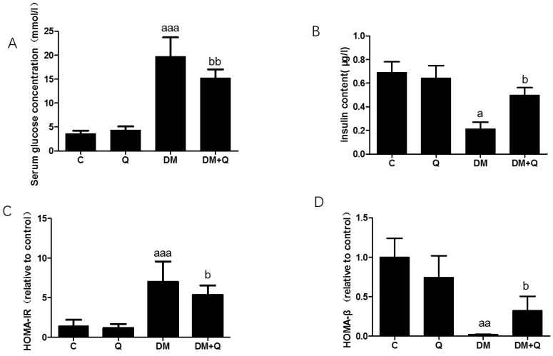
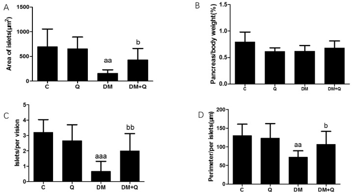
(1) Background: Pancreatic iron deposition has been found in the progression of type 2 diabetes (T2DM); however, whether ferroptosis contributes to the dysfunction of pancreatic β cells (PBC) remains enigmatic. Moreover, the potential protective effect of quercetin is also elusive; (2) Methods: T2DM mice model was established by multiple low dose streptozocin (STZ) injection, after which quercetin was intervened for 4 months; (3) Results: Substantially normalized glucose tolerance, diabetic symptoms, homeostasis model assessment for insulin resistance (HOMA-IR), and homeostasis model assessment for β cell (HOMA-β) index in comparison with the findings of T2DM control. Distorted pancreatic islets and especially shrunken mitochondria with cristae loss in PBC were observed in T2DM mice, which was ameliorated by quercetin. Meanwhile, quercetin lowered the iron level particularly in the islet in T2DM mice. In spite of compensatory xCT up-regulation, T2DM molding depleted glutathione (GSH), down-regulated glutathione peroxidase 4 (GPX4), and induced oxidative stress in pancreatic tissue, which was abolished partially by quercetin. More importantly, insulin secretion was worsened by ferroptosis-inducing erastin or RAS-selective lethal compounds 3 (RSL-3). Quercetin, ferroptosis inhibitor ferrostatin-1 and iron-chelating deferoxamine, rescued cell viability when cells were challenged with high-glucose; (4) Conclusions: Our findings identify that ferroptosis contributes to the PBC loss and dysfunction. Quercetin exerts beneficial effects on T2DM potentially by inhibiting pancreatic iron deposition and PBC ferroptosis, highlighting promising control strategies of T2DM by quercetin.

Keywords: ferroptosis; iron overload; lipid peroxidation; quercetin; type 2 diabetes mellitus.


Conflict of interest statement

The authors declare no conflict of interest.

Figures

Figure 1
Figure 1
Induction of diabetes and measurement of pancreas function following high-fat feeding and streptozotocin injection and quercetin attenuates symptoms of T2DM in mice. (A). serum glucose concentration was measured by standard commercial assays kits. (mmol/L) (n = 10); (B). Insulin levels were determined by mouse-specific ELISA kits (n = 10); (μg/L); (C). The homeostasis model assessment of insulin resistance (HOMA-IR) = fasting insulin × fasting blood glucose/22.5 (n = 10); (D). The homeostasis model assessment of β cell function (HOMA-β) = 20 × fasting insulin/(fasting blood glucose − 3.5 mmol/L) (n = 10). Data were expressed as mean ± SD (a: p < 0.05 vs. C; aa: p < 0.01 vs. C; aaa: p < 0.001 vs. C; b: p < 0.05 vs. DM; bb: p < 0.01 vs. DM). T2DM, type 2 diabetes; DM, Diabetes mellitus; C, control group; Q, quercetin group.
Figure 2
Figure 2
Morphological changes of islets in mice. (A). Fixed pancreatic tissue sections of mice stained with hematoxylin and eosin were observed by light microscope (image magnification: 400×) (n = 3); (B). Representative images of pancreatic sections immunohistochemically stained for insulin (dark brown) (image magnification: 400×) (n = 3); (C). Quantification of insulin level and distribution in islets were detected by immunohistochemically staining in pancreatic tissue (n = 3); Data were expressed as mean ± SD (a: p < 0.05 vs. C; b: p < 0.05 vs. DM).
Figure 3
Figure 3
Morphological analysis of islets in mice. (A). Area of islets calculated by Mingmei camera at magnification: 200× (μm2) (n = 3); (B). Pancreas/body weight (%) (n = 10); (C). Numbers of islets/per vision calculated by Mingmei camera at magnification: 200× (n = 3); (D). Perimeter/per islets calculated by Mingmei camera at magnification: 200× (μm) (n = 3). Data were expressed as mean ± SD (aa: p < 0.01 vs. C; aaa: p < 0.001 vs. C; b: p < 0.05 vs. DM; bb: p < 0.05 vs. DM).
Figure 4
Figure 4
Effect of quercetin on iron levels in the pancreas of mice. (A). Serum iron level was measured by colorimetric kit according to the manufacturer’s instructions (mg/L) (n = 10); (B). Western blot analysis was performed to measure ferritin light chain (FTL) and quantified by Image-Pro Plus 6.0 software. Blotting with β-actin was used as a protein loading control (n = 3); (C). Representative images of pancreatic sections immunohistochemically stained for the ferritin light chain. (dark brown) (n = 3). (image magnification: 400×); (D). Quantification of the ferritin light chain level in islets detected by immunohistochemically staining in pancreatic tissue (n = 3). Data were expressed as mean ± SD. (a: p < 0.05 vs. C; aa: p < 0.01 vs. C; b: p < 0.05 vs. DM).
Figure 5
Figure 5
Effect of quercetin on improving the phenotype of ferroptosis. (A). Glutathione (GSH) content was measured using the homogenates of the pancreas in the light of instructions of reagent kits (n = 10); (B). Malondialdehyde(MDA) level was measured using the homogenates of the pancreas according to reagent kits (n = 10); (C). Superoxide dismutase (SOD) activity was measured using the homogenates of the pancreas according to the manufacturer’s instructions (n = 10); (D). Reactive oxygen species (ROS) level was determined by Dihydroethidium (DHE) probe; (E). Mitochondria (red arrows indicated) were observed from transmission electron microscope (TEM) images (5000×) showing representative mitochondria structure of β cells; (F). Western blot analysis was performed to measure xCT, GPX4, and VAC2 and quantified by Image-Pro Plus 6.0 software. Blotting with β-actin was used as a protein loading control. Data were expressed as mean ± SD (a: p < 0.05 vs. C; aaa: p < 0.001 vs. C; b: p < 0.05 vs. DM bb: p < 0.01 vs. DM; bbb: p < 0.001 vs. DM).
Figure 6
Figure 6
Quercetin alleviates ferroptosis-related phenotype in INS-1 cells induced by high glucose. (A). Cell viability was measured in INS-1 cells treated for 48 h with high glucose (33.3 mmol/L), quercetin (50 μM), Fer-1 (2 μM), Z-VAD-FMK (10 μM), Nec-1 (10 μM), deferoxamine (DFO) (25 μM); (B). GSH level was determined in INS-1 cells treated for 48 h with high glucose (33.3 mmol/L), quercetin (50 μM). (C). Ptgs2 mRNA level was determined in INS-1 cells treated for 48 h with high glucose (33.3 mmol/L), quercetin (50 μM), and normalized to β-actin mRNA; (D). Fluorescence micrographs of INS-1 labeled for 30 min with 1 μM C11-BODIPY581/591 (n = 3); (E). Quantification of fluorescence intensity calculated by dividing the green image by the sum of the green and red images (n = 3); (F). Glucose stimulated insulin secretion when treated for 48 h with erastin (1 μM) and RSL-3(1 μM). Data were expressed as mean ± SEM (a: p < 0.05 vs. C; aa: p < 0.01 vs. C; b: p < 0.05 vs. high glucose (HG)).

References

    1. International Diabetes Federation . IDF Diabetes Atlas. 9th ed. International Diabetes Federation; Brussels, Belgium: 2019.
    1. Roden M., Shulman G.I. The integrative biology of type 2 diabetes. Nature. 2019;576:51–60. doi: 10.1038/s41586-019-1797-8. - DOI - PubMed
    1. Fernández-Real J.M., McClain D., Manco M. Mechanisms Linking Glucose Homeostasis and Iron Metabolism Toward the Onset and Progression of Type 2 Diabetes. Diabetes Care. 2015;38:2169–2176. doi: 10.2337/dc14-3082. - DOI - PubMed
    1. Van Haeften T.W. Early disturbances in insulin secretion in the development of type 2 diabetes mellitus. Mol. Cell. Endocrinol. 2002;197:197–204. doi: 10.1016/S0303-7207(02)00267-8. - DOI - PubMed
    1. Marselli L., Suleiman M., Masini M., Campani D., Bugliani M., Syed F., Martino L., Focosi D., Scatena F., Olimpico F., et al. Are we overestimating the loss of beta cells in type 2 diabetes? Diabetologia. 2014;57:362–365. doi: 10.1007/s00125-013-3098-3. - DOI - PubMed

MeSH terms

LinkOut - more resources