. 2020 Sep 25;12(10):1075.
doi: 10.3390/v12101075.

Binding Mechanism Elucidation of the Acute Respiratory Disease Causing Agent Adenovirus of Serotype 7 to Desmoglein-2

Affiliations

Binding Mechanism Elucidation of the Acute Respiratory Disease Causing Agent Adenovirus of Serotype 7 to Desmoglein-2

Marc-André Hograindleur et al. Viruses. .

Abstract

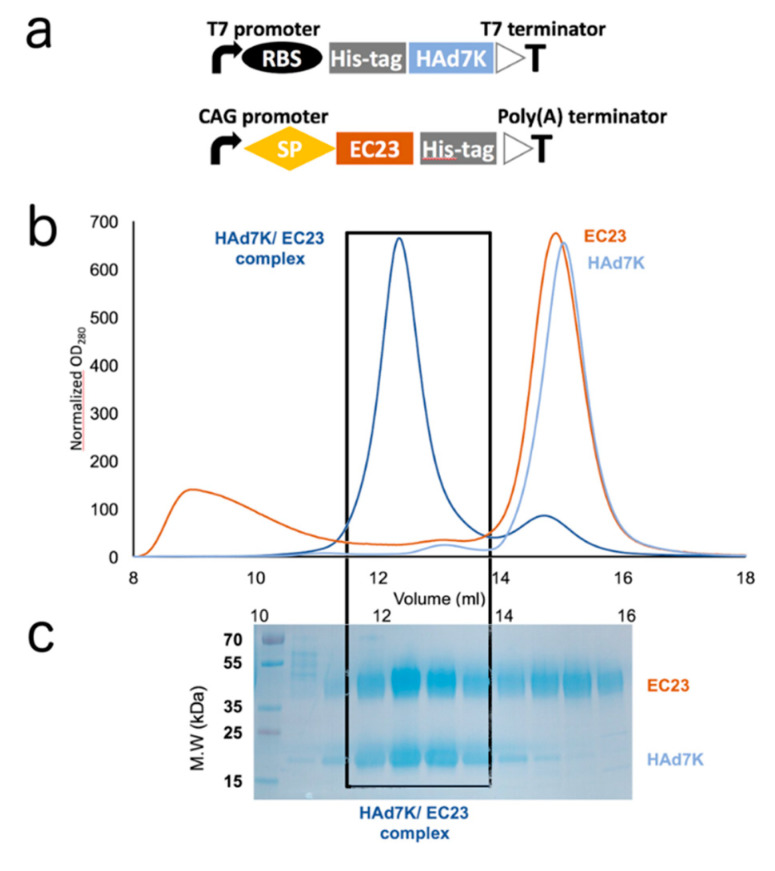
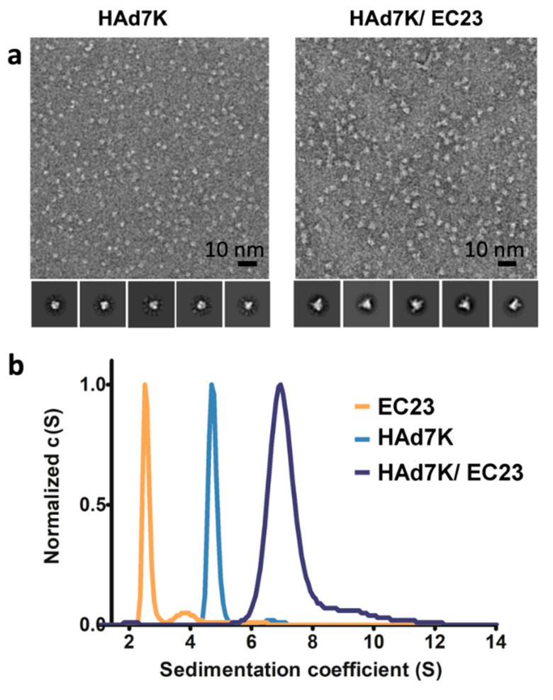
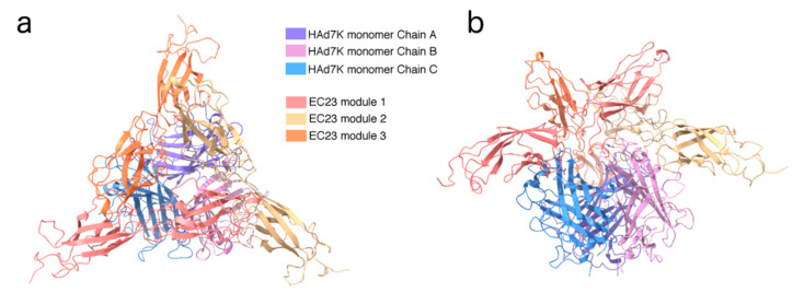
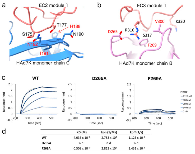
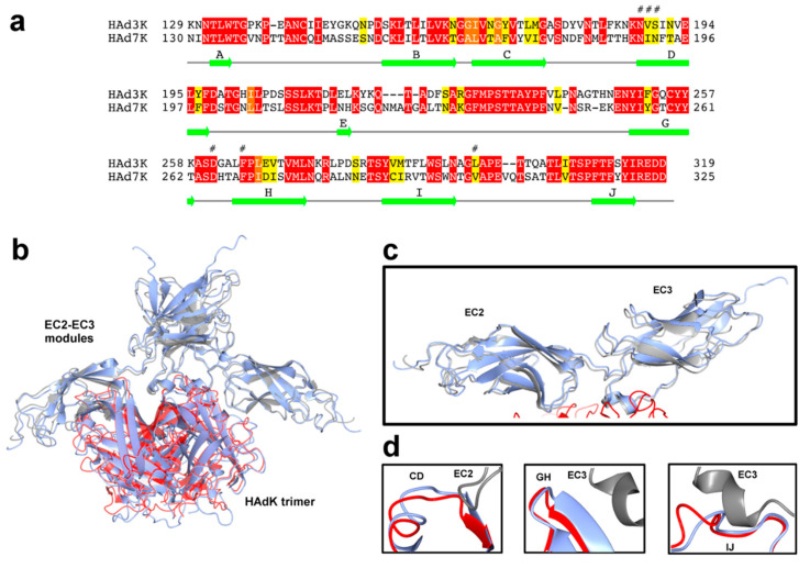
The study of viruses causing acute respiratory distress syndromes (ARDS) is more essential than ever at a time when a virus can create a global pandemic in a matter of weeks. Among human adenoviruses, adenovirus of serotype 7 (HAdV7) is one of the most virulent serotypes. This virus regularly re-emerges in Asia and has just been the cause of several deaths in the United States. A critical step of the virus life cycle is the attachment of the knob domain of the fiber (HAd7K) to the cellular receptor desmoglein-2 (DSG2). Complexes between the fiber knob and two extracellular domains of DSG2 have been produced. Their characterization by biochemical and biophysical methods show that these two domains are sufficient for the interaction and that the trimeric HAd7K could accommodate up to three DSG2 receptor molecules. The cryo-electron microscopy (cryo-EM) structure of these complexes at 3.1 Å resolution confirmed the biochemical data, and allowed the identification of the critical amino acid residues for this interaction, which shows similarities with other DSG2 interacting adenoviruses, despite a low homology in the primary sequences.

Keywords: acute respiratory distress syndromes; adenoviruses; cryo-electron microscopy; desmoglein-2; virus-host interactions.


Conflict of interest statement

The authors declare no conflict of interest.

Figures

Figure 1
Figure 1
(a) Schematic representation of human adenoviruses, adenovirus of serotype 7 (HAd7K) pETDuet™-1 vector and EC23 pHL-sec vector constructs. RBS: ribosome binding site. SP: signal peptide; (b) elution profiles from size exclusion chromatography. HAd7K (light blue curve) and EC23 (red curve) were either loaded separately or co-incubated overnight (dark blue curve) before injection to the column. Complex is depicted by a box. (c) SDS-PAGE analysis of the eluted fractions.
Figure 2
Figure 2
(a) Negative-staining electron microscopy analysis. Left: electron micrograph of HAd7K. Right: electron micrograph of HAd7K in complex with EC23. Representative class averages are shown on the galleries below each micrograph. (b) Analytical ultracentrifugation (AUC) analysis. Normalized sedimentation coefficient distribution profiles obtained from sedimentation velocity experiments for HAd7K (in blue), EC23 (in orange) and for HAd7K in complex with EC23 (in grey).
Figure 3
Figure 3
Structure of HAd7K-(EC23)3 viewed along the 3-fold axis (a) or from the side (b). Each monomer is colored differently according to the legend.
Figure 4
Figure 4
(a,b) Zoomed views of the EC2 (module 1)–HAd7K monomer chain C (a) and EC3 (module 1)–HAd7K monomer chain B (b) interactions. Side chains of key residues are represented and their names are indicated. The residues in red were found by mutagenesis to reduce by more than 80% the binding of EC23 to HAd3K. (c,d) Binding analysis of WT and mutants (D265A and F269A) HAd7K to the cellular receptor desmoglein-2 (DSG2) using bio-layer interferometry (BLI). WT or mutants His HAd7K were immobilized on a Ni-NTA biosensor, and different concentrations of DSG2 were applied in the mobile phase. The dotted lines, representing fits of the raw data (solid lines), were used to obtain equilibrium dissociation constant (KD), association (kon), and dissociation (koff) values.
Figure 5
Figure 5
(a) Pairwise Sequence Alignment is used to identify regions of similarity between HAdV7 and HAdV3 fiber. For each pair of sequences the best global alignment is found using BLOSUM62 as the scoring matrix. A conservation index between 0 and 11 is calculated for each amino acid position on the alignment. Amino acids with a score above 9 are colored from red→orange→yellow corresponding to sequence conservation from high to low. (b) Superposition of HAd7K-(EC23)2 and HAd3K-(EC23)2: EC2-EC3 modules interacting with HAd7K (in red) are colored in grey. HAd3K-(EC23)2 complex is colored in blue. (c,d) Close-up views of superposed EC2-EC3 modules and superposed CD, GH, and IJ loops of HAdK, are shown individually.

References

    1. Human Adenovirus Working Group. [(accessed on 23 April 2020)]; Available online: http://hadvwg.gmu.edu/
    1. Lion T. Adenovirus Infections in Immunocompetent and Immunocompromised Patients. Clin. Microbiol. Rev. 2014;27:441–462. doi: 10.1128/CMR.00116-13. - DOI - PMC - PubMed
    1. Chen X., Lv J., Qin L., Zou C., Tang L. Severe Adenovirus Pneumonia Requiring Extracorporeal Membrane Oxygenation Support in Immunocompetent Children. Front. Pediatr. 2020;8:162. doi: 10.3389/fped.2020.00162. - DOI - PMC - PubMed
    1. Kim Y.-J., Hong J.-Y., Lee H.-J., Shin S.-H., Kim Y.K., Inada T., Hashido M., Piedra P.A. Genome Type Analysis of Adenovirus Types 3 and 7 Isolated during Successive Outbreaks of Lower Respiratory Tract Infections in Children. J. Clin. Microbiol. 2003;41:4594–4599. doi: 10.1128/JCM.41.10.4594-4599.2003. - DOI - PMC - PubMed
    1. Kajon A.E., Lynch J.P. Adenovirus: Epidemiology, Global Spread of Novel Serotypes, and Advances in Treatment and Prevention. Semin. Respir. Crit. Care Med. 2016;37:586–602. doi: 10.1055/s-0036-1584923. - DOI - PMC - PubMed

Publication types

MeSH terms

LinkOut - more resources