Review
. 2020 Sep 25;12(10):2760.
doi: 10.3390/cancers12102760.

Cancer and pH Dynamics: Transcriptional Regulation, Proteostasis, and the Need for New Molecular Tools

Affiliations
Review

Cancer and pH Dynamics: Transcriptional Regulation, Proteostasis, and the Need for New Molecular Tools

Brandon J Czowski et al. Cancers (Basel). .

Abstract

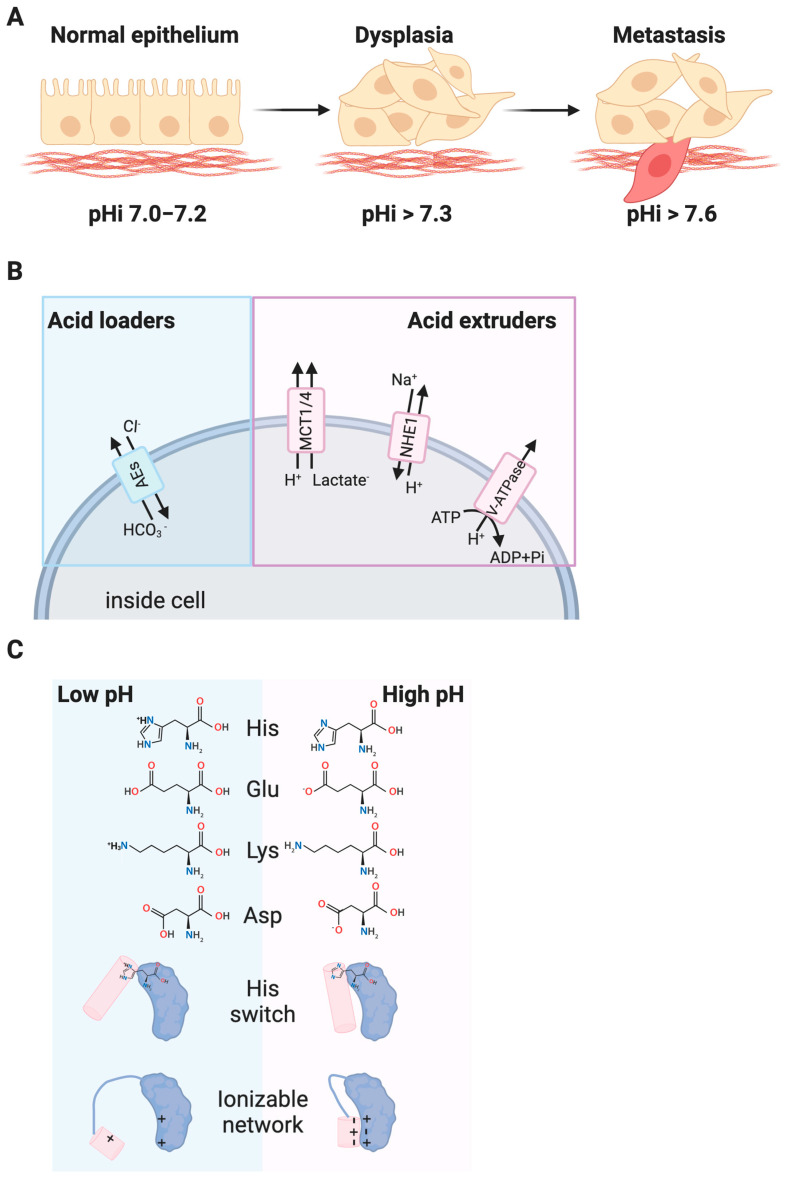
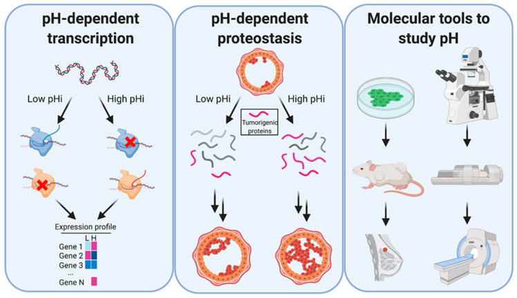
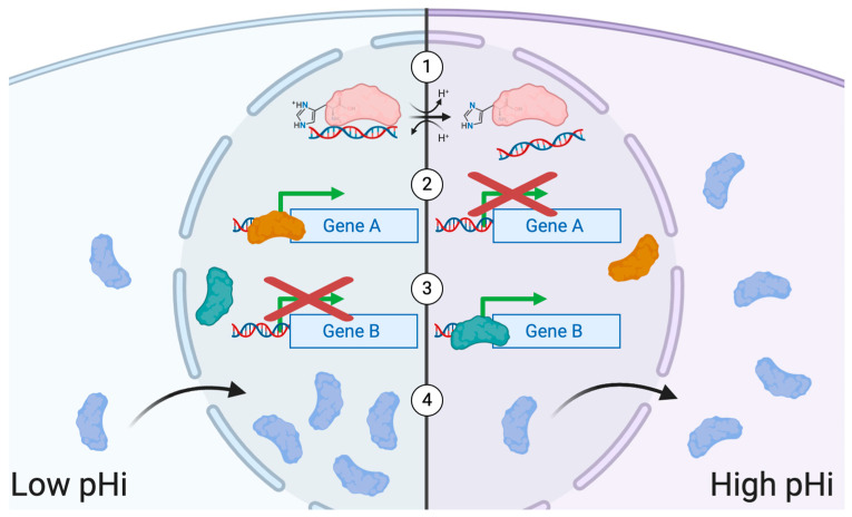
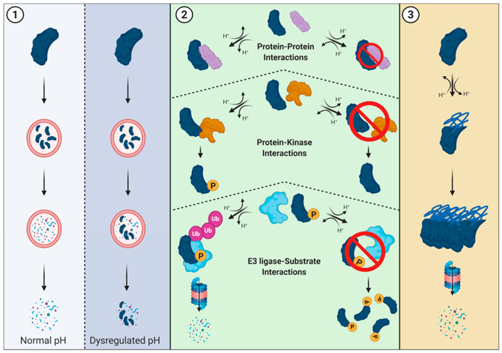
An emerging hallmark of cancer cells is dysregulated pH dynamics. Recent work has suggested that dysregulated intracellular pH (pHi) dynamics enable diverse cancer cellular behaviors at the population level, including cell proliferation, cell migration and metastasis, evasion of apoptosis, and metabolic adaptation. However, the molecular mechanisms driving pH-dependent cancer-associated cell behaviors are largely unknown. In this review article, we explore recent literature suggesting pHi dynamics may play a causative role in regulating or reinforcing tumorigenic transcriptional and proteostatic changes at the molecular level, and discuss outcomes on tumorigenesis and tumor heterogeneity. Most of the data we discuss are population-level analyses; lack of single-cell data is driven by a lack of tools to experimentally change pHi with spatiotemporal control. Data is also sparse on how pHi dynamics play out in complex in vivo microenvironments. To address this need, at the end of this review, we cover recent advances for live-cell pHi measurement at single-cell resolution. We also discuss the essential role for tool development in revealing mechanisms by which pHi dynamics drive tumor initiation, progression, and metastasis.

Keywords: biosensors; cancer; molecular tools; pH regulation; proteostasis; transcription; tumorigenesis.


Conflict of interest statement

The authors declare no direct or perceived conflict of interest.

Figures

Figure 1
Figure 1
Increased intracellular pH (pHi) in cancer. (A) Normal epithelial cells have an intracellular pH (pHi) of 7.0–7.2 while pHi is constitutively increased in dysplastic and metastatic cancer cells. (B) Dysregulation of acid loaders (anion exchangers (AE1)) and acid extruders (the sodium proton exchanger (NHE1), monocarboxylate transporters (MCTs), and plasma-membrane resident vacuolar ATPases (V-ATPases) have been linked to the dysregulated pHi in cancer. (C) Cellular pH dynamics affect R-group titration of key residues like histidine (His, pKa 6.5) and residues like aspartate (Asp), glutamate (Glu), and lysine (Lys) that can have up- or downshifted pKas depending on protein environment. Changes in protonation of single residues, or networks of ionizable residues can alter protein structure and function.
Figure 2
Figure 2
Dysregulated pHi dynamics in cancer can regulate transcription and proteostasis, but better tools are needed to study molecular effects. Left: Altered pH dynamics can affect transcript abundance, where gene 1 transcription is activated by high pHi, gene 2 transcription is activated by low pH, and gene 3 is unaffected. Middle: Altered pH dynamics can also play a role in stabilizing tumorigenic proteins, enabling cancer establishment or progression. Right: Currently, tools exist that allow researchers to study pHi dynamics in living cells by optical microscopy and magnetic resonance imaging (MRI). However, experiments are limited by the constraints of the individual tools, and better tools are required to study the role of pHi dynamics at the cellular, tissue, and organismal level.
Figure 3
Figure 3
Roles for pHi dynamics in regulating transcription. (1) The protonation states of charge-bearing residues can affect the ability of transcription factors to bind DNA under different intracellular pH conditions. (2) Transcription factors that actively promote transcription of certain genes at low pHi can lose their ability to promote gene expression at high pHi. (3) Conversely, transcription factors that do not promote the transcription of certain genes at low pHi may gain the ability to promote gene expression at high pHi. (4) Proteins may exhibit pH-dependent subcellular localization. In the case of transcription factors, moving from the cytoplasm to the nucleus (or vice versa) depending on pHi.
Figure 4
Figure 4
Roles for pH dynamics in regulating proteostasis. (1) Lysosomal-mediated degradation. Lysosomes have an acidic pH (~4.5–5.0) that allows for optimal protease activity and protein degradation. However, dysregulation of pH within the cytosol (and subsequent lysosomal pH changes) can result in failed or partial degradation. (2) Overview of protein-protein interactions required for proteasome-mediated degradation. If pH-sensitive residues play critical roles in protein interaction interfaces, dysregulated pH may prevent (or enhance) an interaction from occurring. When the pH-dependent interaction occurs between a kinase and substrate, dysregulated pH may alter phosphorylation, and potential downstream signaling for protein-kinase interactions. Similarly, when the pH-dependent interaction occurs between an E3 ubiquitin ligase and its substrate, dysregulated pH may alter ubiquitination and protein degradation. (3) Aggregate clearance is the process of removing proteins that have aggregated. Since pH dynamics can also alter the three-dimensional structure of proteins and cause complete misfolding or localized disorder, pH-dependent aggregation is another potential way pH regulates proteostasis.

References

    1. Greaves M., Maley C.C. Clonal evolution in cancer. Nature. 2012;481:306–313. doi: 10.1038/nature10762. - DOI - PMC - PubMed
    1. Gillies R.J., Verduzco D., Gatenby R.A. Evolutionary dynamics of carcinogenesis and why targeted therapy does not work. Nat. Rev. Cancer. 2012;12:487–493. doi: 10.1038/nrc3298. - DOI - PMC - PubMed
    1. Ramachandran S., Ient J., Gottgens E.L., Krieg A.J., Hammond E.M. Epigenetic Therapy for Solid Tumors: Highlighting the Impact of Tumor Hypoxia. Genes. 2015;6:935–956. doi: 10.3390/genes6040935. - DOI - PMC - PubMed
    1. Cairns R.A., Harris I.S., Mak T.W. Regulation of cancer cell metabolism. Nat. Rev. Cancer. 2011;11:85–95. doi: 10.1038/nrc2981. - DOI - PubMed
    1. Brahimi-Horn M.C., Bellot G., Pouyssegur J. Hypoxia and energetic tumour metabolism. Curr. Opin. Genet. Dev. 2011;21:67–72. doi: 10.1016/j.gde.2010.10.006. - DOI - PubMed

LinkOut - more resources