Review
. 2020 Sep 25;12(10):1078.
doi: 10.3390/v12101078.

Twelve Children with Varicella Vaccine Meningitis: Neuropathogenesis of Reactivated Live Attenuated Varicella Vaccine Virus

Affiliations
Review

Twelve Children with Varicella Vaccine Meningitis: Neuropathogenesis of Reactivated Live Attenuated Varicella Vaccine Virus

Ethan H Heusel et al. Viruses. .

Abstract

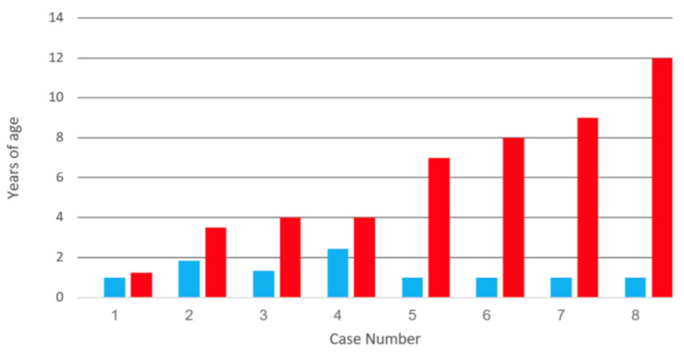
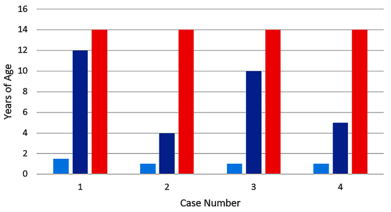
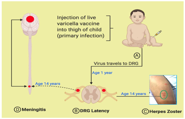
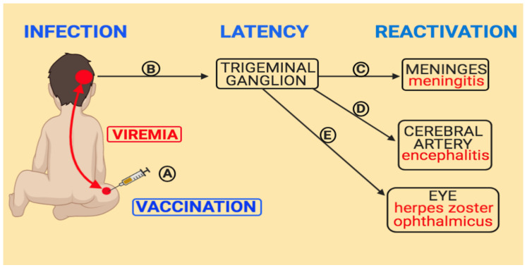
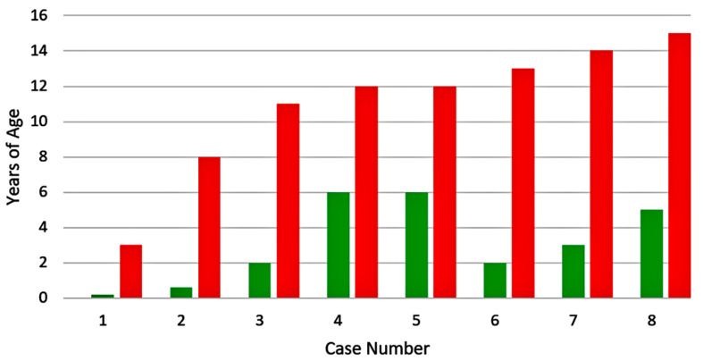
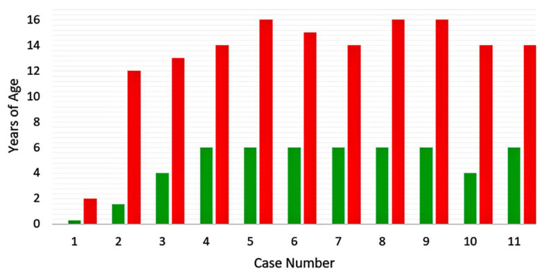
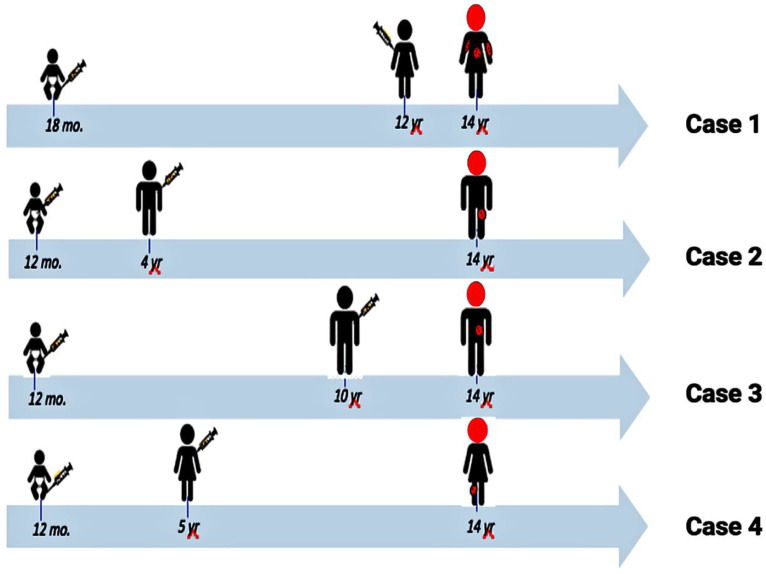
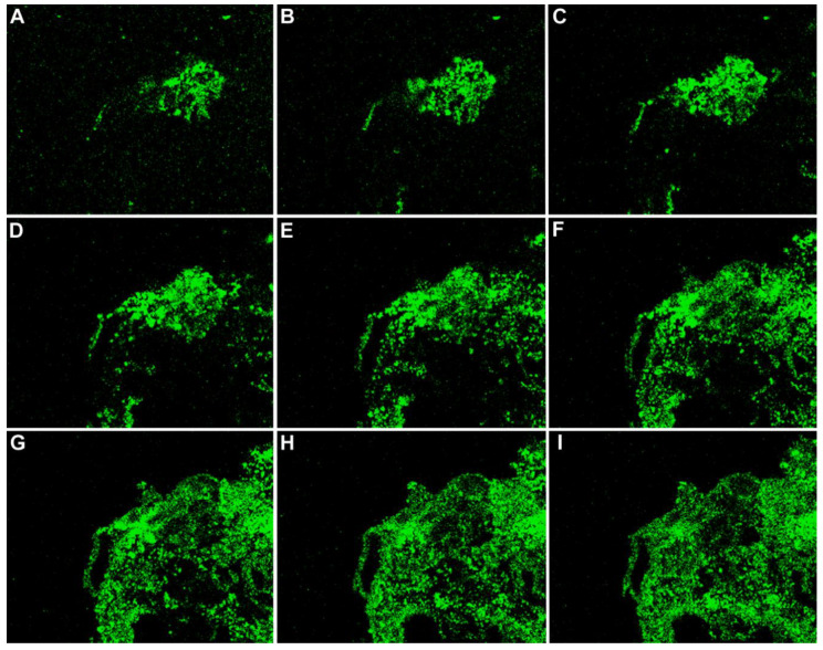
Varicella vaccine is a live attenuated varicella-zoster virus (VZV). Like its parental strain called VZV pOka, the vaccine virus vOka retains some neurotropic properties. To better understand vOka neuropathogenesis, we reassessed 12 published cases of vOka meningitis that occurred in once-immunized and twice-immunized children, all of whom had bouts of herpes zoster preceding the central nervous system infection. Eight of the 12 meningitis cases occurred in children who had received only one immunization. There was no pattern to the time interval between varicella vaccination and the onset of herpes zoster with meningitis. Four of the meningitis cases occurred in children who had received two immunizations. Since all four children were 14 years old when meningitis was diagnosed, there was a strong pattern to the interval between the first vaccination at age 1 year and onset of meningitis, namely, 13 years. Knowledge of pathogenesis requires knowledge of the location of herpes zoster; the majority of dermatomal rashes occurred at sites of primary immunization on the arm or thigh, while herpes zoster ophthalmicus was uncommon. Based on this literature review, currently there is no consensus as to the cause of varicella vaccine meningitis in twice-immunized children.

Keywords: RNA polymerase III; VZV gC; herpes vaccine; herpes zoster; latency; trigeminal ganglion; varicella-zoster virus; viral meningitis.


Conflict of interest statement

The authors declare no potential conflict of interest with respect to the research, authorship, and/or publication of this article.

Figures

Figure 1
Figure 1
Duration between varicella vaccination and varicella vaccine meningitis in eight children. All children had received one varicella vaccination, after which they later developed herpes zoster and meningitis. Data from references [8,12,13]. Note the absence of pattern to the time intervals between vaccination and meningitis. Age of varicella vaccination: blue bar; age of meningitis: red bar.
Figure 2
Figure 2
Duration between varicella vaccination and varicella vaccine meningitis in four children. All children had received two varicella vaccinations. All four children developed herpes zoster and meningitis 13 years after their first vaccination. Data from references [1,10,11]. Age of first varicella vaccination: light blue bar; age of second vaccination: dark blue bar; age of meningitis: red bar.
Figure 3
Figure 3
Varicella antibody in the cerebrospinal fluid. Varicella antibody was detected by a fluorescent antibody to membrane antigen test. The test was carried out by confocal microscopy imaging of varicella-zoster virus (VZV) infected monolayers. Two confocal images are shown (A and B). Clusters of nuclei (Nu; blue circles) within the cytoplasm (Cy) of an infected cell are encircled by a dashed white line. Arrows point to positive fluorescent antibody attaching to VZV antigens in the plasma membrane. The majority of the positivity resides in the plasma membranes of the infected cells because that is where the majority of viral glycoproteins are located. One viral target is VZV glycoprotein gE.
Figure 4
Figure 4
Pathogenesis of varicella meningitis in an immunized child. After a varicella vaccination, the virus is transported via sensory nerves from the skin (A) to the lumbar dorsal root ganglion (DRG) (B). When the virus reactivates from the DRG, the virus is carried to the lumbar dermatome in the thigh (C). The rash in Panel C is the herpes zoster rash of case #4 in Table 2. The virus is also carried into the spinal cord, where meningitis ensues (D).
Figure 5
Figure 5
Pathogenesis of varicella vaccine meningitis following herpes zoster ophthalmicus. Following varicella vaccination of about 50% of young children, virus is carried within lymphocytes throughout the body (A). Some virus enters and become latent in the trigeminal ganglion (B). When this virus reactivates, it can be carried via afferent fibers to the meninges (C), cerebral arteries (D) and eye (herpes zoster ophthalmicus (E).
Figure 6
Figure 6
Eight cases of varicella meningitis caused by wild type virus. Each child had herpes zoster prior to developing meningitis. Data from references [36,37]. Age of wild type varicella: green bar; age of meningitis: red bar.
Figure 7
Figure 7
Eleven cases of varicella meningitis caused by wild type virus. None of the children had herpes zoster prior to developing meningitis. Data from reference [38]. Age of wild type varicella: green bar; age of meningitis: red bar.
Figure 8
Figure 8
Similarities among the four twice-immunized adolescents who developed varicella vaccine meningitis. All four were given their first varicella vaccination between 12–18 months of age. All four manifested herpes zoster with meningitis at the age of 14 years (heads colored red). Thus, there was a pattern between time of first varicella vaccination and onset of meningitis (~13 years). Data from reference [1].
Figure 9
Figure 9
Vesicle of wild type herpes zoster. The VZV glycoprotein gC is a component in the envelope of a VZ virion; every vesicle is filled with virions. The protein is immunolabeled green by a monoclonal antibody probe. The nine micrographs show images of the abundant VZV gC protein from serial sections cut through a herpes zoster vesicle from an edge to the center (AI). Magnification = 200×.

References

    1. Ramachandran V., Elliott S.C., Rogers K.L., Cohrs R.J., Weinberger M., Jackson W., Carpenter J.E., Grose C., Bonthius D.J. Varicella Vaccine Meningitis as a Complication of Herpes Zoster in Twice-Immunized Immunocompetent Adolescents. J. Child Neurol. 2020:883073820938597. doi: 10.1177/0883073820938597. - DOI - PMC - PubMed
    1. Gershon A. Varicella vaccine: Its past, present and future. Pediatr. Infect. Dis. J. 1995;14:742–744. doi: 10.1097/00006454-199509000-00003. - DOI - PubMed
    1. Takahashi M. Effectiveness of live varicella vaccine. Expert Opin. Biol. Ther. 2004;4:199–216. doi: 10.1517/14712598.4.2.199. - DOI - PubMed
    1. Varela F.H., Pinto L.A., Scotta M.C. Global impact of varicella vaccination programs. Hum. Vaccines Immunother. 2018;15:645–657. doi: 10.1080/21645515.2018.1546525. - DOI - PMC - PubMed
    1. Grose C. Varicella vaccination of children in the United States: Assessment after the first decade 1995–2005. J. Clin. Virol. 2005;33:89–95. doi: 10.1016/j.jcv.2005.02.003. - DOI - PubMed

Publication types

LinkOut - more resources