Review
. 2020 Sep 25;12(10):2765.
doi: 10.3390/cancers12102765.

Three-Dimensional Cell Culture Systems in Radiopharmaceutical Cancer Research

Affiliations
Review

Three-Dimensional Cell Culture Systems in Radiopharmaceutical Cancer Research

Alina Doctor et al. Cancers (Basel). .

Abstract

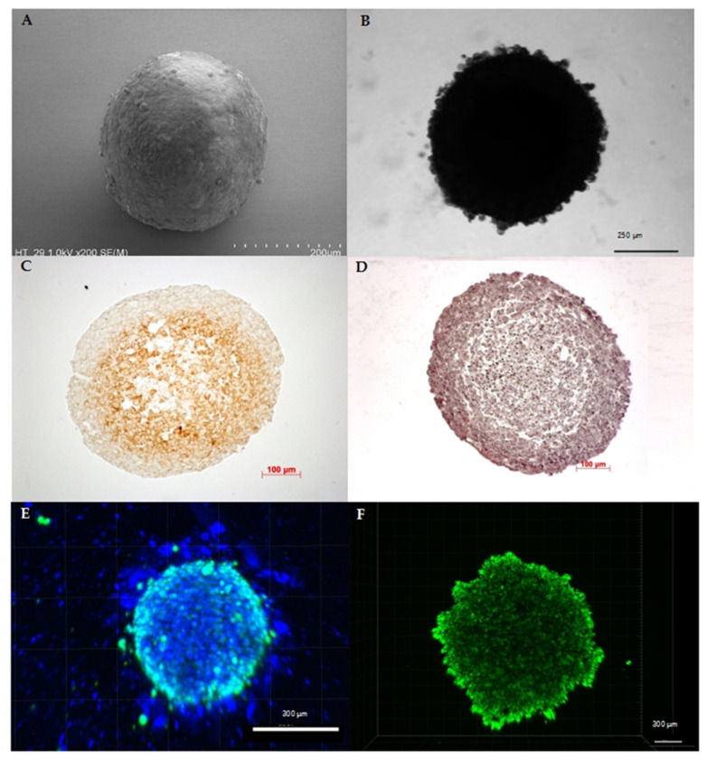
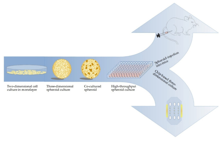
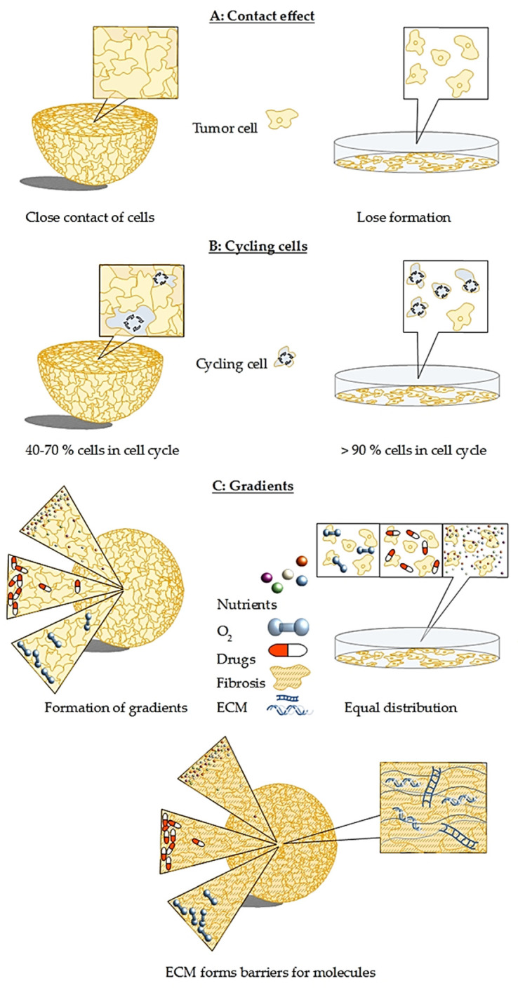
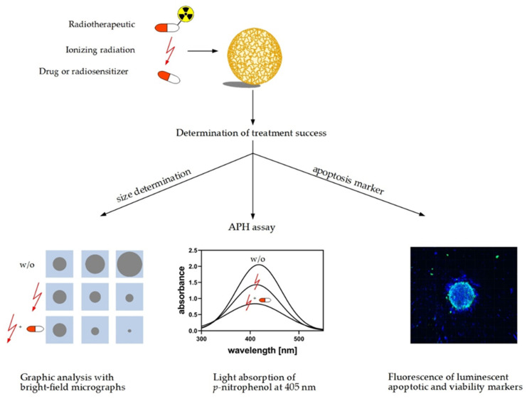
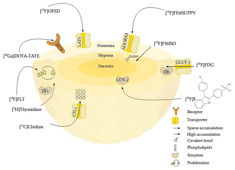
In preclinical cancer research, three-dimensional (3D) cell culture systems such as multicellular spheroids and organoids are becoming increasingly important. They provide valuable information before studies on animal models begin and, in some cases, are even suitable for reducing or replacing animal experiments. Furthermore, they recapitulate microtumors, metastases, and the tumor microenvironment much better than monolayer culture systems could. Three-dimensional models show higher structural complexity and diverse cell interactions while reflecting (patho)physiological phenomena such as oxygen and nutrient gradients in the course of their growth or development. These interactions and properties are of great importance for understanding the pathophysiological importance of stromal cells and the extracellular matrix for tumor progression, treatment response, or resistance mechanisms of solid tumors. Special emphasis is placed on co-cultivation with tumor-associated cells, which further increases the predictive value of 3D models, e.g., for drug development. The aim of this overview is to shed light on selected 3D models and their advantages and disadvantages, especially from the radiopharmacist's point of view with focus on the suitability of 3D models for the radiopharmacological characterization of novel radiotracers and radiotherapeutics. Special attention is paid to pancreatic ductal adenocarcinoma (PDAC) as a predestined target for the development of new radionuclide-based theranostics.

Keywords: 3D model; co-culture; organoids; pancreatic cancer; radiotherapeutics; radiotracer; spheroids; stromal cells; tumor microenvironment.


Conflict of interest statement

The authors declare no conflict of interest. The funders had no influence on the conception of the review, on the interpretation of literature data, on the conclusions drawn, or in the decision to publish this review.

Figures

Figure 1
Figure 1
Characteristics of spheroids originating from human cancer cell lines: (A) SEM image of a colorectal cancer cell line (HT29) spheroid depicting the spherical shape as the main characteristic of spheroids. (B) Bright-field microscopy image of a pancreatic cancer cell line (HPAF II) spheroid confirming stability of the 3D cell assembly. (C) Like tumors in vivo, spheroids show physiological zoning, consisting of a rim of viable cells and, depending on their size, of hypoxic or necrotic core zone. Exemplarily, the hypoxic core of a melanoma cell line (A2508) spheroid with a diameter of approximately 0.55 mm is shown by pimonidazole staining. (D) Hematoxylin and eosin staining of a melanoma cell line (A2508) spheroid showing the heterogeneous composition that can vary in cell and extracellular matrix arrangement depending on cell line and treatment. (E) Distribution of viable cells and, as a further characteristic feature, the possible active migration of cells from the 3D cell assembly: here, exemplarily shown by fluorescence staining with DAPI (4′,6-diamidino-2-phenylindole) (blue) and calcein (green) in a 10-day-old co-cultivated spheroid of pancreatic carcinoma cell line (SW1990) and pancreatic stellate cells (HPaSteC) with an initial seeded cell number of 16,000 cells (ratio 1:3). (F) Spheroids are characterized by the formation of gradients, which, among other things, modify the transport of nutrients, metabolites, and drugs similar to the situation in vivo, as shown here in the passive transport of the fluorescent marker calcein in a 10-day-old pancreatic cancer cell line (PanC-1) spheroid with an initial seeded cell number of 16,000 cells.
Figure 2
Figure 2
Increment of complexity in cell culture methods for PDAC research: cancer research is moving forward from monolayer cultures towards the use of spheroids. Including a second cell line, e.g., fibroblasts, into the model further increases the complexity. One-step further, spheroids are injected into mice for an advanced model in vivo with the possibility to mimic the progression of small metastases to tumors. On the other hand, using biotechnological state-of-the-art, for example HepaChip®, allows for measurement of cell reactions directly with electrodes.
Figure 3
Figure 3
Major differences between spheroid and monolayer culture that cause challenges for therapy development: (A) in contrast to monolayer culture, cells in a spheroid are tightly packed. This leads to the contact effect that induces radioresistance observed in 3D models as well as in patients. (B) The percentage of cycling cells in spheroids is size-dependent and varies between 40 to 70%, while in monolayer culture, more than 90% of cells are cycling. That makes the monolayer cells much more prone to medication and leads to misleading results, explaining the high number of failed clinical trials. (C) Oxygen, nutrient, and drug gradients are present in spheroids with concentration and penetration of all three decreasing towards the spheroid core, whereas in a monolayer culture, oxygen, nutrients, and drugs can reach each cell with equal efficiency. This leads again to false expectations on the true effectiveness of a drug candidate. In PDAC, the situation is even more severe, as fibrosis leads to a dense mesh-like network that hampers nutrients, drugs, as well as oxygen from entering the spheroid, leading to pronounced gradient formation. All three challenges are present and even more complex in co-culture spheroids.
Figure 4
Figure 4
Methods for measuring response of spheroids to anticancer treatment: reactions of spheroids to treatments, e.g., irradiation, radiopharmaceuticals, or nonradioactive compounds, can be determined using these methods. The first option is to measure spheroid size using bright-field microscopy. Treatment effects are evaluated referring to shrinkage in size, reduction of growth rate, and capacity for regrowth (w/o: without). The second option is the acid phosphatase assay (APH) assay utilizing the activity of acid phosphatase in viable cells. The hydrolysis of p-nitrophenyl-phosphate to p-nitrophenol is detected as increase in absorption at 405 nm. The third option is to estimate the fraction of viable cells in a spheroid referring to staining of nucleic acids and viability markers such as DAPI and calcein, respectively.
Figure 5
Figure 5
Schematic overview of molecular or pathophysiologic targets for well-established and novel radiotracers: some of them address membrane-bound cell receptors or transporters like somatostatin receptors (SSTR), adenosine receptors A3 (ADORA3), or L-amino-acid transporters (LAT1). Other tracers such as [11C]choline are taken up by transporters as the choline transporter-like protein (CTL1) and incorporated into the phospholipids of proliferating cells. The enzyme substrates [3H]thymidine and [18F]FLT target proliferating cells by incorporation into the DNA via for example the cytosolic thymidine kinase (TK-1). Enzyme inhibitors, portrayed here by [18F]3, a radiolabeled selective cyclooxygenase 2 inhibitor, in turn accumulate in areas of high enzyme concentration and activity. The radiolabeled glucose derivative [18F]FDG visualizes glucose turnover by glucose transport (GLUT-1) and hexokinase (HK) reaction and consequently the metabolic activity and viability of cells, which is high in the normoxic proliferating regions of the spheroid and lower in the hypoxic core. In contrast, [18F]FMISO binds to macromolecules in a hypoxic environment and thus accumulates in the hypoxic core (Table 4).

References

    1. Friedrich J., Ebner R., Kunz-Schughart L.A. Experimental anti-tumor therapy in 3-D: Spheroids—Old hat or new challenge? Int. J. Radiat. Biol. 2007;83:849–871. doi: 10.1080/09553000701727531. - DOI - PubMed
    1. Nath S., Devi G.R. Three-dimensional culture systems in cancer research: Focus on tumor spheroid model. Pharmacol. Ther. 2016;163:94–108. doi: 10.1016/j.pharmthera.2016.03.013. - DOI - PMC - PubMed
    1. Hirschhaeuser F., Menne H., Dittfeld C., West J., Mueller-Klieser W., Kunz-Schughart L.A. Multicellular tumor spheroids: An underestimated tool is catching up again. J. Biotechnol. 2010;148:3–15. doi: 10.1016/j.jbiotec.2010.01.012. - DOI - PubMed
    1. Large T.Y.S.L., Bijlsma M.F., Kazemier G., van Laarhoven H.W.M., Giovannetti E., Jimenez C.R. Key biological processes driving metastatic spread of pancreatic cancer as identified by multi-omics studies. Semin. Cancer Biol. 2017;44:153–169. doi: 10.1016/j.semcancer.2017.03.008. - DOI - PubMed
    1. Guo X.-Z., Cui Z.-M., Liu X. Current developments, problems and solutions in the non-surgical treatment of pancreatic cancer. World J. Gastrointest. Oncol. 2013;5:20. doi: 10.4251/wjgo.v5.i2.20. - DOI - PMC - PubMed

LinkOut - more resources