. 2020 Sep 29;20(1):317.
doi: 10.1186/s12876-020-01442-5.

Factors contributing to diagnostic delay of Caroli syndrome: a single-center, retrospective study

Affiliations

Factors contributing to diagnostic delay of Caroli syndrome: a single-center, retrospective study

Wen Shi et al. BMC Gastroenterol. .

Abstract

Background: Caroli syndrome (CS) is a rare congenital disorder without pathognomonic clinical symptoms or laboratory findings; therefore, the diagnosis is often delayed. The objective of this study was to investigate the diagnostic delay and associated risk factors in CS patients.

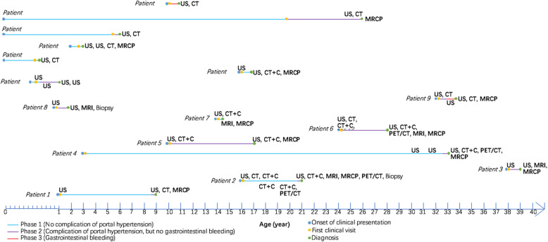
Methods: This was a retrospective analysis of 16 CS patients admitted to a single tertiary medical center on mainland China. The diagnostic timelines of CS patients were reviewed to demonstrate the initial findings of CS at diagnosis, the risk factors associated with diagnostic delay, and potential clues leading to early diagnosis.

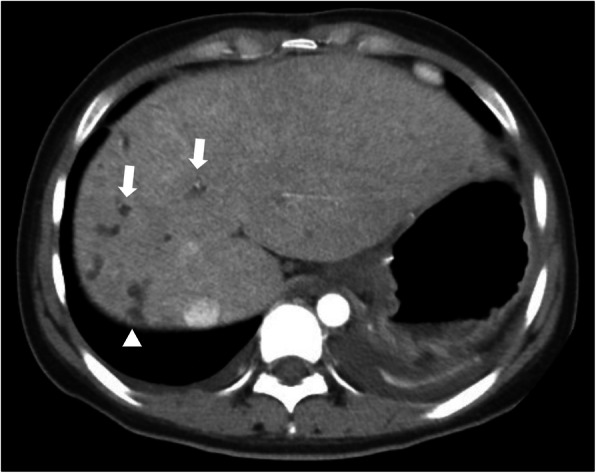
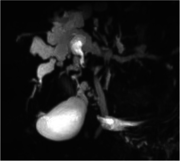
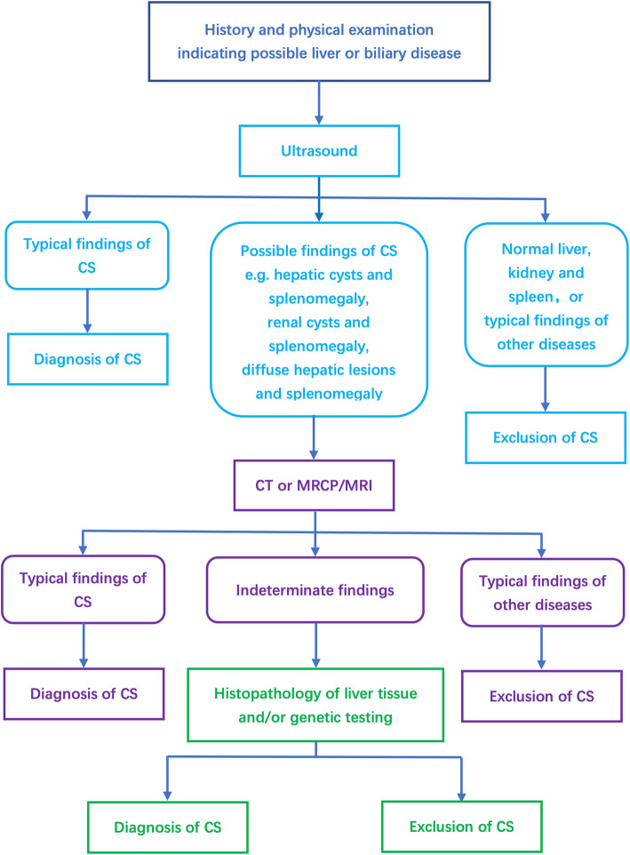
Results: The median diagnostic delay was 1.75 years (range: 1 month to 29 years, interquartile range: 6.2 years) in 16 enrolled CS patients. Sex, age, and initial symptoms were not associated with diagnostic delay. 87.5% of CS patients were diagnosed by imaging, and the accuracies of ultrasonography, computed tomography (CT), and magnetic resonance cholangiopancreatography were 25, 69.2, and 83.3%, respectively. The median diagnostic delays for patients with or without CT performed at the first hospital visited according to physician and radiologist suspicion of the diagnosis were 7.4 months and 6 years, respectively (p = 0.021). Hepatic cysts with splenomegaly were detected by ultrasound in over half of CS patients.

Conclusions: The majority of CS patients were not diagnosed until complications of portal hypertension had already developed. Recognition and early suspicion of the disease were important factors influencing diagnostic delay of CS. Hepatic cysts plus splenomegaly detected by US might raise the clinical suspicion to include CS in the differential diagnosis.

Keywords: Caroli syndrome; Diagnosis; Imaging.


Conflict of interest statement

The authors declare that they have no competing interests.

Figures

Fig. 1
Fig. 1
Diagnostic timelines of 16 CS patients. Important time points including onset of clinical presentation, first clinical visit, and diagnosis are depicted for each CS patient along the timeline. The course of the disease was divided into three phases: phase 1 (blue), defined as no proof of complication of portal hypertension (i.e., hypersplenism); phase 2 (purple), defined as discovered complication of portal hypertension without gastrointestinal variceal bleeding; and phase 3 (red), defined as at least one recorded variceal bleed. Imaging modalities and/or biopsy used before diagnosis are marked along the timelines. US: ultrasonography; CT: computed tomography; CT + C: computed tomography with contrast; MRI: magnetic resonance imaging; MRCP: magnetic resonance cholangiopancreatography; PET/CT: positron emission tomography/computed tomography
Fig. 2
Fig. 2
CT scan of a CS patient revealing multiple dilatations of intrahepatic bile ducts (arrow) and tiny dots of strong contrast within, which is the central dot sign (arrowhead)
Fig. 3
Fig. 3
MRCP in a CS patient showing dilatation of intrahepatic bile ducts
Fig. 4
Fig. 4
A proposed clinical practice workflow when there is a suspicion of CS

References

    1. Yonem O, Bayraktar Y. Clinical characteristics of Caroli's syndrome. World J Gastroenterol. 2007;13(13):1934–1937. doi: 10.3748/wjg.v13.i13.1934. - DOI - PMC - PubMed
    1. Bakoyiannis A, Delis S, Triantopoulou C, Dervenis C. Rare cystic liver lesions: a diagnostic and managing challenge. World J Gastroenterol. 2013;19(43):7603–7619. doi: 10.3748/wjg.v19.i43.7603. - DOI - PMC - PubMed
    1. Shorbagi A, Bayraktar Y. Experience of a single center with congenital hepatic fibrosis: a review of the literature. World J Gastroenterol. 2010;16(6):683–690. doi: 10.3748/wjg.v16.i6.683. - DOI - PMC - PubMed
    1. Wang ZX, Li YG, Wang RL, Li YW, Li ZY, Wang LF, Yang HY, Zhu Y, Wang Y, Bai YF, et al. Clinical classification of Caroli’s disease: an analysis of 30 patients. HPB (Oxford) 2015;17(3):278–283. doi: 10.1111/hpb.12330. - DOI - PMC - PubMed
    1. Zhang DY, Ji ZF, Shen XZ, Liu HY, Pan BJ, Dong L. Caroli’s disease: a report of 14 patients and review of the literature. J Dig Dis. 2012;13(9):491–495. doi: 10.1111/j.1751-2980.2012.00619.x. - DOI - PubMed

LinkOut - more resources